Abstract
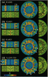
Kinesin and ncd motor proteins are homologous in sequence yet move in opposite directions along microtubules. We have previously shown that monomeric kinesin and ncd bind in the same orientation on equivalent sites relative to the ends of tubulin sheets of known polarity. We now report cryoelectron microscope images of 16-protofilament microtubules decorated with both single- and double-headed kinesin and double-headed ncd. Three-dimensional density maps and difference maps show that, in adenosine 5'-[beta,gamma-imido]triphosphate, both dimeric motors bind tightly to microtubules via one head, leaving the other free, though apparently in a fixed position. The attached heads of dimers bind to tubulin in the same way as single kinesin heads. The second heads are connected to the tops of the first but, whereas the second kinesin head is closely associated with the first, pairs of ncd heads are splayed apart. There is also a distinct difference in orientation: the second kinesin head is tilted toward the microtubule plus end, while the second head of ncd points toward the minus end.
Full text
PDF

Images in this article
Selected References
These references are in PubMed. This may not be the complete list of references from this article.
- Berliner E., Young E. C., Anderson K., Mahtani H. K., Gelles J. Failure of a single-headed kinesin to track parallel to microtubule protofilaments. Nature. 1995 Feb 23;373(6516):718–721. doi: 10.1038/373718a0. [DOI] [PubMed] [Google Scholar]
- Chandra R., Salmon E. D., Erickson H. P., Lockhart A., Endow S. A. Structural and functional domains of the Drosophila ncd microtubule motor protein. J Biol Chem. 1993 Apr 25;268(12):9005–9013. [PubMed] [Google Scholar]
- Crevel I. M., Lockhart A., Cross R. A. Weak and strong states of kinesin and ncd. J Mol Biol. 1996 Mar 22;257(1):66–76. doi: 10.1006/jmbi.1996.0147. [DOI] [PubMed] [Google Scholar]
- Goldstein L. S. With apologies to scheherazade: tails of 1001 kinesin motors. Annu Rev Genet. 1993;27:319–351. doi: 10.1146/annurev.ge.27.120193.001535. [DOI] [PubMed] [Google Scholar]
- Hackney D. D. Evidence for alternating head catalysis by kinesin during microtubule-stimulated ATP hydrolysis. Proc Natl Acad Sci U S A. 1994 Jul 19;91(15):6865–6869. doi: 10.1073/pnas.91.15.6865. [DOI] [PMC free article] [PubMed] [Google Scholar]
- Hackney D. D. Highly processive microtubule-stimulated ATP hydrolysis by dimeric kinesin head domains. Nature. 1995 Oct 5;377(6548):448–450. doi: 10.1038/377448a0. [DOI] [PubMed] [Google Scholar]
- Harrison B. C., Marchese-Ragona S. P., Gilbert S. P., Cheng N., Steven A. C., Johnson K. A. Decoration of the microtubule surface by one kinesin head per tubulin heterodimer. Nature. 1993 Mar 4;362(6415):73–75. doi: 10.1038/362073a0. [DOI] [PubMed] [Google Scholar]
- Hirokawa N., Pfister K. K., Yorifuji H., Wagner M. C., Brady S. T., Bloom G. S. Submolecular domains of bovine brain kinesin identified by electron microscopy and monoclonal antibody decoration. Cell. 1989 Mar 10;56(5):867–878. doi: 10.1016/0092-8674(89)90691-0. [DOI] [PubMed] [Google Scholar]
- Hirose K., Fan J., Amos L. A. Re-examination of the polarity of microtubules and sheets decorated with kinesin motor domain. J Mol Biol. 1995 Aug 18;251(3):329–333. doi: 10.1006/jmbi.1995.0437. [DOI] [PubMed] [Google Scholar]
- Hirose K., Lockhart A., Cross R. A., Amos L. A. Nucleotide-dependent angular change in kinesin motor domain bound to tubulin. Nature. 1995 Jul 20;376(6537):277–279. doi: 10.1038/376277a0. [DOI] [PubMed] [Google Scholar]
- Hoenger A., Sablin E. P., Vale R. D., Fletterick R. J., Milligan R. A. Three-dimensional structure of a tubulin-motor-protein complex. Nature. 1995 Jul 20;376(6537):271–274. doi: 10.1038/376271a0. [DOI] [PubMed] [Google Scholar]
- Howard J., Hudspeth A. J., Vale R. D. Movement of microtubules by single kinesin molecules. Nature. 1989 Nov 9;342(6246):154–158. doi: 10.1038/342154a0. [DOI] [PubMed] [Google Scholar]
- Howard J. The mechanics of force generation by kinesin. Biophys J. 1995 Apr;68(4 Suppl):245S–255S. [PMC free article] [PubMed] [Google Scholar]
- Huang T. G., Hackney D. D. Drosophila kinesin minimal motor domain expressed in Escherichia coli. Purification and kinetic characterization. J Biol Chem. 1994 Jun 10;269(23):16493–16501. [PubMed] [Google Scholar]
- Huang T. G., Suhan J., Hackney D. D. Drosophila kinesin motor domain extending to amino acid position 392 is dimeric when expressed in Escherichia coli. J Biol Chem. 1994 Jun 10;269(23):16502–16507. [PubMed] [Google Scholar]
- Kikkawa M., Ishikawa T., Wakabayashi T., Hirokawa N. Three-dimensional structure of the kinesin head-microtubule complex. Nature. 1995 Jul 20;376(6537):274–277. doi: 10.1038/376274a0. [DOI] [PubMed] [Google Scholar]
- Kull F. J., Sablin E. P., Lau R., Fletterick R. J., Vale R. D. Crystal structure of the kinesin motor domain reveals a structural similarity to myosin. Nature. 1996 Apr 11;380(6574):550–555. doi: 10.1038/380550a0. [DOI] [PMC free article] [PubMed] [Google Scholar]
- Lockhart A., Crevel I. M., Cross R. A. Kinesin and ncd bind through a single head to microtubules and compete for a shared MT binding site. J Mol Biol. 1995 Jun 16;249(4):763–771. doi: 10.1006/jmbi.1995.0335. [DOI] [PubMed] [Google Scholar]
- Lockhart A., Cross R. A. Kinetics and motility of the Eg5 microtubule motor. Biochemistry. 1996 Feb 20;35(7):2365–2373. doi: 10.1021/bi952318n. [DOI] [PubMed] [Google Scholar]
- Ma Y. Z., Taylor E. W. Mechanism of microtubule kinesin ATPase. Biochemistry. 1995 Oct 10;34(40):13242–13251. doi: 10.1021/bi00040a040. [DOI] [PubMed] [Google Scholar]
- Milligan R. A., Flicker P. F. Structural relationships of actin, myosin, and tropomyosin revealed by cryo-electron microscopy. J Cell Biol. 1987 Jul;105(1):29–39. doi: 10.1083/jcb.105.1.29. [DOI] [PMC free article] [PubMed] [Google Scholar]
- Peskin C. S., Oster G. Coordinated hydrolysis explains the mechanical behavior of kinesin. Biophys J. 1995 Apr;68(4 Suppl):202S–211S. [PMC free article] [PubMed] [Google Scholar]
- Ray S., Meyhöfer E., Milligan R. A., Howard J. Kinesin follows the microtubule's protofilament axis. J Cell Biol. 1993 Jun;121(5):1083–1093. doi: 10.1083/jcb.121.5.1083. [DOI] [PMC free article] [PubMed] [Google Scholar]
- Romberg L., Vale R. D. Chemomechanical cycle of kinesin differs from that of myosin. Nature. 1993 Jan 14;361(6408):168–170. doi: 10.1038/361168a0. [DOI] [PubMed] [Google Scholar]
- Sablin E. P., Kull F. J., Cooke R., Vale R. D., Fletterick R. J. Crystal structure of the motor domain of the kinesin-related motor ncd. Nature. 1996 Apr 11;380(6574):555–559. doi: 10.1038/380555a0. [DOI] [PubMed] [Google Scholar]
- Scholey J. M., Heuser J., Yang J. T., Goldstein L. S. Identification of globular mechanochemical heads of kinesin. Nature. 1989 Mar 23;338(6213):355–357. doi: 10.1038/338355a0. [DOI] [PubMed] [Google Scholar]
- Shimizu T., Sablin E., Vale R. D., Fletterick R., Pechatnikova E., Taylor E. W. Expression, purification, ATPase properties, and microtubule-binding properties of the ncd motor domain. Biochemistry. 1995 Oct 10;34(40):13259–13266. doi: 10.1021/bi00040a042. [DOI] [PubMed] [Google Scholar]
- Song Y. H., Mandelkow E. Recombinant kinesin motor domain binds to beta-tubulin and decorates microtubules with a B surface lattice. Proc Natl Acad Sci U S A. 1993 Mar 1;90(5):1671–1675. doi: 10.1073/pnas.90.5.1671. [DOI] [PMC free article] [PubMed] [Google Scholar]
- Stewart R. J., Thaler J. P., Goldstein L. S. Direction of microtubule movement is an intrinsic property of the motor domains of kinesin heavy chain and Drosophila ncd protein. Proc Natl Acad Sci U S A. 1993 Jun 1;90(11):5209–5213. doi: 10.1073/pnas.90.11.5209. [DOI] [PMC free article] [PubMed] [Google Scholar]
- Trachtenberg S., DeRosier D. J. Three-dimensional structure of the frozen-hydrated flagellar filament. The left-handed filament of Salmonella typhimurium. J Mol Biol. 1987 Jun 5;195(3):581–601. doi: 10.1016/0022-2836(87)90184-7. [DOI] [PubMed] [Google Scholar]
- Wade R. H., Chrétien D., Job D. Characterization of microtubule protofilament numbers. How does the surface lattice accommodate? J Mol Biol. 1990 Apr 20;212(4):775–786. doi: 10.1016/0022-2836(90)90236-F. [DOI] [PubMed] [Google Scholar]
- Walker R. A., Salmon E. D., Endow S. A. The Drosophila claret segregation protein is a minus-end directed motor molecule. Nature. 1990 Oct 25;347(6295):780–782. doi: 10.1038/347780a0. [DOI] [PubMed] [Google Scholar]
- Walker R. A., Sheetz M. P. Cytoplasmic microtubule-associated motors. Annu Rev Biochem. 1993;62:429–451. doi: 10.1146/annurev.bi.62.070193.002241. [DOI] [PubMed] [Google Scholar]
- Yang J. T., Laymon R. A., Goldstein L. S. A three-domain structure of kinesin heavy chain revealed by DNA sequence and microtubule binding analyses. Cell. 1989 Mar 10;56(5):879–889. doi: 10.1016/0092-8674(89)90692-2. [DOI] [PubMed] [Google Scholar]
- Yang J. T., Saxton W. M., Stewart R. J., Raff E. C., Goldstein L. S. Evidence that the head of kinesin is sufficient for force generation and motility in vitro. Science. 1990 Jul 6;249(4964):42–47. doi: 10.1126/science.2142332. [DOI] [PubMed] [Google Scholar]
- Young E. C., Berliner E., Mahtani H. K., Perez-Ramirez B., Gelles J. Subunit interactions in dimeric kinesin heavy chain derivatives that lack the kinesin rod. J Biol Chem. 1995 Feb 24;270(8):3926–3931. doi: 10.1074/jbc.270.8.3926. [DOI] [PubMed] [Google Scholar]


