Abstract
Recent work has suggested the value of electroencephalographic (EEG) measures in the study of infants’ processing of human action. Studies in this area have investigated desynchronization of the sensorimotor mu rhythm during action execution and action observation in infancy. Untested but critical to theory is whether the mu rhythm shows a differential response to actions which share similar goals but have different motor requirements or sensory outcomes. By varying the invisible property of object weight, we controlled for the abstract goal (reach, grasp, and lift the object), while allowing other aspects of the action to vary. The mu response during 14-month-old infants’ own executed actions showed a differential hemispheric response between acting on heavier and lighter objects. EEG responses also showed sensitivity to “expected object weight” when infants simply observed an experimenter reach for objects that the infants’ prior experience indicated were heavier versus lighter. Crucially, this neural reactivity was predictive – during the observation of the other reaching toward the object, before lifting occurred. This suggests that infants’ own self-experience with a particular object’s weight influences their processing of others’ actions on the object, with implications for developmental social-cognitive neuroscience.
Keywords: EEG, mu rhythm, action, infant, goals, neural mirroring
Research from multiple perspectives supports the notion that an observed action activates similar processes in the human observer’s brain as would be activated if the action was performed or planned by the observer (e.g., Hari & Kujala, 2009). Developmental neuroscience studies in this area have focused on the electroencephalographic (EEG) mu rhythm, a sensorimotor rhythm which occurs in the alpha frequency range over central electrode sites (for a review see Marshall & Meltzoff, 2011; Saby & Marshall, 2012). Findings that the mu rhythm is desynchronized (reduced in amplitude) during both the execution and observation of actions have provided initial evidence of neural linkages between action production and action perception in infants (Marshall, Young, & Meltzoff, 2011; Southgate, Johnson, Osborne, & Csibra, 2009; Warreyn et al., in press).
A variety of recent studies have begun to uncover properties of the infant mu rhythm, particularly its reactivity during action observation (Marshall & Meltzoff, 2011). For instance, it has been suggested that the infant mu rhythm is more responsive during the observation of movements with which infants have more prior experience (van Elk, van Schie, Hunnius, Vesper, & Bekkering, 2008) and that it shows greater desynchronization to goal-directed actions compared with “empty” or mimed actions without a clear goal (Nyström, Ljunghammar, Rosander, & von Hofsten, 2011; Southgate, Johnson, El Karoui, & Csibra, 2010; although see Warreyn et al., in press). Other work has examined the intermodal properties of the infant mu rhythm (Paulus, Hunnius, van Elk, & Bekkering, 2012) as well as its responsivity to being imitated by a social other (Reid, Striano, & Iacoboni, 2011; Saby, Marshall, & Meltzoff, 2012).
However, as outlined by Marshall and Meltzoff (2011) there are a number of further questions that need to be addressed in order to build a more comprehensive picture of the utility of the infant mu rhythm in the study of early action processing. For instance, is mu rhythm desynchronization during the observation of goal-directed actions sensitive to variation in the specific requirements or anticipated consequences of those actions, or does it show a relatively nonspecific response? Developmentally-oriented exploration of this question can inform ongoing debates about the nature and evolutionary/psychological function of neural systems involved in action processing and understanding (Csibra, 2007; Meltzoff, Kuhl, Movellan, & Sejnowski, 2009; Rizzolatti & Sinigaglia, 2010).
In the current work we chose to study this question in 14-month-olds, since this age provides an ideal confluence of infant attention and social motivation for carrying out interactive EEG protocols with live actions that require the continuation of back-and-forth social action sequences (see also Marshall et al., 2011; Saby et al., 2012). We recorded EEG data while allowing infants to reach, grasp, and lift a pair of objects which were the same size and shape, but differed in weight. Through this familiarization, infants learned that the relative weight of the two objects (an invisible property) could be predicted by the visible property of color (Mash, 2007). Our initial question was how the mu rhythm response varied during the execution of infants’ own actions on lighter versus heavier objects. Our working hypothesis was that the mu rhythm at central sites would be sensitive to the force required, with greater desynchronization during actions on heavier than lighter objects. In adults, the magnitude of mu rhythm desynchronization during action execution is proportional to the force exerted (Mima, Simpkins, Oluwatimilehin, & Hallett, 1999).
We also examined EEG responses while infants simply observed an adult acting on objects identical to the ones that the infants had received self-experience with. While infant and adult actions shared the same overall goal, the experimental variation of actual object weight allowed us to examine how this factor influenced mu rhythm activity at central sites for observed actions. It also allowed us to ask whether infants’ first-person experiences with the invisible properties of objects affected their EEG responses to observing someone else act on these same objects. This question is rooted in the “Like-Me” framework of social-cognitive development which proposes that infants’ processing and interpretation of the actions of others is deeply colored by their own prior self-experiences (e.g., Meltzoff, 2007; Meltzoff & Brooks, 2008; Meltzoff, Williamson, & Marshall, in press).
Our initial expectation was that any differences in mu rhythm desynchronization at central sites that were apparent during infants’ acting on heavier versus lighter objects would also be reflected in mu responses to the observation of others’ actions on those objects. While we are not aware of prior EEG work on this question in either adults or infants, studies using transcranial magnetic stimulation (TMS) with adults have shown increased facilitation of motor cortex activity during the observation of grasping and lifting actions on objects expected to be heavier rather than lighter (Alaerts et al., 2010; Senot et al., 2011). Especially relevant to the current study is the finding that this motor facilitation effect in adults was present during the observation of reaching towards objects that were expected to be heavy or light (Alaerts, de Beukelaar, Swinnen, & Wenderoth, 2012). We were interested in whether a similar effect for the mu rhythm was apparent while infants watched an experimenter reach towards the objects, prior to any contact of the adult’s hand with the object. Not only might the mu rhythm respond to the observation of actual actions executed by others, but also to the “expected weight” that the other person would encounter – in an anticipated or prospective action plan not yet fully carried out.
Our examination of the EEG response during the observation of reaching also relates to suggestions that the infant mu rhythm has a predictive or anticipatory component (e.g., Southgate et al., 2010; Stapel, Hunnius, van Elk, & Bekkering, 2010). However, our specific question of whether the mu rhythm would be sensitive to different expectancies about unfolding object-directed actions (rather than a general response simply to the presence versus absence of an object) has not been previously examined in any age group, including adults. Given the growing interest in the utility of neuroscience methods to help investigate how children come to interpret the actions, goals and intentions of other people (Meltzoff et al., in press), addressing this question from a developmental perspective – and more specifically, with preverbal infants – is particularly important.
Method
Participants
Thirty-five 14-month-old infants participated in the study (M = 61.7 weeks, SD = 1.4, 20 male). Families were recruited from a diverse urban environment using commercially available mailing lists. Families were not invited to participate if their infant was born preterm, if both parents were left-handed, if the infant had experienced chronic developmental problems, or if the infant was on long-term medication. A number of infants did not have useable EEG data because of technical problems (n = 2) or because they became excessively fussy during cap preparation (n = 3). Further exclusions (e.g., infants with insufficient numbers of artifact-free trials) are detailed below.
Stimuli
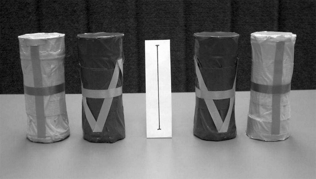
Two pairs of same-sized cylindrical objects were created which varied only by color (blue/yellow) and weight (40g/260g). In order to facilitate infants’ attention to actions on the objects, all four objects were constructed such that they made a similar rattling sound when shaken, and each one was decorated with a striped pattern that was fashioned from ribbon material (Figure 1).
Figure 1.
Photograph of the two pairs of objects created for the study. The length of the reference line in the center is 10 cm.
The presentation of the stimuli to the infants involved two experimenters. The first experimenter (E1) sat at a table opposite to the infant and was responsible for presenting the objects to the infants and for demonstrating the actions of grasping and lifting of the objects during the action observation trials. The second experimenter (E2) sat to the side of E1 but was hidden from the infant’s view by a partition. E2 coordinated the experimental sequence by placing and removing the objects from the table according to the randomized experimental protocol specified below.
Procedure
While sitting on the caregiver’s lap at a table, infants were fitted with the EEG cap (see below), and were then given experience with one pair of the objects. Infants were randomly assigned to receive experience with only one of the two object pairs: Pair 1 consisted of the 40g blue and 260g yellow object; Pair 2 consisted of the 40g yellow and 260g blue object. The experiment began with a series of six familiarization trials (three of each weight) in which infants were presented with the heavy and light objects alternately for 10s/trial (as in Mash, 2007). To begin each familiarization trial, one object was placed on the table by E1 (who had been given the object by E2 according to the experimental protocol). Infants were allowed to reach for, grasp, and lift the object, which occurred on more than 95% of trials. Infants then typically began to shake the object in order to elicit the rattling sound. After 10s had elapsed, the object was retrieved from the infant by E1, and the other object from the pair was presented.
During these familiarization trials, the infant’s initial grasp of the object was typically followed by a lift of the object within 1–2 video frames following contact (approximately 70% of trials). For the remaining trials, the lift occurred slightly later, usually within 1 s after contact with the object. While the infant was acting on one object, the other object was hidden from the infant’s view. After six familiarization trials (three with each object) the familiarization period was terminated and the first series of observation trials was commenced.
In each observation trial, the infant first viewed a card (55 × 30cm) onto which attractive visual patterns had been attached. This card was held upright by E2, who slid it into view of the infant. The infant’s viewing of the card constituted the baseline condition for the EEG analyses (3s duration). During the baseline presentation, one object was selected by E2 (out of the view of E1) and E2 placed it behind the (vertical) card on the table so that the infant could not yet see it. E2 followed a random order for object selection, and the object could be any one of the four objects from the two pairs of stimuli (Pair 1 and Pair 2). The card was then removed by E2 to reveal the object. After ensuring that the infant was attending to her, E1 reached for, lifted, and shook the object to elicit the rattling sound. The experimenter’s lift of the object always occurred within 1–2 video frames of the initial contact between her hand and the object. After shaking the object for 10s, E1 placed the object in its starting position on the table and moved her hand to her lap. At that point, the baseline card was held up again by E2 who then removed the object and placed the next one on the table behind the vertical card.
The use of all four objects in the observation condition (in contrast to the execution trials, which only utilized one fixed pair) as well as the selection and placement of the objects by E2 meant that the experimenter (E1) who was demonstrating the reaches to the infant did not know whether the object that she was reaching for was heavy or light, based on vision alone. In this way we attempted to equate for the kinematics of the approach reaches for each object by E1, allowing us to examine the effect of infants’ self-experience with the objects on the EEG response during observation of this approach reach, before actual lifting occurred.
After four observation trials, a pair of execution trials occurred in which infants were presented with each object from the original familiarization pair (one object per trial). On each execution trial, E1 reached behind a partition and was given one of the objects (by E2). E1 then presented this object to the infant, who was given 30s/trial to act on the object. As in the familiarization trials, infants readily reached for, grasped, and lifted the objects.
The remainder of the experiment comprised alternations of four observation trials and two execution trials, which continued as long as the infant remained actively interested. Each session was videotaped, with a vertical interval time code (VITC) being placed on the video signal during recording. Calibration procedures had ensured that the VITC signal was synchronized with EEG collection, such that the video was aligned with the EEG data to the precision of one NTSC video frame (33ms). Videos were coded offline using the Video Coding System from James Long Company (Caroga Lake, NY). The onset and offset of the baseline epochs were marked as well as the frames in which the experimenter or infant grasped (made contact with) the object. Other frames that were marked included the onset and offset of the reaches by the infant and experimenter towards the objects, as well as the frames in which the object was first lifted. Across the entire sample, this coding resulted in a mean of 14.23 (SD = 3.9) execution epochs and 14.1 (SD = 4.5) observation epochs. The videos were also coded for infant motor movement: Baseline and observation epochs containing infant arm and hand movements resembling reaching, grasping, or shaking motions were not included in subsequent EEG analyses. Because our main analyses concerned the mu rhythm at central electrode sites overlying the sensorimotor hand areas, we focused on the exclusion of epochs containing infant arm and hand movements, as described in our prior work (Marshall et al. 2011; Saby et al., 2012).
EEG Collection and Processing
Collection and processing of the EEG signal mirrored the methods used in Marshall et al. (2011). EEG was recorded using a lycra stretch cap (Electro-Cap International, Inc.) from the following sites: Fp1, Fp2, F3, F4, Fz, F7, F8, C3, C4, T7, T8, P3, P4, Pz, P7, P8, O1, O2, and the left and right mastoids. Electro-Gel conducting gel was utilized and scalp electrode impedances were accepted if they were below 25 kilohms. The signal from each site was amplified using optically isolated, high input impedance (> 1 GΩ) custom bioamplifiers (SA Instrumentation, San Diego) and was digitized using a 16-bit A/D converter (+/− 5 V input range). Bioamplifier gain was 4000 and the hardware filter (12 db/octave rolloff) settings were. 1 Hz (high-pass) and 100 Hz (low-pass). The signals were collected referenced to the vertex (Cz) with an AFz ground.
Offline processing and analysis of the EEG data was carried out using software from James Long Company as well as MATLAB and EEGLAB (Delorme & Makeig, 2004). The digitized signals were first re-referenced offline to an average mastoids reference. As in Marshall et al. (2011), a procedure involving independent component analysis (ICA) was then used to clear the EEG data of ocular and muscle artifact. The ICA procedure was an automation of the method described by Jung et al. (2000). After this had been carried out, any epochs in which the EEG signal for any channel exceeded +/− 250 µV were excluded from further analysis.
Event-related changes in band power between the baseline and the execution or observation epochs were computed using established methods for computing event-related desynchronization (ERD; Pfurtscheller, 2003). Based on previous work on the frequency of the mu rhythm in infants of this age (Marshall, Bar-Haim, & Fox, 2002), the band used in the analyses was 6–9 Hz. For the computation of ERD scores, the following sequence from Marshall et al. (2011) and Saby et al. (2012) was used: (a) Bandpass filtering of the EEG signals between 6 and 9 Hz using a two-way least squares finite impulse response filter; (b) Squaring of the filtered signals to convert to a power metric; (c) Computation of event-related averages for each condition within each participant; (d) Computation of the mean power of the event-related signal in 125ms epochs, in line with Pfurtscheller (2003). Desynchronization values were computed for each 125ms epoch as ([A–R]/R)*100, where A is band power during action observation or execution, and R is band power during the baseline condition (Pfurtscheller & Lopes da Silva, 1999). Negative ERD values reflect desynchronization (i.e., a decrease in band power relative to the baseline), while positive values reflect synchronization (i.e., an increase in band power relative to the baseline).
The reported results focus on analyses from the left and right central electrode sites (C3 and C4) overlying the sensorimotor hand areas. Supplementary analyses examined results from other scalp regions in order to clarify regional specificity of effects. Mean ERD was computed separately for the 500ms epochs prior to and immediately following the first frame of contact between the object and the hand of the infant or the experimenter. Inspection of the timing of events derived from the video coding showed that the 500ms prior to the onset of the grasp was encompassed by the experimenter’s or infant’s reach toward the object. For example, the infants’ reaches began on average 878ms prior to the first video frame of the grasp, while the experimenter’s reach began an average of 568ms prior to grasp onset.
Exclusionary Criteria for EEG Analyses
A number of infants were excluded from further analyses based on criteria used in Marshall et al. (2011). Specifically, nine infants had fewer than three artifact-free trials in any of the execution or observation conditions and five of the remaining 21 participants had extreme ERD values (more than 1.5 times the interquartile range from the median) in one or more conditions. For the remaining 16 infants used in the final analyses there were an average of 5.6 (SD = 2.2, range 3–10) artifact-free trials per participant for the action execution conditions and an average of 6.2 trials (SD = 1.8, range 3–9) for the action observation conditions. These numbers of trials are lower than in our previous study of the infant mu rhythm of imitative actions (Marshall et al., 2011), mainly because that study only included one execution condition and one observation condition. The numbers of trials in the current study are more comparable to studies of infant mu which have included more conditions (e.g., Saby et al., 2012). Also, the duration of each trial in the current protocol was much longer than in our prior work, which resulted in a lower overall number of trials.
As in Mash (2007), infants’ reaches were primarily bimanual with one hand (typically the right hand) being used to grasp the object and the other hand being used to support or stabilize the object during grasping and lifting. The EEG patterns reported below for the final sample of 16 infants were not significantly altered when a further six infants were excluded who used either a relatively equal combination of left or right hands (n = 3) to initially grasp the object or who predominantly used their left hand (n = 3) as the primary grasping hand.
Results
The results are reported in two sub-sections. The first reports results for the action execution trials, in which the infants acted on the objects, and the second reports from the action observation trials in which they simply observed the adult’s actions.
Action Execution
Repeated measures analyses of variance (ANOVAs) were carried out using the following factors: Weight (light; 40g vs. heavy; 260g), and Hemisphere (left vs. right), with separate analyses being conducted for the reaching (hand transport) epoch and the grasping/lifting epoch. Probability values have been adjusted using the Greenhouse-Geisser correction factor.
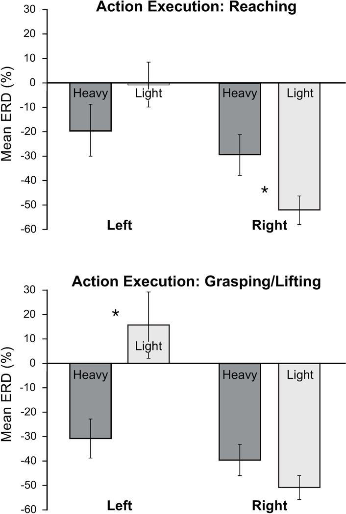
For the reaching epoch there was a significant main effect of Hemisphere, F(1,15) = 11.30, p < .01, ηp2 = .43, with desynchronization being greater at the right central site (C4) compared with the left central site (C3). There was a significant interaction between Weight and Hemisphere, F(1,15) = 7.81, p < .05, η2 = .34. Follow-up comparisons of the reaching epoch (see Figure 2 for means) showed that at the right central electrode, the lighter object was associated with a significantly larger desynchronization than the heavier object, t(15) = 2.30, p < .05, d = 0.78. There was no significant difference between the lighter and heavier objects at the left central electrode, t(15) = 1.42, p = .18, d = .47.
Figure 2.
Action execution: Mean event-related desynchronization (% ERD relative to baseline) in the 6–9 Hz band at the left and right central electrodes during infants directly acting on the light (40g) versus the heavy (260g) object. The upper panel shows the means from the reaching epoch prior to contact with the object and the lower panel shows the means from the grasping/lifting epoch. Error bars indicate ±1 S.E and asterisks denote significant comparisons (p < .05).
For the grasping/lifting epoch there was a significant main effect of Weight, F(1,15) = 4.64, p < .05, ηp2 = .24, with the heavier object eliciting greater desynchronization compared with the lighter object. The main effect for Hemisphere was also significant, F(1,15) = 26.28, p < .001, ηp2 = .64, with desynchronization being greater at the right central site. There was also a significant interaction between Weight and Hemisphere, F(1,15) = 9.09, p < .01,ηp2 = .38. At the left central electrode, the heavier object was associated with a significantly larger desynchronization than the lighter object, t(15) = 2.83, p < .05, d = 1.04. There was no significant difference between the lighter and heavier objects at the right central electrode, t(15) = 1.65, p = .12, d = .50.
In order to examine regional specificity of effects, similar ANOVAs were also conducted for mid-frontal (F3/F4), mid-parietal (P3/P4), and occipital (O1/O2) regions. For the reaching epoch, there were near-significant main effects of Hemisphere at mid-frontal sites, F(1,15) = 4.07, p=.06,ηp2 = .21 and mid-parietal sites, F(1,15) = 4.39, p=.05, ηp2=.23. For the grasping/lifting epoch, there was a significant main effect of Hemisphere at mid-frontal sites, F(1,15) = 6.49, p < .05, ηp2 = .30 and at occipital sites, F(1,15) = 4.70, p < .05, ηp2 = .24, with a marginal effect at mid-parietal sites, F(1,15) = 3.97, p = .07, ηp2 = .21. These results all reflected greater desynchronization over right-sided electrodes. No significant main effects or interactions involving the Weight factor were found for these other scalp regions.
Action Observation
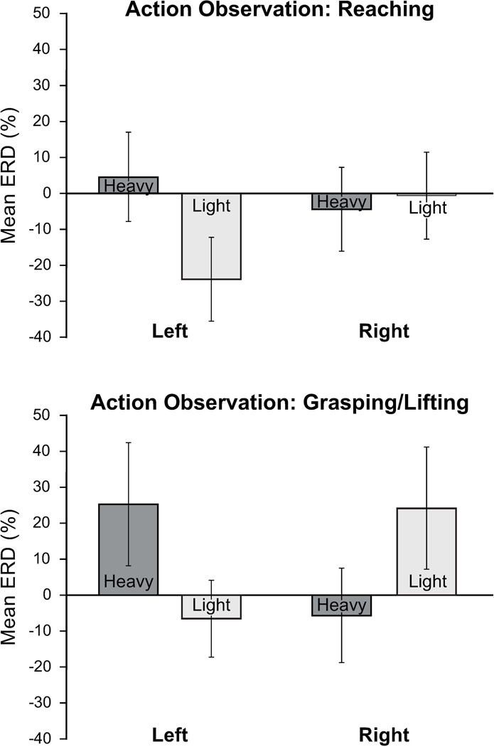
The basic ANOVA structure for action observation was identical to that for action execution, with the Weight factor now meaning experienced/expected weight, based on the color-weight linkage that the infant was exposed to. During the observation of the experimenter’s reach there were no significant main effects of Weight or Hemisphere, but there was a significant interaction between Weight and Hemisphere, F(1,15) = 5.12, p < .05, ηp2 = .26. Follow-up analyses within each hemisphere did not reveal significant differences between conditions either at the left or right central electrode when examined in isolation, t(15) = 1.54, p = .14, d = .59 and t(15) = .25, p = .80, d = .08 respectively. As shown in Figure 3, the significant interaction effect is most likely due to a combination of non-significant differences, particularly the different patterning of the means between the left and right electrodes within each of the two weight conditions.
Figure 3.
Action observation: Mean event-related desynchronization (% ERD relative to baseline) in the 6–9 Hz band at the left and right central electrodes during infants’ observation of other people acting on objects that infants had learned during familiarization were light (40g) versus heavy (260g). The upper panel shows the means from the reaching epoch prior to contact with the object and the lower panel shows the means from the grasping/lifting epoch. Error bars indicate ±1 S.E.
For the observation of the experimenter’s grasping/lifting there were no significant main effects of Weight or Hemisphere but there was again a significant interaction between Weight and Hemisphere, F(1,15) = 7.92, p < .05, ηp2 = .35. At the right central electrode, observing actions on the object that was experienced by the infant as being heavier was associated with greater desynchronization than observing actions on the object that was experienced as being lighter, with this effect approaching statistical significance t(15) = 2.02, p = .06, d = .49. This pattern was reversed at the left central electrode, where there was a trend towards statistical significance for the lighter object to be associated with greater desynchronization, t(15) = 1.81, p = .09, d = .56. This interaction effect appears to be partly driven by the related presence of synchronization (an increase in power relative to baseline) for the heavier object at the left central site and for the lighter object at the right central site (Figure 3).
In order to examine regional specificity of effects, similar ANOVAs were also conducted for mid-frontal (F3/F4), mid-parietal (P3/P4), and occipital (O1/O2) regions. No significant main effects or interactions were noted for these scalp regions.
Discussion
There is burgeoning interest in using neuroscience approaches to investigate action processing in infancy, but work in this area is still at an early stage, and there are a number of critical questions that need to be addressed (for a review see Marshall & Meltzoff, 2011). The current study focused on a key question for theory in this emerging field – whether the EEG mu rhythm response to the execution and observation of actions in the context of similar overall goals is sensitive to variation in the specific requirements or anticipated consequences of those actions. To our knowledge, this question has not been examined in prior work in infants, children, or adults. Addressing it with preverbal infants provided a particular opportunity to begin unpacking the ways in which neuroscience approaches can inform the study of action processing and social-cognitive understanding, particularly with respect to predicting the consequences of the actions of other people. The work also enriches our understanding of infants’ physical knowledge about abstract and invisible object properties, such as weight, by documenting neural correlates of the active manipulation and “hefting” of heavy versus light objects.
We recorded EEG during infants’ manipulation of differently-weighted objects, and examined patterns of mu rhythm desynchronization when infants observed another person reaching for and grasping similar objects that the infant believed, based on prior self-experience, to be of differing weights. When carrying out their actions, the adult experimenter and the infant had the same overall simple goal (to reach, grasp and lift the objects), but by systematically varying the object weight we prompted variations in other aspects of the action sequence. These include the force required to perform the action as well as proprioceptive and kinesthetic differences involved in lifting objects of different weights.
Our hypothesis concerning EEG data during action execution was that acting on the heavier object would be associated with greater mu rhythm desynchronization compared with the lighter object. This prediction drew on a finding from the adult literature that the magnitude of mu rhythm desynchronization during execution is proportional to the force exerted (Mima et al., 1999). The results showed partial support for the hypothesis. Over the left central region, actions on the heavier object were associated with greater mu rhythm desynchronization relative to actions on the lighter object. This effect of object weight was most apparent in the epoch in which the infant grasped and began to lift the object, rather than while they were reaching toward the objects during arm transport. During the reaching epoch, a different pattern was evident at the right central electrode, where greater desynchronization was elicited by the lighter than the heavier object. These hemispheric effects need further exploration both developmentally and in the context of adult work showing complex changes across different EEG bands during grasping and lifting actions on differently-weighted objects (Quandt, Marshall, Shipley, Beilock, & Goldin-Meadow, 2012). However, the presence of a weight-related difference in mu desynchronization during infants’ action execution connects with detailed behavioral work showing that infants at this age differentially prepare action plans for picking up objects which they expect to be heavy or light as indicated by color (Mash, 2007). On a related note, other behavioral work with infants has examined the ways in which infants come to guide their actions to differently-weighted objects based on other properties of those objects such as their compressibility and texture (Hauf & Paulus, 2011; Hauf, Paulus, & Baillargeon, 2012).
One general finding from the action execution analyses was that overall EEG desynchronization (collapsing across weight) was greater in the right than the left hemisphere while infants acted on the objects. Results from prior infant EEG studies have not presented a consistent picture of hemispheric asymmetries during action execution, although in a related study we found a similar right-sided desynchronization across the scalp during infants’ execution of button-pressing and grasping actions (Saby et al., 2012). The current findings are also consistent with that work in that a right-sided asymmetry was seen across other scalp regions, and not only at central sites. However, in the current study the significant interaction of hemisphere with object weight was only apparent at central sites. This suggests that while infant action execution may be associated with broad hemispheric differences across the scalp, specific qualities of the objects being acted on (e.g., their weight) may affect the extent of mu rhythm desynchronization at central sites.
Concerning the EEG response during observation of the experimenter’s acting on the objects, we found evidence for subtle, interesting effects of infants’ prior self-experience. The experiment was intentionally designed so that the infants had learned particular color-weight correspondences (as in Mash, 2007), and so the stage was set to test how they would react to observing others interacting with objects that were thought to be heavy or light. While the action observation condition was not associated with an overall desynchronization of the mu rhythm, the effects of object weight were manifested in hemispheric differences in the EEG response to the (expected) heavier and lighter objects. These hemispheric differences were specific to central electrode sites, since we did not observe interaction effects over other regions. In interpreting these asymmetries, we should note that while the hemisphere by weight interaction effect was statistically significant at central sites for the observation condition, post-hoc comparisons failed to reach conventional levels of statistical significance, so the following interpretation of the patterning of means is a cautious one.
The direction of the hemispheric asymmetries for the action observation condition was quite different to that of the action execution condition. Compared with executing reaches and grasps of lighter objects, mu desynchronization during infants’ executing actions on heavier objects was greater over the left hemisphere, contralateral to the hand (right) that infants primarily used to grasp the object. During action observation, a similar direction of means was apparent in the right hemisphere, but not in the left hemisphere. Work in adults suggests that hemispheric asymmetries in cortical activity during observation of hand actions depend on a variety of factors, including whether the actor uses the left or right hand (Perry & Bentin, 2009) and which side of the visual field the hand appears in (Kilner, Marchant, & Frith, 2006). It is also possible that patterns of activation could vary according to the perspective from which the participant views the actor’s actions (e.g. first vs third person; Jackson, Meltzoff, & Decety, 2006). However, the influence of such factors on the infant mu rhythm response is not well understood, and this should be a topic for future investigation. Such work would also help in addressing the inconsistencies in prior infant EEG studies concerning hemispheric asymmetries during action observation (Nyström et al., 2011; Reid et al., 2011; Southgate et al., 2010; Stapel et al., 2010).
Although there was clearly a good deal of between-subjects variability in the data, the patterning of means during action observation indicated that the heavier object tended to be associated with greater desynchronization over the right central site, with an opposing effect being seen for the left central site. These effects were partly the result of synchronization in power (an increase in power over baseline) for the heavier object at the left central electrode and for the lighter object at the right central electrode. In terms of interpretation of the presence of synchronization rather than desynchronization, it is notable that this effect was mainly observed during in the second half of the observation epoch (i.e., during grasping/lifting). In other recent work with 14-month-olds we noted a tendency for mean ERD be stronger during the observation of reaching and then become significantly less pronounced (i.e., moving towards synchronization) after the experimenter’s hand had contacted the object (Saby et al., 2012). Further work on the time course of infant mu activity during action observation is needed to better clarify the nature of this effect, perhaps in the context of other work on increases in alpha-range activity in infants that have been linked to changes in attentional processing (Orekhova, Stroganova, & Posikera, 2001).
In further discussing the lack of an overall desynchronization in the action observation condition, it is notable that the context of this condition was quite different to that of prior studies of the infant mu rhythm response, for two reasons: First, in the current study, infants were given experience with specific objects prior to observing another person acting on them. Second, the variation in weight between the two objects made the invisible property of weight particularly (and perhaps unusually) salient for the infants. Prior EEG studies involving actions on objects have focused on everyday objects with straightforward properties, and have not generally given infants experience with those objects prior to the action observation trials (Nyström et al., 2011; Southgate et al., 2010; Southgate et al., 2009). It is possible that these differences contributed to the particular pattern of EEG responses in the current study, although it will take future work to fully address this question.
Although the effect of expected object weight on the mu response to action observation was subtle, it is notable that this effect was present during the observation of the experimenter’s reaches as well as during the observation of her grasp/lift of the object. In order to equate the kinematics of the observed reaches to the different color objects, the experimenter doing the reaching was unaware whether the object to be picked up was heavy or light. Although the reaching experimenter was also responsible for handling the object pair that was presented to the infants, it is unlikely that doing so resulted in implicit color-weight associations that would have differentially altered the kinematics of her own reaches to the objects. Given that the experimenter was unaware of object weight, the EEG differences during observation of her reaches are therefore likely to be a function of infants’ own self-experience with (and their related expectancies/predictions about) the weight of the objects that the experimenter was reaching for.
A recent study in adults reported increased excitability of motor cortex during observation of reaches toward objects that were expected to be heavy versus objects that were expected to be light (Alaerts et al., 2012). Our findings suggest a related developmental phenomenon which bears further investigation, not least because it bears on how infants interpret and prospectively anticipate the detailed actions of social partners, as measured using neuroscience tools. The findings also make a case that infant EEG methods (as well as newer MEG techniques) are very relevant for work in this area, since their high temporal resolution allows investigators a good deal of temporal precision in parsing action sequences and social-cognitive predictions.
The current study provides an important starting point for the continued exploration of how infant brain responses to goal-directed actions may vary as a function of the way that those goals are achieved. This question is particularly relevant for developmental theory in the area of action processing (Marshall & Meltzoff, 2011). Our findings also suggest that at the neural level, infants’ reactions to simply observing another person reaching to differently weighted objects is conditioned by the infants’ beliefs about these objects, as derived from their prior first-person “hefting” of them. Along with other recent work (Saby et al., 2012), this contributes developmental neuroscience evidence to a range of behavioral findings supporting the notion that infants’ own self-experiences with objects, including the invisible or abstract properties of objects, influences infants’ processing and expectancies about others’ engagements with these objects (Meltzoff & Brooks, 2008; Williamson, Meltzoff, & Markman, 2008).
Acknowledgements
This work was supported by NIH (HD-68734) and NSF (BCS-0642404) awards to PJM and NSF (SMA-0835854) and ONR (N00014-0910097) awards to ANM. We are grateful to Christina Comalli, Hayley Haaf, Jamie Matthews, Kate Ridge, and Sarah Sanders for assistance with data collection and coding. We also acknowledge Thomas Young for his assistance with EEG processing and analysis.
Contributor Information
Peter J. Marshall, Temple University.
Joni N. Saby, Temple University
Andrew N. Meltzoff, University of Washington
References
- Alaerts K, de Beukelaar TT, Swinnen SP, Wenderoth N. Observing how others lift light or heavy objects: Time-dependent encoding of grip force in the primary motor cortex. Psychological Research. 2012;76:503–513. doi: 10.1007/s00426-011-0380-1. [DOI] [PubMed] [Google Scholar]
- Alaerts K, Senot P, Swinnen SP, Craighero L, Wenderoth N, Fadiga L. Force requirements of observed object lifting are encoded by the observer's motor system: A TMS study. European Journal of Neuroscience. 2010;31:1144–1153. doi: 10.1111/j.1460-9568.2010.07124.x. [DOI] [PubMed] [Google Scholar]
- Csibra G. Action mirroring and action understanding: An alternative account. In: Haggard P, Rosetti Y, Kawato M, editors. Sensorimotor foundations of higher cognition: Attention and performance XXII. Oxford, UK: Oxford University Press; 2007. pp. 435–459. [Google Scholar]
- Delorme A, Makeig S. EEGLAB: An open source toolbox for analysis of single-trial EEG dynamics including independent component analysis. Journal of Neuroscience Methods. 2004;134:9–21. doi: 10.1016/j.jneumeth.2003.10.009. [DOI] [PubMed] [Google Scholar]
- Hari R, Kujala MV. Brain basis of human social interaction: From concepts to brain imaging. Physiological Reviews. 2009;89:453–479. doi: 10.1152/physrev.00041.2007. [DOI] [PubMed] [Google Scholar]
- Hauf P, Paulus M. Experience matters: 11-month-old infants can learn to use material information to predict the weight of novel objects. Infant Behavior and Development. 2011;34:467–471. doi: 10.1016/j.infbeh.2011.05.002. [DOI] [PubMed] [Google Scholar]
- Hauf P, Paulus M, Baillargeon R. Infants use compression information to infer objects' weights: Examining cognition, exploration, and prospective action in a preferential-reaching task. Child Development. 2012;83:1978–1995. doi: 10.1111/j.1467-8624.2012.01824.x. [DOI] [PMC free article] [PubMed] [Google Scholar]
- Jackson PL, Meltzoff AN, Decety J. Neural circuits involved in imitation and perspective-taking. Neuroimage. 2006;31:429–439. doi: 10.1016/j.neuroimage.2005.11.026. [DOI] [PMC free article] [PubMed] [Google Scholar]
- Jung TP, Makeig S, Humphries C, Lee TW, McKeown MJ, Iragui V, Sejnowski TJ. Removing electroencephalographic artifacts by blind source separation. Psychophysiology. 2000;37:163–178. [PubMed] [Google Scholar]
- Kilner JM, Marchant JL, Frith CD. Modulation of the mirror system by social relevance. Social Cognitive and Affective Neuroscience. 2006;1:143–148. doi: 10.1093/scan/nsl017. [DOI] [PMC free article] [PubMed] [Google Scholar]
- Marshall PJ, Bar-Haim Y, Fox NA. Development of the EEG from 5 months to 4 years of age. Clinical Neurophysiology. 2002;113:1199–1208. doi: 10.1016/s1388-2457(02)00163-3. [DOI] [PubMed] [Google Scholar]
- Marshall PJ, Meltzoff AN. Neural mirroring systems: Exploring the EEG mu rhythm in infancy. Developmental Cognitive Neuroscience. 2011;1:110–123. doi: 10.1016/j.dcn.2010.09.001. [DOI] [PMC free article] [PubMed] [Google Scholar]
- Marshall PJ, Young T, Meltzoff AN. Neural correlates of action observation and execution in 14-month-old infants: An event-related EEG desynchronization study. Developmental Science. 2011;14:474–480. doi: 10.1111/j.1467-7687.2010.00991.x. [DOI] [PMC free article] [PubMed] [Google Scholar]
- Mash C. Object representation in infants' coordination of manipulative force. Infancy. 2007;12:329–341. [Google Scholar]
- Meltzoff AN. The 'like me' framework for recognizing and becoming an intentional agent. Acta Psychologica. 2007;124:26–43. doi: 10.1016/j.actpsy.2006.09.005. [DOI] [PMC free article] [PubMed] [Google Scholar]
- Meltzoff AN, Brooks R. Self-experience as a mechanism for learning about others: A training study in social cognition. Developmental Psychology. 2008;44:1257–1265. doi: 10.1037/a0012888. [DOI] [PMC free article] [PubMed] [Google Scholar]
- Meltzoff AN, Kuhl PK, Movellan J, Sejnowski TJ. Foundations for a new science of learning. Science. 2009;325:284–288. doi: 10.1126/science.1175626. [DOI] [PMC free article] [PubMed] [Google Scholar]
- Meltzoff AN, Williamson RA, Marshall PJ. Developmental perspectives on action science: Lessons from infant imitation and cognitive neuroscience. In: Prinz W, Beisert M, Herwig A, editors. Action Science: Foundations of an Emerging Discipline. Cambridge, MA: MIT Press; (in press). [Google Scholar]
- Mima T, Simpkins N, Oluwatimilehin T, Hallett M. Force level modulates human cortical oscillatory activities. Neuroscience Letters. 1999;275:77–80. doi: 10.1016/s0304-3940(99)00734-x. [DOI] [PubMed] [Google Scholar]
- Nyström P, Ljunghammar T, Rosander K, von Hofsten C. Using mu rhythm desynchronization to measure mirror neuron activity in infants. Developmental Science. 2011;14:327–335. doi: 10.1111/j.1467-7687.2010.00979.x. [DOI] [PubMed] [Google Scholar]
- Orekhova EV, Stroganova TA, Posikera IN. Alpha activity as an index of cortical inhibition during sustained internally controlled attention in infants. Clinical Neurophysiology. 2001;112:740–749. doi: 10.1016/s1388-2457(01)00502-8. [DOI] [PubMed] [Google Scholar]
- Paulus M, Hunnius S, van Elk M, Bekkering H. How learning to shake a rattle affects 8-month-old infants' perception of the rattle's sound: Electrophysiological evidence for action-effect binding in infancy. Developmental Cognitive Neuroscience. 2012;2:90–96. doi: 10.1016/j.dcn.2011.05.006. [DOI] [PMC free article] [PubMed] [Google Scholar]
- Perry A, Bentin S. Mirror activity in the human brain while observing hand movements: A comparison between EEG desynchronization in the μ-range and previous fMRI results. Brain Research. 2009;1282:126–132. doi: 10.1016/j.brainres.2009.05.059. [DOI] [PubMed] [Google Scholar]
- Pfurtscheller G. Induced oscillations in the alpha band: Functional meaning. Epilepsia. 2003;44(Suppl 12):2–8. doi: 10.1111/j.0013-9580.2003.12001.x. [DOI] [PubMed] [Google Scholar]
- Pfurtscheller G, Lopes da Silva FH. Event-related EEG/MEG synchronization and desynchronization: Basic principles. Clinical Neurophysiology. 1999;110:1842–1857. doi: 10.1016/s1388-2457(99)00141-8. [DOI] [PubMed] [Google Scholar]
- Quandt LC, Marshall PJ, Shipley TF, Beilock SL, Goldin-Meadow S. Sensitivity of alpha and beta oscillations to sensorimotor characteristics of action: An EEG study of action production and gesture observation. Neuropsychologia. 2012;50:2745–2751. doi: 10.1016/j.neuropsychologia.2012.08.005. [DOI] [PMC free article] [PubMed] [Google Scholar]
- Reid VM, Striano T, Iacoboni M. Neural correlates of dyadic interaction during infancy. Developmental Cognitive Neuroscience. 2011;1:124–130. doi: 10.1016/j.dcn.2011.01.001. [DOI] [PMC free article] [PubMed] [Google Scholar]
- Rizzolatti G, Sinigaglia C. The functional role of the parieto-frontal mirror circuit: Interpretations and misinterpretations. Nature Reviews Neuroscience. 2010;11:264–274. doi: 10.1038/nrn2805. [DOI] [PubMed] [Google Scholar]
- Saby JN, Marshall PJ. The utility of EEG band power analysis in the study of infancy and early childhood. Developmental Neuropsychology. 2012;37:253–273. doi: 10.1080/87565641.2011.614663. [DOI] [PMC free article] [PubMed] [Google Scholar]
- Saby JN, Marshall PJ, Meltzoff AN. Neural correlates of being imitated: An EEG study in preverbal infants. Social Neuroscience. 2012;7:650–661. doi: 10.1080/17470919.2012.691429. [DOI] [PMC free article] [PubMed] [Google Scholar]
- Senot P, D'Ausilio A, Franca M, Caselli L, Craighero L, Fadiga L. Effect of weight-related labels on corticospinal excitability during observation of grasping: A TMS study. Experimental Brain Research. 2011;211:161–167. doi: 10.1007/s00221-011-2635-x. [DOI] [PubMed] [Google Scholar]
- Southgate V, Johnson MH, El Karoui I, Csibra G. Motor system activation reveals infants' on-line prediction of others' goals. Psychological Science. 2010;21:355–359. doi: 10.1177/0956797610362058. [DOI] [PubMed] [Google Scholar]
- Southgate V, Johnson MH, Osborne T, Csibra G. Predictive motor activation during action observation in human infants. Biology Letters. 2009;5:769–772. doi: 10.1098/rsbl.2009.0474. [DOI] [PMC free article] [PubMed] [Google Scholar]
- Stapel JC, Hunnius S, van Elk M, Bekkering H. Motor activation during observation of unusual versus ordinary actions in infancy. Social Neuroscience. 2010;5:451–460. doi: 10.1080/17470919.2010.490667. [DOI] [PubMed] [Google Scholar]
- van Elk M, van Schie HT, Hunnius S, Vesper C, Bekkering H. You'll never crawl alone: Neurophysiological evidence for experience-dependent motor resonance in infancy. Neuroimage. 2008;43:808–814. doi: 10.1016/j.neuroimage.2008.07.057. [DOI] [PubMed] [Google Scholar]
- Warreyn P, Ruysschaert L, Wiersema JR, Handl A, Pattyn G, Roeyers H. Infants’ mu suppression during the observation of real and mimicked goal-directed actions. Developmental Science. doi: 10.1111/desc.12014. (in press). [DOI] [PubMed] [Google Scholar]
- Williamson RA, Meltzoff AN, Markman EM. Prior experiences and perceived efficacy influence 3-year-olds' imitation. Developmental Psychology. 2008;44:275–285. doi: 10.1037/0012-1649.44.1.275. [DOI] [PMC free article] [PubMed] [Google Scholar]


