To the Editor:
Contrast-enhanced ultrasound (CEUS) examination is based on the use of a second-generation, blood-pool contrast agent and contrast harmonic imaging technology. It is widely used for the study of parenchymal microvascularization and is an accurate and very sensitive method for the detection of ischemic lesions of the kidneys [3]. Pathologic studies have clearly demonstrated that gross kidney infarction is common in patients with infective endocarditis [1]. We recently observed a case of this type in a 63-year-old male with aortic valve stenosis, who was admitted to our hospital for fever. Blood cultures obtained prior to the initiation of empiric antibiotic therapy were positive for Streptococcus anginosus. Transthoracic echocardiography was negative, but a subsequent transesophageal echocardiogram revealed vegetations on the native aortic valve.
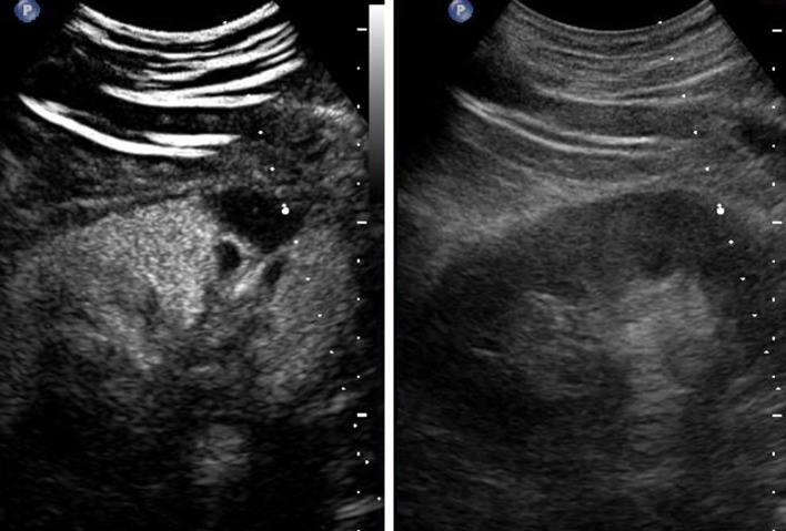
Infective endocarditis was definitively diagnosed in accordance with the revised Duke criteria [2], and a course of antibiotic therapy was initiated. Four weeks later, the patient developed acute left flank pain. The gray-scale ultrasound examination showed no abnormalities involving the left kidney. A CEUS study of the left kidney was then performed with the patient’s written, informed consent. SonoVue® (Bracco, Milan, Italy) was used as the contrast agent. During the venous phase of the examination (an interval of 4 min, beginning approximately 1 min after the intravenous injection of Sonovue), a single, wedged-shaped area of hypovascularity, 30 mm long, was observed in the inferior portion of the kidney (Fig. 1), findings that were consistent with kidney infarction.
Fig. 1.
Sonographic depictions of the left kidney (longitudinal view). The split screen display shows (left) CEUS findings in the venous phase: in the inferior portion of the kidney, a single, subcapsular wedge-shaped area of hypovascularity with a longitudinal diameter of 30 mm was seen. Gray-scale ultrasound images are shown on the right side of the screen
In the presence of left-sided infective endocarditis, peripheral embolization at any site may represent either a complication or a diagnostic clue; in either case, they are often difficult to detect. There are currently no bedside methods for identifying kidney embolization, but our experience with this case suggests that CEUS may be an effective means for filling this diagnostic gap.
Conflict of interest
G. Menozzi, V. Maccabruni and E. Gabbi declare that they have no conflict of interest.
Contributor Information
Guido Menozzi, Phone: +39-05-22296407, FAX: +39-05-22296961, Email: pelikan128@gmail.com, Email: menozzi.guido@asmn.re.it.
Valeria Maccabruni, Email: valeria.maccabruni@ausl.re.it.
Ermanno Gabbi, Email: ermanno.gabbi@asmn.re.it.
References
- 1.Fernández Guerrero ML, Álvarez B, Manzarbeitia F, Renedo G. Infective endocarditis at autopsy: a review of pathologic manifestations and clinical correlates. Medicine (Baltimore) 2012;9:152–164. doi: 10.1097/MD.0b013e31825631ea. [DOI] [PubMed] [Google Scholar]
- 2.Li JS, Sexton DJ, Mick N, et al. Proposed modifications to the Duke criteria for the diagnosis of infective endocarditis. Clin Infect Dis. 2000;30:633–638. doi: 10.1086/313753. [DOI] [PubMed] [Google Scholar]
- 3.Piscaglia F, Nolsøe C, Dietrich CF, et al. The EFSUMB Guidelines and Recommendations on the Clinical Practice of Contrast Enhanced Ultrasound (CEUS): Update 2011 on non-hepatic applications. Ultraschall Med. 2012;33:33–59. doi: 10.1055/s-0031-1281676. [DOI] [PubMed] [Google Scholar]


