Abstract
Ectopic activation of the Wnt signaling pathway is highly oncogenic for many human tissues. Here, we show that ectopic Wnt signaling increases the effective stem cell activity in mouse mammary glands in vivo. Furthermore, Wnt effectors induce the accumulation of mouse mammary epithelial progenitors (assayed by Hoechst dye exclusion, a surrogate stem cell marker, side population cells) both in vivo and in vitro. The longevity of stem cells makes them good candidate tumor precursors, and we propose that Wnt-induced progenitor amplification is likely to be key to tumor initiation. In support of this notion, mammary glands from a tumor-resistant strain of mice (carrying a null mutation in syndecan-1) contain fewer side population cells. When this strain is crossed to mice that overexpress effectors of the β-catenin/T cell factor Wnt pathway, the amplification of progenitors is reduced, together with all subsequent events of tumor development. We propose that the growth dynamic of the stem cell fraction is a major determinant of tumor susceptibility.
Keywords: Wnt-1, β-catenin, syndecan-1, heparan sulfate proteoglycan, mouse mammary epithelial cells
The majority of cells in adult epithelia are thought to have a limited intrinsic division potential. They are the daughters of a minor population of progenitor cells that are relatively undifferentiated and long-lived (or immortal). This longevity is thought to facilitate the accumulation of oncogenic genetic mutations and make transformation more likely.
In mouse mammary gland, the presence of primitive progenitors has been implied by serial transplantation studies of mammary epithelial cells (1). Thus, outgrowth of a full mammary branching tree from limiting dilutions of mammary epithelial transplants is considered to be an assay of clonal stem cell function (2), and this activity is depleted after four to six transplants. Data obtained from the study of hematopoietic stem cells has served to define molecular and functional properties that are shared by other somatic stem cells. Thus, a progenitor-enriched fraction [so-called side population (SP) cells] was defined by Hoechst-33342 dye efflux (conferred by the ABC transporter Bcrp1) (3, 4). When SP fractions were purified from mammary epithelial cell populations, they were shown to be enriched for BrdUrd label-retaining cells, to show low expression of differentiated markers (such as cytokeratins 14 and 19) and to express higher telomerase activity. These SP cells, when transplanted into cleared mammary fat pads, could reconstitute both myoepithelial and luminal mammary lineages in vivo (5, 6).
It is likely that stem cells have unique modes of growth factor reception and signal transduction (7–9), especially with regard to pathways that regulate asymmetric cell division, a property associated with stem cell self-renewal. In model organisms, such as Caenorhabditis elegans and Drosophila, Wnt, Notch, and Hedgehog pathways are key to regulating asymmetric divisions. These same pathways are oncogenic when misregulated in mammalian tissues (10). Thus, many human tumors contain mutations that serve to stabilize the Wnt effector β-catenin (11). Clues to the mechanism underlying the oncogenicity of ectopic Wnt signaling have been gleaned from the reciprocal effects that loss- and gain-of-function Wnt signaling mutations have on morphologic compartments thought to contain somatic stem cells. More specifically, a mutation in the Wnt signaling pathway in intestine (Tcf4–/–) eliminates the crypt-associated stem cell population (12), whereas gain-of-function mutations of Wnt signaling [(Apcmin or ΔN89β-catenin (ΔNβcat)] induce hyperplastic development and tumors (13, 14). Similarly, if Wnt signaling is inhibited in mouse skin, hair follicle development is eliminated (hair follicles are a source of epidermal stem cell activity) (15). On the other hand, gain of function of Wnt signaling (ΔNβcat expressed in basal keratinocytes) induces follicular hyperplasia and pilomatricoma tumors (16, 17). More recently, the size of the hematopoietic stem cell pool has been shown to be directly regulated by Wnt signaling (18).
Here, we test whether mammary progenitors are modulated by Wnt signaling. This hypothesis was based on the observation that Wnt-1-induced tumors comprise cells from both mammary lineages, suggesting that the tumor precursor is a bipotential progenitor. We find that functional progenitor activity is indeed elevated, and that the degree of accumulation of SP cells directly correlates with the risk of tumor development.
Materials and Methods
Transgenic Mouse Strains. FVB mice expressing the oncogenes Wnt1 or ΔNβcat in mammary gland under the control of the mouse mammary tumor virus (MMTV)-LTR and Sdc1–/– mice were as described in refs. 19 and 20.
Immunocytochemistry of Mammary Tissues. Mammary tissues were paraformaldehyde-fixed and embedded, and tissues were processed for antigen retrieval as specified by Zymed (heat-induced epitope retrieval). Sections were incubated in antibodies as follows: anti-Muc1 [CT2 Armenian Hamster monoclonal antibody; gift from S. Gendler (Mayo Clinic, Scottsdale, AZ)], FITC secondary antibody (catalog no. 127-095-160, Jackson Immuno Research), and Cy3 anti-smooth muscle actin (SMA) (clone 1C4, Sigma).
Preparation of Primary Mammary Epithelial Cells (MECs) and Assay of SP Fractions. MECs were prepared essentially as described in ref. 21 with modifications as described in ref. 19. MECs (1 × 10⁶) were preplated (to remove fibroblasts) for 20 min in DMEM/F12 plus 10% FBS and insulin (10 μg/ml), washed twice with PBS, and incubated with proteolytic enzymes once more to enrich the yield of single cells [2 ml of F12 media plus 4 mg/ml collagenase/150 μg/ml hyaluronidase (catalog no. H-4272, Sigma)/5 mg/ml dispase type II (catalog no. 165859, Roche)/50 units/ml DNase I (catalog no. 104132, Roche) for 5 min at 37°C with trituration, followed by 1 ml of 0.05% trypsin plus 0.05% EDTA (GIBCO/BRL) for 5 min at 37°C]. Cells were washed and resuspended in DMEM plus 2% FBS and 5 μg of Hoechst-33342 dye per 1 × 10⁶ cells (catalog no. B-2261, Sigma), incubated at 37°C for exactly 90 min, and analyzed by flow cytometry.
Primary Culture of MECs with Purified Wnt/Dkk Proteins. To test the response of MEC cultures to purified Wnt3A and/or dickkhopf-1 (Dkk1), BALB/c MECs from mice 5–6 months old were plated at 0.5 × 10⁶ per 60-mm plate in F12 media plus 2% FBS, insulin, hydrocortisone, PenStrep, and gentamycin. Purified Wnt-3A (100 μg/ml; catalog no. 1324-wn, R & D Systems) and/or Dkk-1 (100 μg/ml; catalog no. 1096-dk-010, R & D Systems) were added to the F12 culture media 12 h after plating. For cells treated with both Wnt-3A and Dkk-1, Dkk-1 was added 8 h before Wnt-3A to allow Dkk-1 to bind and down-regulate lipoprotein receptor-related protein receptors (22). Cells were cultured with purified proteins for 60 h before they were harvested and labeled with Hoecsht dye as described above.
Transplantation of Cleared Mammary Fat Pads. MECs from ΔNβcat mice or FVB control littermates, 2–3 months old, were preplated and used to inoculate fat pads either with (experiment 1) or without (experiment 2) further enzymatic digestion (as above). Cells for transplantation were counted, suspended in DMEM plus 2% FBS with 5 μg/ml Matrigel (catalog no. 354234, Becton Dickinson) at 4°C together with loading dye (final concentration: 5% glycerol/0.5% trypan blue/25 mM Hepes), and inoculated in a 1-μl volume containing 1,000–50,000 cells per μl. Eight to 12 weeks after transplantation, fat pads were dissected, processed, and stained with carmine as described in ref. 19.
Results
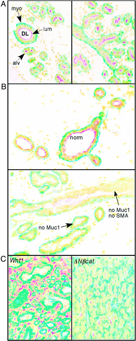
Wnt-1-Induced Tumors Are Aggressive but Contain Differentiated Cells of Both Mammary Lineages. Differentiated adenocarcinomas emerge in a highly concerted manner as solitary, rapidly growing tumors within mammary glands that are systemically hyperplastic in response to the expression of the [MMTV-LTR]-driven Wnt1 oncogene (23, 24). Other groups have noticed that SMA-positive cells coexist with another cytokeratin 8-positive epithelial cell type in these tumors (23, 25), implying that both myoepithelial and luminal cell lineages persist. We tested for expression of molecular markers of these two lineages in hyperplastic [MMTV-Wnt1] transgenic mammary glands and found that their expression was inhibited in overtly hyperplastic ductal zones (Fig. 1B). In contrast, in Wnt1-induced tumors, differentiated cells from both lineages were represented throughout (Fig. 1C). The number of SMA-positive cells varied between 10% and 65%; the number of Muc1-positive cells was between 20% and 55%. No cell appeared to express both markers, and a minor subpopulation expressed neither (7–17%). Tumors induced by ΔNβcat also contain both mammary lineages (Fig. 1C). Tumors that arise in the TgN(Wnt1)1Hev strain have been shown to be clonal (or pseudoclonal) by retroviral tagging (26, 27), and more recently loss of heterozygosity at the PTEN locus was shown to be shared by both myoepithelial and luminal lineages (28). We conclude that the most likely precursor of Wnt1 and ΔNβcat-induced tumors is a bipotential stem cell, and we propose that dysregulation of this subpopulation will be evident early during tumor initiation.
Fig. 1.
Wnt1- and ΔNβcat-induced tumors contain at least three types of cells, including cells that express markers of luminal or myoepithelial cells and some that express neither. Samples of normal mammary gland from a 12-day pregnant mouse (A), hyperplastic mammary gland from an 11-week-old virgin Wnt1-transgenic mouse (in estrus) (B), and Wnt1- or ΔNβcat-induced mammary tumor (C) were fixed and paraffin-embedded, and the sections were stained with antibodies to Muc-1 (stains the apical membrane of luminal cells; red) or to SMA (stains the cytoskeleton of myoepithelial cells; green). Nuclei were counterstained with Hoecsht (shown here in yellow). (A) Mature mammary glands comprised of hollow ducts (ductal lumen; DL) lined by an inner luminal layer (lum) and a basal myoepithelial cell layer (myo). During growth and differentiation, alveoli (alv) develop that have smaller lumens and an incomplete basal layer. (B) Hyperplastic glands are characterized by core ductal zones that have relatively normal architecture (norm; Upper), surrounded by pseudolobuloalveolar elements with multilayered epithelia that sometimes entirely fill the ductal lumen. There is no accumulation of apical Muc-1 in these zones of hyperplasia, and the basal layer expresses SMA only in patches. (C) One representative example each of a Wnt1- and a ΔNβcat-induced tumor are shown. Cells are organized into cohesive sheets of myoepithelia interspersed with bands of luminal derivatives. This pattern is typical of the majority of tumors; the remaining 15% of tumors were undifferentiated, in papillary form, and infiltrated with highly reactive stroma.
SP-Enriched Fractions Are Increased in Mammary Glands of MMTV-Wnt-1- and MMTV-ΔNβcat-Transgenic Mice by 3- and 9-Fold, Respectively. To assay mammary stem/progenitor-enriched fractions, we tested the Hoechst-dye efflux properties of MECs prepared from glands from [MMTV-Wnt-1] and [MMTV-ΔNβcat] transgenic mice. The regulation of dye pump Bcrp1 activity is not yet well understood. Bcrp1 activity is known to be modified by signaling pathways such as Akt/PTEN, and it is not absolutely required for stem cell function (29, 30). Therefore, although SP cell fractions from several tissues are enriched in progenitors and there is no example to date of stem cell activity residing outside this fraction, all SP cells do not have equal (or perhaps any) stem cell potential (4). With these caveats in mind, we applied dye efflux as a surrogate stem cell marker.
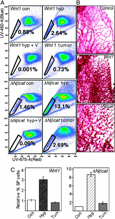
MECs from virgin, 2- to 3-month-old females were isolated, trypsinized to obtain single cells, labeled with Hoechst dye, and analyzed by flow cytometry (Fig. 2). We found that control littermate glands contained between 0.5% and 2.0% SP cells; whereas in the Wnt-1-induced hyperplastic gland, that fraction increased by 2.5- to 3.0-fold (Fig. 2 A and C). Similarly, but more dramatically, the accumulation of SP cells in MMTV-ΔNβcat transgenic mammary glands was 9-fold (Fig. 2 A and C). Although the MMTV promoter is not entirely mammary epithelial cell-specific, we showed that the relative accumulation of SP cells was not hematopoietic in origin (supporting information, which is published on the PNAS web site). The glandular hyperplasia that was associated with these SP profiles is shown in Fig. 2B. Interestingly, in tumors from both mouse strains, the progenitor fraction reproducibly declined to relatively normal levels (Fig. 2 A and C), although the mitotic index increased (data not shown).
Fig. 2.
Progenitor cell-enriched SP fractions are increased in Wnt1- and ΔNβcat-transgenic mammary glands. MECs were prepared from mammary glands from 2- to 3-month-old FVB MMTV-Wnt1- and MMTV-ΔNβcat-transgenic mice, together with control littermates (this experiment shows pooled MECs from 7 Wnt1:FVB compared with 11 control FVB mice and 11 ΔNβcat:FVB compared with 12 FVB controls). (A) Cells were labeled with Hoecsht dye and assayed by flow cytometry for fluorescence in the red and blue channels. The side population was gated as a spur of cells with less fluorescence. Representative scans are shown for two sets of samples. The first shows control Wnt1+/– hyperplastic glands (Wnt1 hyp), the hyperplastic cells labeled in the presence of verapamil (an inhibitor of Hoechst dye efflux), and a Wnt1-induced tumor. The second set shows the same sample set for ΔNβcat-induced glands and a tumor. (B) The morphogenesis of control, Wnt1-transgenic glands and a [ΔNβcat]-transgenic gland (3 months old) is shown in carmine-stained whole-mount preparations. (C) The results of SP assays were quantified. Average values for the relative proportion of SP cells, together with their SDs, were derived from three independent sorts, and these results were repeated with a separate set of MECs isolated on different days. Analysis of SP fractions of [MMTV-Wnt]:C57BL6 transgenic mammary glands gave similar results. Con, control; hyp, hyperplastic; tum, tumor.
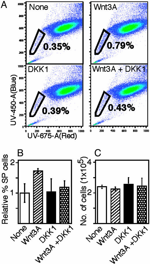
Ectopic Wnt Ligands Increase the SP Fraction in the MEC Population After 3 Days of Primary Culture. In transgenic glands after 10 weeks of development in vivo, measurement of progenitor fractions can only be considered to be a distal effect of ectopic Wnt effector treatment. To test whether Wnt effectors were inducing this progenitor accumulation as a proximal, direct effect, we tested whether incubation of short-term, primary MEC cultures in soluble Wnt ligands affected the SP fraction. This experiment was performed by using two sources of the soluble Wnt ligand Wnt3A. Wnt3A falls into the transforming class of Wnt ligands (like Wnt1) and induces transactivation of T cell factor/β-catenin reporters in most cell types (including these primary mammary epithelial cell cultures (19). Either a crude source of Wnt3A (conditioned medium from Wnt3A-transfected L cells; data not shown) or a relatively pure source of Wnt3A (a commercial source derived by expression in baculovirus; Fig. 3) were added to cultures in the presence or absence of the canonical Wnt-signaling pathway inhibitor dkk1 (31). Wnt3A increased the fraction of SP cells by 70% but did not affect the growth of the total mammary cell population (Fig. 3 B and C). Epidermal growth factor also maintains the SP cell fraction in these MEC cultures, and the addition of Wnt3A in the presence of epidermal growth factor had no additional effect, suggesting that these two factors are functionally redundant (data not shown). We could also show that the Wnt3A-induced increase was specifically inhibited by the addition of dkk1, and we conclude that Wnt3A-induced canonical signaling specifically induces the growth or survival of SP cells in primary culture.
Fig. 3.
The progenitor cell fraction is increased in primary cultures of MECs incubated with soluble Wnt3A. (A) MECs from BALB/c mice were transferred to primary culture and incubated in purified Wnt3A protein, canonical Wnt inhibitor Dkk1, or both; cultures were then analyzed by flow cytometry. (B) Quantitation shows that the SP fractions increase by 70% in the presence of Wnt3A, and this increase is reversed by the addition of the Dkk1 inhibitor. (C) The growth of the total MEC population was not, however, affected by these supplements. Averages and SDs were from triplicate determinations.
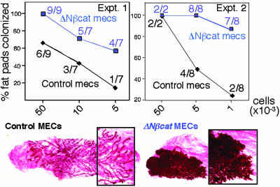
Cells from ΔNβcat Transgenic Glands Colonize Cleared Mammary Fat Pads More Efficiently Than Control Cells. The progenitor activity of SP cells is difficult to test directly, probably because of the toxicity of Hoechst dye binding and flow cytometric MECs. Therefore, we chose to transfer cells from 10-week-old ΔNβcat transgenic glands by limiting dilution (1,000–50,000) into cleared fat pads of 3-week-old congenic recipients to measure their functional progenitor capacity (Fig. 4). At this age, the donor glands were not substantially hyperplastic. At all cell numbers tested, more glands were colonized after transfer of ΔNβcat-expressing MECs, and the outgrowths showed exacerbated hyperplastic development. We conclude that the increased SP fraction observed in ΔNβcat transgenic MECs does indeed reflect increased functional stem cell activity.
Fig. 4.
Transgenic MECs expressing the oncogene ΔNβcat colonized mammary fat pads more efficiently than control cells. MECs were isolated from [ΔNβcat]-transgenic and control mice and counted; then several inocula containing different numbers of cells were transferred to cleared fat pads to test their progenitor potential. The fraction of cleared mammary fat pads colonized by cells was scored. For experiment 1, cells were transferred after the cell preparation was substantially reduced to single cells by enzymatic dissociation, a procedure that tends to impair viability. For experiment 2, cells were transferred in the clusters typical of our standard preparation of MECs. The results were substantially the same and showed that the progenitor activity in MEC populations from [ΔNβcat]-glands was higher than normal. Numbers in parentheses represent the number of glands colonized per total number of glands transplanted. The morphogenesis of outgrowths from 50,000 cell inocula is also shown.
Sdc1–/– Mammary Glands Are Tumor-Resistant, Show Hypomorphic Development, and Contain 40% of the Normal Progenitor Fraction. Sdc1–/– mice resist Wnt1- and ΔNβcat-induced tumor development (19, 20). Although we predicted that the Sdc1 heparan sulfate proteoglycan would be a coreceptor for the Wnt-Fz signaling complex (32), we showed instead that these gene products interacted only indirectly (19). In the absence of Sdc1, a Wnt-responsive cell population was missing. Furthermore, we noticed that not only was the hyperplastic response to Wnt1 or ΔNβcat reduced but also that the normal adult Sdc1–/– mammary tree was hypomorphic. These data suggested that the effect of Sdc1 deficiency was to alter mammary development and to create a mammary population resistant to Wnt effector-dependent tumor induction.
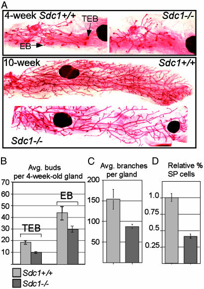
To better describe the hypomorphic development in Sdc1–/– mammary glands, we scored mammary gland complexity in juvenile and adult mice. Morphometric analysis showed that the branching complexity of adult glands was decreased (Fig. 5 A and C). Because adult branching complexity is a function of terminal end bud activity during juvenile ductal extension, we assessed mammary morphogenesis at 4 weeks of age, and found an ≈50% decrease in terminal end bud number (Fig. 5 A and B).
Fig. 5.
Sdc1–/– glands have reduced mammary gland complexity and SP cells. (A) Carmine-stained whole-mount preparations of mammary glands from 4- and 10-week-old control and Sdc1–/– mice are shown. The arrows indicate a typical terminal end bud (TEB) and end bud (EB) structures. (B) The results of morphometric analysis of the average number of TEBs and EBs per mammary gland of 4-week-old Sdc1+/+ and Sdc1–/– mice are shown. The number of TEBs is reduced by 47% (P = 4 × 10⁻⁵; 2-tailed Wilcoxon rank test). (C) The number of branches per gland of virgin 10-week-old Sdc1–/– mice was reduced by 43% (P = 0.01). (D) The relative fraction of SP cells was determined from three independent sorts of different MEC preparations from 6- to 7-month-old BALB/c Sdc1+/+ (30) and Sdc1–/– (25) mice. Younger mice (4–5 months old) or C57BL6 Sdc1–/– mice gave the same result.
We propose that hypomorphic development is likely to be associated with changes in the progenitor fraction of Sdc1–/– glands and, therefore, assayed SP cells from 4- to 6-month-old virgins. The SP fraction was reduced by 60% (Fig. 5D). Thus, hyperplastic development and progenitor number are raised in mammary glands that overexpress Wnt effectors and are reduced in Sdc1–/– mice that resist Wnt-induced tumor development.
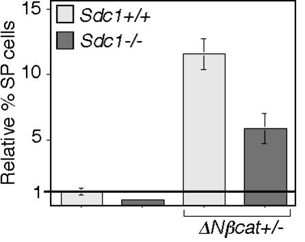
The Null Allele of Sdc1 and Wnt Effector Transgenes Oppose Each Other to Regulate Mammary Progenitor Division. We propose, therefore, that Sdc1 and Wnt signaling both regulate mammary progenitor cell function. To test this hypothesis, we assayed the progenitor population in ΔNβcat+/– glands in the absence of Sdc1 expression (Fig. 6). The SP population was reduced by 66% in Sdc1–/– glands even on this mixed-strain background. When ΔNβcat was expressed in the absence of Sdc1, the fold increase in the SP fraction was also reduced by half, and this decrease correlated with reduced tumor incidence and increased tumor latency (19). This finding is consistent with the proposal that both Sdc1 and Wnt modulate the growth or differentiation of this progenitor population.
Fig. 6.
The absence of Sdc1 reduces the amplification of progenitors induced by ΔNβcat. MECs were isolated from five 2.5- to 3-month-old female mice from each of four genotypes produced by a cross of [ΔNβcat]-transgenic and Sdc1–/– mice (19). This cohort was of mixed-strain background (a BALB/c/FVB backcross). SP cells were separated by flow cytometry, and the results are presented as percent SP cells relative to ΔNβcat–/–; Sdc1+/+ mice. The average and SD were generated from three independent sorts.
Discussion
We have found that mammary stem cell activity is increased in transgenic glands from [MMTV-ΔNβcat] mice. The SP cell fraction (a progenitor-enriched fraction) is increased in both [MMTV-Wnt1] and [MMTV-ΔNβcat] mammary glands in vivo and after just 60 h of treatment of primary cultures of MECs with the Wnt3A ligand. This specific growth/survival stimulus is inhibited by the addition of the canonical (lipoprotein receptor-related protein-dependent) Wnt pathway inhibitor dkk1 (31), suggesting that β-catenin/TCF-induced transactivation is responsible for the progenitor-specific responses. Data from two other mouse strains suggests that Wnt signaling is involved in the division and survival of lobuloalveolar progenitors. Thus, when β-catenin/T cell factor signaling was inhibited by expression of a dominant negative β-catenin (a β catenin-engrailed fusion protein driven by MMTV or whey acidic protein) (33) or the Wnt canonical pathway inhibitor axin (driven by an inducible MMTV construct) (34), there was transient apoptosis followed by significant developmental attrition, consistent with the loss of a progenitor population. Another group (28) has recently shown that a keratin 6-, Sca-1-positive cell fraction likely to be progenitor-enriched is amplified only in transgenic mammary glands from strains expressing Wnt effectors and not other oncogenes, such as polyoma middle T, H-Ras, or Neu. Thus, although these other oncogenes also induce hyperplasia and dysregulated growth, the preneoplastic mammary glands are not associated with an increased proportion of immature cells.
Data from other groups also suggests that the continuous presence of Wnt effectors is required to maintain Wnt-induced cells. Gunther et al. (35) showed that if Wnt1 expression was withdrawn from a transgenic mouse with a doxycycline-inducible, MMTV-driven Wnt1 construct, tumors regressed within 10 days. Furthermore, when Wnt signaling was inhibited in LS174T colon cancer cells, their growth was arrested (36). We suggest that the transcriptional context of stem cells is unique and that the mitogenic effects of β-catenin/T cell factor-induced transactivation are only expressed in this context. We propose that mammary progenitor dysregulation initiates Wnt-induced tumor development and that the tumor precursor cell is drawn from this subpopulation.
In support of this hypothesis, a tumor-resistant mouse strain with a null mutation in Sdc1 contains just 50% of the normal SP fraction, and this reduction correlates with reduced branching complexity at all stages of outgrowth of the mammary tree. This implies that during ductal extension, mammary branching may be regulated by stem cell number/activity. We do not yet understand why mammary development in Sdc1–/– mice is hypomorphic, but it is unlikely to be related directly to the activity of the Wnt-receptor signaling complex (19). However, when these SP-deficient mice are crossed with those that accumulate mammary progenitors, the SP fraction is intermediate, the preneoplastic hyperplasia is reduced, and the onset of tumor development is slowed. Thus, the higher the fraction of progenitor/stem cells, the more likely subsequent mutation will induce tumor development. Note that for 90% of human breast tumor samples tested, Al-Hajj et al. (37) showed that the tumor-forming potential resides in a morphologically undifferentiated, karyotypically diploid subfraction that contains the tumor growth potential and recreates the karyotypically abnormal majority after xenografting at limiting dilution. This group concluded that tumor growth is likely to be driven by the division of tumor stem cells that are related in some way to the normal stem cell compartment.
The accumulation of stem cells in Wnt-effector-induced mammary glands is associated with morphogenic hyperplasia. We propose that this hyperplasia, or disrupted ductal architecture, is the result of the accumulation of relatively undifferentiated, progenitor and transit-amplifying cells. However, the ductal density does not exceed the normal range, and we deduce that the mechanisms that govern mammary cell density during normal development are still intact (38). Genetic changes that have been shown to cooperate during progression in this mouse model include the up-regulation of mitogens, such as fibroblast growth factor (26, 27) or tumor growth factor α (39). We propose that mutations in bipotential progenitor cells that allow differentiated progeny to escape growth inhibition will cooperate to transform these hyperplastic outgrowths into a tumor. Thus, the brakes that slow the division of mature cells in virgin glands would be removed and the proliferation of all cells in this lineage maximized. In support of this idea, we find that the SP fraction in tumors declines to a fraction typical of a normal population profile. This trend was also apparent for the tumor progression profiles reported by Li et al. (28).
Few human breast tumors have the same properties of rapid growth together with bilineal differentiation that is typical of tumors that evolve in this mouse model. However, tumor evolution in [MMTV-Wnt1]-mice was different in a p53–/– background; tumors arose earlier, grew faster, were more anaplastic, and showed reduced expression of differentiated markers (25). Thus, they resembled their human counterparts more faithfully. Furthermore, myoepithelial “basal” markers are expressed by some of the cells in up to one-fifth of human breast cancers, implying that there is at least residual expression of both mammary lineages. These tumors fell into a separate class according to transcriptional profiling data (40, 41), and we propose that this phenotype is consistent with a bipotential stem cell origin.
In summary, we propose that factors that regulate progenitor division also regulate susceptibility to tumor development. This hypothesis is consistent with a model that includes a relatively mutable, long-lived progenitor cell. We infer that measurement of progenitor fractions of human breast samples (and especially of lesions considered to be preneoplastic) may be an excellent prognostic for assessing risk of progression. We also infer that stem cell technologies that rely on amplification of these mutable progenitor cell types may be especially prone to subsequent tumor development and that this risk factor should be carefully evaluated before the widespread adoption of, for example, neural stem cell transplants.
Supplementary Material
Acknowledgments
We thank Joel Puchalski and members of the University of Wisconsin Flow Cytometry facility for their expertise. This work was supported by National Institutes of Health Grant CA 90877–01, American Cancer Society Grant IRG-58–011-43–01 and University of Wisconsin Medical School, Department of Cancer Biology, Training Award T32 CA09135 (to B.L.).
Abbreviations: ΔNβcat, ΔN89β-catenin; SP, side population cells from a flow cytometric assay of Hoecsht dye efflux; MEC, mammary epithelial cell; MMTV, mouse mammary tumor virus; SMA, smooth muscle actin.
References
- 1.Daniel, C. W. (1973) Experientia 29, 1422–1424. [DOI] [PubMed] [Google Scholar]
- 2.Kordon, E. C. & Smith, G. H. (1998) Development (Cambridge, U.K.) 125, 1921–1930. [DOI] [PubMed] [Google Scholar]
- 3.Zhou, S., Schuetz, J. D., Bunting, K. D., Colapietro, A.-M., Sampath, J., Morris, J. J., Lagutina, I., Grosveldd, G. C., Osawa, J., Nakauchi, H. & Sorrentino, B. (2001) Nat. Med. 7, 1028–1034. [DOI] [PubMed] [Google Scholar]
- 4.Goodell, M., Rosenzweig, M., Kim, H., Marks, D. F., DeMaria, M., Paradis, G., Grupp, S. A., Sieff, C. A. & Mulligan, R. C. (1997) Nat. Med. 3, 1337–1345. [DOI] [PubMed] [Google Scholar]
- 5.Welm, B. E., Tepera, S. B., Venezia, T., Graubert, T. A., Rosen, J. M. & Goodell, M. A. (2002) Dev. Biol. 245, 42–56. [DOI] [PubMed] [Google Scholar]
- 6.Alvi, A., Clayton, H., Joshi, C., Enver, T., Ashworth, A., Vivanco, M. d. M., Dale, T. C. & Smalley, M. J. (2002) Breast Cancer Res. 5, R1–R8. [DOI] [PMC free article] [PubMed] [Google Scholar]
- 7.Tropepe, V., Sibilia, M., Ciruna, B. G., Rossant, J., Wagner, E. F. & van der Kooy, D. (1999) Development (Cambridge, U.K.) 208, 166–188. [DOI] [PubMed] [Google Scholar]
- 8.de Haan, G., Weersing, E., Dontje, B., van Os, R., Bystrykh, L. V., Vellenga, E. & Miller, G. (2003) Dev. Cell 4, 241–251. [DOI] [PubMed] [Google Scholar]
- 9.Burdon, T., Smith, A. & Savatier, P. (2002) Trends Cell Biol. 12, 432–438. [DOI] [PubMed] [Google Scholar]
- 10.Taipale, J. & Beachy, P. A. (2001) Nature 411, 349–354. [DOI] [PubMed] [Google Scholar]
- 11.Polakis, P. (2000) Genes Dev. 14, 1837–1851. [PubMed] [Google Scholar]
- 12.Korinek, V., Barker, N., Moerer, P., van Donselaar, E., Huls, G., Peters, P. J. & Clevers, H. (1998) Nat. Genet. 19, 379–383. [DOI] [PubMed] [Google Scholar]
- 13.Bienz, M. & Clevers, H. (2000) Cell 103, 311–320. [DOI] [PubMed] [Google Scholar]
- 14.Harada, N., Tamai, Y., Ishikawa, T., Sauer, B., Takaku, K., Oshima, M. & Taketo, M. M. (1999) EMBO J. 18, 5931–5942. [DOI] [PMC free article] [PubMed] [Google Scholar]
- 15.Andl, T., Reddy, S. T., Gaddapara, T. & Millar, S. E. (2002) Dev. Cell 2, 643–653. [DOI] [PubMed] [Google Scholar]
- 16.Gat, U., DasGupta, R., Degenstein, L. & Fuchs, E. (1998) Cell 95, 605–614. [DOI] [PubMed] [Google Scholar]
- 17.Alonso, L. & Fuchs, E. (2003) Genes Dev. 17, 1189–1200. [DOI] [PubMed] [Google Scholar]
- 18.Reya, T., Duncan, A. W., Ailles, L., Domen, J., Schere, D. C., Willert, K., Hintz, L., Nusse, R. & Weissman, I. L. (2003) Nature 423, 409–414. [DOI] [PubMed] [Google Scholar]
- 19.Liu, B., Kim, Y.-C., Leatherberry, V., Cowin, P. & Alexander, C. M. (2003) Oncogene 22, 9243–9253. [DOI] [PubMed] [Google Scholar]
- 20.Alexander, C. M., Reichsman, F., Hinkes, M. T., Lincecum, J., Becker, K. A., Cumberledge, S. & Bernfield, M. (2000) Nat. Genet. 25, 329–332. [DOI] [PubMed] [Google Scholar]
- 21.Emerman, J. T. & Bissell, M. J. (1988) Adv. Cell Cult. 6, 137–159. [Google Scholar]
- 22.Mao, B., Wu, W., Davidson, G., Marhold, J., Li, M., Mechler, B. M., Delius, H., Hoppe, D., Stannek, P., Walter, C., Glinka, A. & Niehrs, C. (2002) Nature 417, 664–667. [DOI] [PubMed] [Google Scholar]
- 23.Rosner, A., Miyoshi, K., Landesman-Bollag, E., Xu, X., Seldin, D. C., Moser, A. R., MacLeod, C. L., Shyamala, G., Gillgrass, A. E. & Cardiff, R. D. (2002) Am. J. Path. 161, 1087–1097. [DOI] [PMC free article] [PubMed] [Google Scholar]
- 24.Tsukamoto, A. S., Grosschedl, R., Guzman, R. C., Parslow, T. & Varmus, H. E. (1988) Cell 55, 619–625. [DOI] [PubMed] [Google Scholar]
- 25.Cui, X. S. & Donehower, L. A. (2000) Oncogene 19, 5988–5996. [DOI] [PubMed] [Google Scholar]
- 26.Shackleford, G. M., MacArthur, C. A., Kwan, H. C. & Varmus, H. E. (1993) Proc. Natl. Acad. Sci. USA 90, 740–744. [DOI] [PMC free article] [PubMed] [Google Scholar]
- 27.Kapoun, A. M. & Shackleford, G. M. (1997) Oncogene 14, 2985–2989. [DOI] [PubMed] [Google Scholar]
- 28.Li, Y., Welm, B., Podsypanina, K., Huang, S., Chamorro, M., Zhang, X., Rowlands, T., Egeblad, M., Cowin, P., Werb, Z., et al. (2003) Proc. Natl. Acad. Sci. USA 100, 15853–15858. [DOI] [PMC free article] [PubMed] [Google Scholar]
- 29.Mogi, M., Yang, J., Lambert, J. F., Colvin, G. A., Shiojima, I., Skurk, C., Summer, R., Fine, A., Quesenberry, P. J. & Walsh, K. (2003) J. Biol. Chem. 278, 39068–39075. [DOI] [PubMed] [Google Scholar]
- 30.Zhou, S., Morris, J. J., Barnes, Y., Lan, L., Schuetz, J. D. & Sorrentino, B. P. (2002) Proc. Natl. Acad. Sci. USA 99, 12339–12344. [DOI] [PMC free article] [PubMed] [Google Scholar]
- 31.Semenov, M. V., Tamai, K., Brott, B. K., Kuhl, M., Sokol, S. & He., X. (2001) Curr. Biol. 11, 951–961. [DOI] [PubMed] [Google Scholar]
- 32.Perrimon, N. & Bernfield, M. (2000) Nature 404, 725–728. [DOI] [PubMed] [Google Scholar]
- 33.Tepera, S. B., McCrea, P. D. & Rosen, J. M. (2002) J. Cell Sci. 116, 1137–1149. [DOI] [PubMed] [Google Scholar]
- 34.Hsu, W., Shakya, R. & Costantini, F. (2001) J. Cell Biol. 155, 1055–1064. [DOI] [PMC free article] [PubMed] [Google Scholar]
- 35.Gunther, E. J., Moody, S. E., Belka, G. K., Hahn, K. T., Innocent, N., Dugan, K. D., Cardiff, R. D. & Chodosh, L. A. (2003) Genes Dev. 17, 488–501. [DOI] [PMC free article] [PubMed] [Google Scholar]
- 36.van de Wetering, M., Sancho, E., Verweij, C., de Lau, W., Oving, I., Hurlstone, A., van der Horn, K., Batlle, E., Coudreuse, D., Haramis, A. P., et al. (2002) Cell 111, 241–250. [DOI] [PubMed] [Google Scholar]
- 37.Al-Hajj, M., Wicha, M. S., Benito-Hernandez, A., Morrison, S. J. & Clarke, M. F. (2003) Proc. Natl. Acad. Sci. USA 100, 3983–3988. [DOI] [PMC free article] [PubMed] [Google Scholar]
- 38.Daniel, C. W., Robinson, S. & Silberstein, G. B. (1996) J. Mammary Gland Biol. Neoplasia 1, 331–341. [DOI] [PubMed] [Google Scholar]
- 39.Schroeder, J. A., Troyer, K. L. & Lee, D. C. (2000) Oncogene 19, 3193–3199. [DOI] [PubMed] [Google Scholar]
- 40.Perou, C., Sorlie, T., Eisen, M. B., Van de Jirn, M., Jeffrey, S. S., Rees, C. A., Pollack, J. R., Ross, D. T., Johnsen, H., Akslen, L. A., et al. (2000) Nature 406, 747–752. [DOI] [PubMed] [Google Scholar]
- 41.Sorlie, T., Perou, C. M., Tibshirani, R., Aas, T., Geisler, S., Johnsen, H., Hastie, T., Eisen, M. B., van de Rijn, M., Jeffrey, S. S., et al. (2001) Proc. Natl. Acad. Sci. USA 98, 10869–10874. [DOI] [PMC free article] [PubMed] [Google Scholar]
Associated Data
This section collects any data citations, data availability statements, or supplementary materials included in this article.


