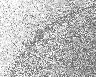
Trypanosomatids are protozoan parasites responsible for important tropical diseases. One example, Trypanosoma brucei, causes African sleeping sickness, and related parasites cause Chagas disease and leishmaniasis. Because they are among the earliest-branching eukaryotes, trypanosomatids have unusual biological properties. One of their most curious features is a unique mitochondrial DNA network known as kinetoplast DNA (kDNA) (1). The kDNA network is composed of several thousand minicircles that are interlocked like the links in medieval chain mail. Also intertwined in the network are a few dozen maxicircles. See Fig. 1 for an electron micrograph of a segment of an isolated kDNA network from Crithidia fasciculata, a trypanosomatid often studied because it is nonpathogenic and easy to cultivate. The function of kDNA maxicircles, like mitochondrial DNA in conventional eukaryotes, is to encode a few gene products such as rRNA and subunits of respiratory complexes. However, the mechanism of gene expression is highly unconventional in that maxicircle transcripts must be edited to form a functional mRNA. Editing is an amazing form of RNA processing in which uridine residues are inserted or deleted at precise internal sites within the maxicircle transcripts, generating ORFs. Minicircles encode guide RNAs that are templates for editing of maxicircle transcripts. See ref. 2 for a review of editing and ref. 3 for a discussion of the evolution of kDNA and the significance of the network structure.
Fig. 1.
Electron micrograph of a segment of a kDNA network from C. fasciculata. Small loops are minicircles. [Reproduced with permission from ref. 1 (Copyright 2001, Elsevier Science).]
In this issue of PNAS, Sinha et al. (4) describe a novel C. fasciculata mitochondrial DNA ligase. This enzyme is distinct from the nuclear ligase I from the same organism (5) and from all other eukaryotic DNA ligases reported to date. Its small size (56 kDa) and sequence are reminiscent of virally encoded ligases. In yeast, a single gene encodes both a nuclear and a mitochondrial DNA ligase I by using an internal in-frame AUG to produce the nuclear protein (6). Vertebrates also produce a dual-targeted ligase (DNA ligase III) via a similar mechanism, and the mitochondrial version is present in two isoforms that differ at their C termini (reviewed in ref. 7). In contrast, the T. brucei genome encodes multiple ligases that appear to target specifically to the mitochondrion (4) (M. Lindsay and M.M.K., unpublished data) and which are likely involved in kDNA replication or repair. As discussed below, the novel intramitochondrial localization of the C. fasciculata ligase suggests a role in repairing gaps in newly replicated kDNA minicircles (e.g., joining Okazaki fragments) (4).
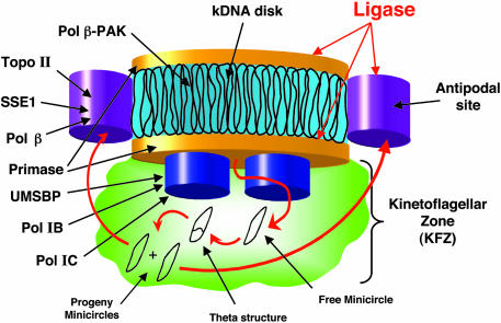
Each trypanosomatid cell contains one kDNA network in the matrix of its single mitochondrion. The kDNA is condensed into a disk-shaped structure, and its synthesis is facilitated by the fact that its replication machinery is precisely organized around the disk. The first reported localization of a kDNA replication protein by immunofluorescence was for a topoisomerase (topo) II, which is positioned in antipodal sites flanking the kDNA disk (8) (see diagram in Fig. 2 for the localization of kDNA-associated proteins). Subsequent reports revealed the same locations for a DNA polymerase (pol) β (9) and a structure-specific endonuclease I (SSE1), an enzyme with RNase H activity (10). It was initially assumed that all replication proteins would be positioned in the two antipodal sites, forming replisomes, but other proteins turned out to have different localizations. Primase is concentrated on the two faces of the disk (11), whereas UMSBP (the minicircle replication origin-binding protein) (12) and two DNA pol I-related enzymes (likely the replicative polymerases) (13) are situated in two foci below the kDNA disk in the kinetoflagellar zone (KFZ). A second DNA polymerase β, pol β-PAK, localizes within the kDNA disk (14). Now, Sinha et al. (4) report that the C. fasciculata DNA ligase has yet another localization pattern, distinct from that of proteins previously studied. It is positioned both in the antipodal sites and on the two faces of the kDNA disk (Fig. 2).
Fig. 2.
Diagram of kDNA disk and surrounding replication proteins. See text for description. Adapted from ref. 1.
The intramitochondrial location of the ligase suggests a role in repairing gaps in kinetoplast DNA.
What do the locations of these proteins reveal about the replication mechanism? The current model, outlined in Fig. 2, focuses on minicircles. Replication commences with release of individual minicircles from the network, vectorially into the KFZ, by an unknown topo II that is likely positioned within the kDNA disk (15). The free minicircles, which are covalently closed, are then bound by UMSBP at their replication origin (16). UMSBP is thought to assemble primase, the replicative polymerases, and other proteins to initiate unidirectional replication as a theta-structure. When progeny minicircles have segregated, these molecules, containing gaps, migrate to the antipodal sites where late stages of replication occur. These steps include primer removal by SSE1, filling of most of the gaps by pol β, and repair of the filled gaps by the newly discovered ligase (4). The fact that ligase and pol β interact (as shown by coimmunoprecipitation) suggests that they work together in gap filling (4). Topo II then attaches the newly replicated minicircles, still containing at least one gap within the replication origin (17), onto the network periphery adjacent to the antipodal sites. As replication proceeds, the network grows until the minicircle copy number has doubled. The last remaining gaps are then filled, probably by pol β-PAK, and the final nick, as discussed in the following paragraph, is sealed by ligase. Once all minicircles are covalently closed, the network is split in two, a mysterious reaction possibly catalyzed by an unknown topo II that unlinks minicircles along a line bisecting the double-size network.
Why is the C. fasciculata ligase localized not only in the antipodal sites but also on the two faces of the kDNA disk? The answer may lie in the sequence organization of the C. fasciculata minicircle. These molecules have two replication origins, positioned 180° apart, although only one or the other is used during each round of replication (17, 18). The final nicks to be closed are located within the origin sequences (17). Sinha et al. (4) speculate that the minicircle sequences are aligned so that the gapped origins are near the two faces of the disk. Then, the ligase would be perfectly positioned to seal the nicks. It may be possible to prove the sequence alignment by high-resolution in situ hybridization.
Discovery of the C. fasciculata DNA ligase adds one more protein to the kDNA replication repertoire, but there must be many more. One reason for the large number is that there is redundancy in enzymatic activities. For example, T. brucei has six different mitochondrial DNA polymerases (13, 14), whereas most other eukaryotes have only one, pol γ. There may be a similar multiplicity of mitochondrial helicases (B. Liu and S. Motyka, unpublished observations). Another reason is that there are key processes in kDNA replication that are not understood mechanistically, and these must require novel proteins. For example, there must be proteins that control the timing of kDNA replication, ensuring its concurrence with the nuclear S phase (19). Other proteins likely facilitate migration of newly replicated minicircles from the KFZ to the antipodal sites (Fig. 2). There must be proteins that catalyze scission of the double-size kDNA network. There may be multiple proteins forming the link between the kDNA and the flagellar basal body, which is thought to control segregation of sister networks during cytokinesis (20). Discovery of new proteins will be greatly facilitated by the newly completed genome sequences of trypanosomes and Leishmania (www.genedb.org), by proteomics analysis, and by reverse and forward genetics approaches involving RNA interference (21–23). The challenge will be to determine exactly what role these proteins play in kDNA network replication.
See companion article on page 4361.
References
- 1.Klingbeil, M. M., Drew, M. E., Liu, Y., Morris, J. C., Motyka, S. A., Saxowsky, T. T., Wang, Z. & Englund, P. T. (2001) Protist 152, 255–262. [DOI] [PubMed] [Google Scholar]
- 2.Madison-Antenucci, S., Grams, J. & Hajduk, S. L. (2002) Cell 108, 435–438. [DOI] [PubMed] [Google Scholar]
- 3.Lukes, J., Guilbride, D. L., Votypka, J., Zikova, A., Benne, R. & Englund, P. T. (2002) Eukaryotic Cell 1, 495–502. [DOI] [PMC free article] [PubMed] [Google Scholar]
- 4.Sinha, K. M., Hines, J. C., Downey, N. & Ray, D. S. (2004) Proc. Natl. Acad. Sci. USA 101, 4361–4366. [DOI] [PMC free article] [PubMed] [Google Scholar]
- 5.Brown, G. W. & Ray, D. S. (1992) Nucleic Acids Res. 20, 3905–3910. [DOI] [PMC free article] [PubMed] [Google Scholar]
- 6.Willer, M., Rainey, M., Pullen, T. & Stirling, C. J. (1999) Curr. Biol. 9, 1085–1094. [DOI] [PubMed] [Google Scholar]
- 7.Martin, I. V. & MacNeill, S. A. (2002) Genome Biol. 3, REVIEWS3005. [DOI] [PMC free article] [PubMed] [Google Scholar]
- 8.Melendy, T., Sheline, C. & Ray, D. S. (1988) Cell 55, 1083–1088. [DOI] [PubMed] [Google Scholar]
- 9.Ferguson, M., Torri, A. F., Ward, D. C. & Englund, P. T. (1992) Cell 70, 621–629. [DOI] [PubMed] [Google Scholar]
- 10.Engel, M. L. & Ray, D. S. (1999) Proc. Natl. Acad. Sci. USA 96, 8455–8460. [DOI] [PMC free article] [PubMed] [Google Scholar]
- 11.Li, C. & Englund, P. T. (1997) J. Biol. Chem. 272, 20787–20792. [DOI] [PubMed] [Google Scholar]
- 12.Abu-Elneel, K., Robinson, D. R., Drew, M. E., Englund, P. T. & Shlomai, J. (2001) J. Cell Biol. 153, 725–734. [DOI] [PMC free article] [PubMed] [Google Scholar]
- 13.Klingbeil, M. M., Motyka, S. A. & Englund, P. T. (2002) Mol. Cell 10, 175–186. [DOI] [PubMed] [Google Scholar]
- 14.Saxowsky, T. T., Choudhary, G., Klingbeil, M. M. & Englund, P. T. (2003) J. Biol. Chem. 278, 49095–49101. [DOI] [PubMed] [Google Scholar]
- 15.Drew, M. E. & Englund, P. T. (2001) J. Cell Biol. 153, 735–744. [DOI] [PMC free article] [PubMed] [Google Scholar]
- 16.Abu-Elneel, K., Kapeller, I. & Shlomai, J. (1999) J. Biol. Chem. 274, 13419–13426. [DOI] [PubMed] [Google Scholar]
- 17.Birkenmeyer, L., Sugisaki, H. & Ray, D. S. (1987) J. Biol. Chem. 262, 2384–2392. [PubMed] [Google Scholar]
- 18.Sugisaki, H. & Ray, D. S. (1987) Mol. Biochem. Parasitol. 23, 253–263. [DOI] [PubMed] [Google Scholar]
- 19.Woodward, R. & Gull, K. (1990) J. Cell Sci. 95, 49–57. [DOI] [PubMed] [Google Scholar]
- 20.Robinson, D. R. & Gull, K. (1991) Nature 352, 731–733. [DOI] [PubMed] [Google Scholar]
- 21.Wang, Z., Morris, J. C., Drew, M. E. & Englund, P. T. (2000) J. Biol. Chem. 275, 40174–40179. [DOI] [PubMed] [Google Scholar]
- 22.Morris, J. C., Wang, Z., Drew, M. E. & Englund, P. T. (2002) EMBO J. 21, 4429–4438. [DOI] [PMC free article] [PubMed] [Google Scholar]
- 23.Motyka, S. A., Zhao, Z., Gull, K. & Englund, P. T. (2004) Mol. Biochem. Parasitol. 134, 163–167. [DOI] [PubMed] [Google Scholar]


