

Abstract
Chilblain lupus erythematosus is a rare form of chronic cutaneous lupus erythematosus. It is characterised by purple plaques/nodules and oedematous skin mainly around the acral regions of the body, which are most exposed to the cold. In this paper we report a case of chilblain lupus erythematosus that was diagnosed using the Mayo Clinic Diagnostic Criteria and its successful treatment with hydroxychloroquine.
Background
There is not much literature on chilblain lupus erythematosus to date, hence the objective of this paper is to create an awareness of this autoimmune disorder including how to investigate and manage it, and the possibility of underlying systemic lupus erythematosus or progression to develop systemic lupus erythematosus, which is known to have an improved prognosis if recognised and managed early.
Case presentation
A 35-year-old Thai woman presented to her general practitioner in December with a 1-month history of an itchy and painful rash on the fingers of her right hand associated with finger swelling (figure 1). The rash started at the distal interphalangeal joint of her right index finger and spread proximally along the finger and in a similar fashion along the other fingers of the right hand. This rash had not improved with a course of oral and topical antibiotics (flucloxacillin, coamoxiclav, daktacort and mupirocin).
Figure 1.
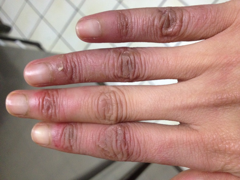
Dorsal view of the right hand.
On examination she had a purpuric scaly rash on the fingers of the right hand and livido reticularis was noted on her legs. There was good range of movement in the right hand, however her grip was impaired due to swelling of the little and ring fingers.
Her medical history included childhood asthma and childhood seborrhoeic dermatitis. She had no significant family history and was not taking any regular medications.
Investigations
Our patient was referred to the emergency dermatology clinic where she underwent a set of standard blood tests including a full blood count, urea and electrolytes, C reactive protein and a clotting screen which were all normal. Her erythrocyte sedimentation rate was raised at 24. The systemic lupus erythematosus screen was negative as per the dilute Russell viper venom screen and dilute activated partial thromboplastin time results. The IgG and IgM anticardiolipin antibodies, IgG, IgA, IgM, rheumatoid factor, neutrophil cytoplasmic antibodies, crithidia DNA immunofluorescence, antistreptolysin O titre, anti-DNase B, protein electrophoresis and a hepatitis screen were all normal. The human epithelial cell line (Hep2) antinuclear antibody was weakly positive and there was a speckled Hep2 nuclear pattern, which is associated with anti-Ro (SS-A)/LA (SS-B) antibodies. Cryoglobulin and cold agglutinin studies were both negative. She had a finger swab and urine culture, which did not grow any organisms and she underwent a skin punch biopsy for immunofluorescence, which showed fibrinogen deposition in the dermal vessels; the histology displayed perivascular lymphocyte infiltrates in the dermis, hyperkeratosis and necrotic keratinocytes. These findings are consistent with the diagnosis of chilblain lupus erythematosus as per the Mayo Clinic Diagnostic Criteria.1
Differential diagnosis
The differential diagnoses of a purpuric skin rash include chilblain lupus erythematosus, idiopathic chilblains and lupus pernio.
Idiopathic chilblains are a skin disorder thought to occur due to the disturbance of dermal blood flow control. They present similarly to chilblain lupus erythematosus with oedematous, erythematous/violaceous plaques/papules commonly affecting acral regions of the body.2
Idiopathic chilblains have similar findings on histology to chilblain lupus erythematosus, however, there is an absence of fibrin deposition on immunofluorescence.3 4
Lupus pernio is a characteristic cutaneous form of sarcoidosis and presents with chronic violaceous/erythematous plaques/papules. It is usually associated with upper respiratory disease and is most commonly found on the face and ears. Histologically, lupus pernio is a specific lesion characterised by non-caseating granulomas.5
Treatment
Our patient was started on a course of hydroxychloroquine and followed up in the dermatology clinic.
Outcome and follow-up
Three months later our patient’s finger lesions have nearly completely resolved with treatment and she has not displayed any other signs of cutaneous or systemic lupus erythematosus.
Discussion
Chilblain lupus erythematosus is a rare skin condition and up to date only a small number of cases have been reported.3
It is a form of chronic cutaneous lupus erythematosus, which is an autoimmune inflammatory disease of the skin and may be present in people with or without systemic lupus erythematosus.6 Approximately 20% of patients go on to develop systemic lupus erythematosus.3
Chilblain lupus erythematosus presents with a rash mainly affecting acral surfaces that are most exposed to the cold such as the toes, fingers, ears and nose. The rash is characterised by tender plaques that have a purple discolouration, nodules (which may have central erosions/ulcerations) and oedematous skin.2 The lesions tend to start off as a pruritic rash and thereafter become tender.7
The underlying pathophysiology is of an autoimmune nature and most cases of chilblain lupus erythematosus are sporadic; however, there have been cases noted of a rare familial form, which is inherited in an autosomal dominant manner.3 Sporadic chilblain lupus erythematosus is thought to be triggered by cold-induced microvascular injury or vasoconstriction.3
Chilblain lupus erythematosus may be diagnosed based on clinical presentation and histopathological examination or direct immunofluorescence study.
A set of diagnostic criteria1 have been proposed by the Mayo Clinic after studying a small number of patients with chilblain lupus erythematosus. This includes the presence of two major criteria: acral skin lesions associated with cold temperature and evidence of lupus erythematosus in skin lesions on histopathology or direct immunofluorescence, and one minor criterion: systemic lupus erythematosus/discoid lupus erythematosus or response to antilupus erythematosus therapy or negative cryoglobulin/cold agglutinin studies.
Histological features of chilblain lupus erythematosus include vacuolar interface dermatitis, perivascular lymphocytic infiltration and papillary oedema.3 4 Immunofluorescence study displays dermoepidermal junction deposition of IgM, IgA, C3 and fibrinogen.3
Patients with chilblain lupus erythematosus may also display hypergammaglobulinaemia (over 2/3), positive rheumatoid factor (in half), antinuclear antibody, antiphospholipid or anti-Ro antibodies. They are usually negative for antidouble-stranded DNA antibodies.3
The managementof chilblain lupus erythematosus is still unclear and more research needs to be conducted in this area. Treatment aims are to reduced disease activity and minimise damage.
The first-line treatment for mild and localised cutaneous lupus erythematosus is topical corticosteroids. Studies8 have also shown benefit from the use of topical tacrolimus and pimecrolimus. If the lesions are more extensive or resistant to topical therapy, the first-line systemic treatment consists of oral corticosteroids or antimalarials.8 9
Second-line systemic treatments consist mainly of immunomodulators and immunosuppresants7 and are used if patients are not responding to or unable to tolerate first-line medications.
The use of antisystemic lupus erythematosus medication was found to be beneficial to the Mayo Clinic study group,3 and studies8 9 conducted on the management of cutaneous lupus erythematosus have also displayed successful management of the disease using antimalarials, steroids and immunomodulators/immunosupressants. In addition to antisystemic lupus erythematosus medication, calcium channel blockers may also play a role in the treatment of chilblain lupus erythematosus,3 however, they have been linked with drug-induced lupus.10
Our patient showed a good response to hydroxychloroquine again reinforcing the benefits of antisystemic lupus erythematosus medication in the management of chilblain lupus erythematosus.
Learning points.
Chilblain lupus erythematosus is a form of cutaneous lupus erythematosus which should be considered as a differential diagnosis when faced with a cold-induced purpuric rash.
Approximately, 20% of cases of chilblain lupus erythematosus may go onto develop systemic lupus erythematosus.
The Mayo Clinic Diagnostic Criteria, which takes into consideration the clinical presentation, histopathological and immunofluorescence studies and treatment response can aid diagnosis.
Antisystemic lupus erythematosus medication is beneficial in the treatment of chilblain lupus erythematosus.
Footnotes
Contributors: SP planned, researched and drafted the case report then it was edited by FH.
Competing interests: None.
Patient consent: Obtained.
Provenance and peer review: Not commissioned; externally peer reviewed.
References
- 1.Su WP, Perniciaro C, Rogers RS, et al. Chilblain lupus erythematosus (lupus pernio): clinical review of the Mayo Clinic experience and proposal of diagnostic criteria. Cutis 1994;2013:395–9 [PubMed] [Google Scholar]
- 2.AlMahameed A, Pinto DS. Pernio (chilblains). Curr Treat Options Cardiovasc Med 2008;2013:128–35 [DOI] [PubMed] [Google Scholar]
- 3.Hedrich CM, Fiebig B, Hauck FH, et al. Chilblain lupus erythematosus-a review of literature. Clin Rheumatol 2008;2013:949–54 [DOI] [PubMed] [Google Scholar]
- 4.Crowson AN, Magro C. The cutaneous pathology of lupus erythematosus: a review. J Cutan Pathol 2001;2013:1–23 [DOI] [PubMed] [Google Scholar]
- 5.Fernandez-Faith E, McDonnell J. Cutaneous sarcoidosis: differential diagnosis. Clin Dermatol 2007;2013:276–87 [DOI] [PubMed] [Google Scholar]
- 6.Kuhn A, Sticherling M, Bonsmann G. Clinical manifestations of cutaneous lupus erythematosus. J Dtsch Dermatol Ges 2007;2013:1124–40 [DOI] [PubMed] [Google Scholar]
- 7.Lin JH, Dutz JP, Sontheimer RD, et al. Pathophysiology of cutaneous lupus erythematosus. Clin Rev Allergy Immunol 2007;2013:85–106 [DOI] [PubMed] [Google Scholar]
- 8.Weizel J, Brahler S, Baler R, et al. Efficacy and safety of methotrexate in recalcitrant cutaneous lupus erythematosus: results of a retrospective study in 43 patients. Br J Dermatol 2005;2013:1365–2133 [DOI] [PubMed] [Google Scholar]
- 9.Chang AY, Werth VP. Treatment of cutaneous lupus. Curr Rheumatol Rep 2011;2013:300–7 [DOI] [PMC free article] [PubMed] [Google Scholar]
- 10.Marzano AV, Vezzoli P, Crosti C. Drug-induced lupus: an update on its dermatologic aspects. Lupus 2009;2013:935–40 [DOI] [PubMed] [Google Scholar]