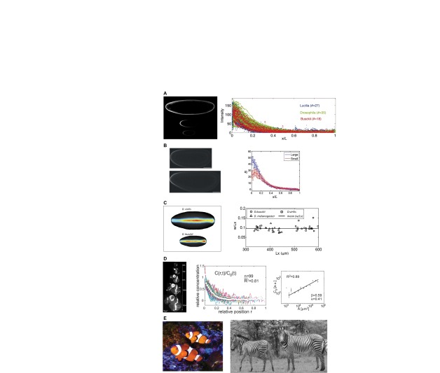
Fig. 1.
Scale invariance of patterns in diverse contexts. (A) Interspecies scaling of the Bcd protein distribution in blastoderm embryos. Image (left) shows Bcd immunofluorescence stains for Lucilia sericata (top), Drosophila melanogaster (middle) and Drosophila busckii (bottom). The plot (right) shows the distribution of fluorescence in each species at relative spatial positions. Reproduced from Gregor et al. (Gregor et al., 2005). (B) Intraspecies scaling of the Bcd gradient in Drosophila melanogaster populations that have undergone rounds of artificial selection based on egg size. The image (left) shows a large embryo (bottom) and a small embryo (top). Image brightness was enhanced for clarity. The plot (right) shows the relative Bcd staining intensity for large and small embryos as a function of relative spatial position. Reproduced with permission (Cheung et al., 2011). (C) Scaling of dorsal surface patterning by bone morphogenetic proteins is an example of a complex, highly non-linear spatial patterning system. The image (left) shows the distribution of pMad in a large Drosophila virilis embryo (top) and a small Drosophila busckii embryo (bottom). The plot (right) shows the ratio of average pattern width to embryo length versus the embryo length. A threshold of signaling intensity of 0.2 was used for comparison of widths. Adapted with permission (Umulis et al., 2010). (D) Scaling of the Dpp gradient during growth of the wing imaginal disc is an example of dynamic scaling. The image (left) shows Dpp-GFP expression at various time points during development. Normalized Dpp-GFP profiles from multiple time-points (center) are shown at relative spatial positions. During growth, the amplitude of the morphogen gradient grows (right) in proportion to the length of the disc squared. Adapted with permission (Wartlick et al., 2011). (E) Pigmentation patterns on the skin of the clownfish Amphiprion percula (left; photo by D. M. Umulis) and coat patterns on zebras (right). Zebra image reprinted with permission (Cordingley et al., 2009).

