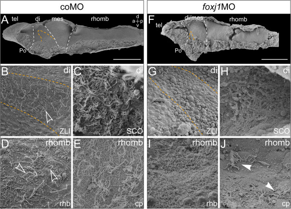
Figure 6.
Reduced CSF flow velocity correlates with defects in ciliogenesis. Scanning electron microscopy (SEM) of dissected brain explants at stage 46. (A-E) Control morpholino (coMO) -injected specimen. (F-J)foxj1 morpholino (foxj1MO) -injected specimen. (A,F) SEM pictures of brain explants dissected sagittally with view on the ventricular surface, the zona limitans intrathalamica (ZLI) is delimited with orange dashed line and boundaries between brain regions indicated by white dashed lines. Bars represent 200 ?m. (B,G) View onto the ZLI in the diencephalon (di). Note the loss of cilia on the ZLI in foxj1 morphants (G). (C,H) Close-up views onto the subcommissural organ (SCO) in frontally dissected specimens. (D,I) Close-up views onto the rhombencephalon in sagittally dissected specimens, depicting the rhombomere boundaries. Outlined arrows in (D) point to elongated monocilia. Note the absence of cilia in (I). (E,J) Close-up view onto the choroid plexus (cp) of the hindbrain roof. Arrowheads in (J) point to the few remaining MCCs in foxj1 morphants. a = anterior; d = dorsal; mes = mesencephalon; tel = telencephalon; rhomb = rhombencephalon; Po = preoptic region; p = posterior; rhb = rhombomere boundary; v = ventral.

