Abstract
We examined temperature dependence in plastic scintillation detectors (PSDs) made of BCF-60 or BCF-12 scintillating fiber coupled to optical fiber with cyanoacrylate. PSDs were subjected to a range of temperatures using a temperature-controlled water bath and irradiated at each temperature while either the dose was measured using a CCD camera or the spectral output was measured using a spectrometer. The spectrometer was used to examine the intensity and spectral distribution of scintillation light emitted by the PSDs, Cerenkov light generated within the PSD, and light transmitted through an isolated optical coupling. BCF-60 PSDs exhibited a 0.50% decrease and BCF-12 PSDs a 0.09% decrease in measured dose per °C increase, relative to dose measured at 22°C. Spectrometry revealed that the total intensity of the light generated by BCF-60 and BCF-12 PSDs decreased by 0.32% and 0.13%, respectively, per °C increase. The spectral distribution of the light changed slightly with temperature for both PSDs, accounting for the disparity between the change in measured dose and total light output. The generation of Cerenkov light was temperature independent. However, light transmitted through optical coupling between the scintillator and the optical fiber also exhibited temperature dependence.
I. Introduction
The plastic scintillation detector (PSD) is a thoroughly studied detector notable for a unique collection of characteristics that make it well suited for dosimetry. For example, previous studies have established that PSDs are water equivalent; exhibit a linear relationship between scintillation light and deposited dose; are energy, dose rate, and angularly independent; have a high spatial resolution; and are temperature independent (Beddar et al 1992a, Beddar et al 1992b). Some of these characteristics, most notably temperature independence, have been accepted as fact without independent validation by other groups.
Twenty years have passed since the initial studies were conducted that established these characteristics, and the design and construction of PSDs has changed in that time. Specifically, the first PSD described in the published literature was constructed with a BC-400 scintillator coupled to a silica light guide using silicon optical coupling grease (Beddar et al 1992a). It is now not uncommon to use different materials; SCSF-3HF(1500), SCSF-78, BCF-12, and BCF-60 scintillating fibers often replace BC-400 owing to their superior light collection and/or spectral properties. Plastic optical fibers are commonly substituted in place of the silica light guide to achieve better water equivalence. Cyanoacrylate or epoxies are regularly used for optical coupling (Archambault et al 2005a, Ayotte et al 2006).
New generations of PSDs have been shown to possess almost all of the dosimetric characteristics of the original PSDs from the 1992 study, including response linearity, water equivalence, and energy, dose rate, and angular independence, as evidenced by their successful use in increasingly advanced dosimetric studies (Archambault et al 2010, Klein et al 2010, Klein et al 2012). However, to the best of our knowledge, temperature independence has not been independently validated or investigated for either the original PSD or any subsequent generations of PSDs.
We were prompted to investigate temperature dependence in response to a systematic error exhibited by PSDs employed in an ongoing in-vivo dosimetry protocol at our institution. These PSDs were regularly subjected to both in-vivo dose measurements in patients and in-phantom validation designed to replicate the in-vivo conditions. The dose measured by the PSDs in the phantom agreed excellently with the calculated dose in the treatment planning system; however, the dose measured in vivo differed from that calculated by the treatment plan. Because the phantom was at room temperature during validation, we concluded that temperature dependence was an important avenue of investigation.
A brief initial investigation, reported in a letter to the editor previously (Beddar 2012), confirmed that the PSDs did indeed exhibit temperature dependence. This investigation indicated that the measured dose decreased by an average of 0.6% per C increase, relative to room temperature, for PSDs made with BCF-60 scintillating fibers. This prompted us to conduct a more thorough systematic investigation of the effects of temperature on PSDs built with BCF-12 and BCF-60 scintillating fibers, which are two of the most common scintillating fibers used in PSDs. In this article, we present our investigation and report the results.
II. Methods and Materials
II.A. Detectors
The PSDs used for this study were constructed according to the following method. A 2-mm length of either BCF-60 or BCF-12 scintillating fiber (Saint-Gobain Crystals, Hiram, OH) was optically coupled to an Eska GH-4001-P clear plastic optical fiber (Mitsubishi Rayon Corporation, Japan) with cyanoacrylate glue. The abutting ends of the scintillating fiber and the optical fiber were polished with fine grit polishing paper to facilitate high optical transmission efficiency (Ayotte et al 2006). The entire assembly was light-shielded in black polyethylene jacketing. Additionally, an opaque, black, alcohol-based adhesive was used to conceal exposed portions of scintillating fiber and optical fiber (e.g. at the end of the PSD where the scintillating fiber terminated) to prevent the admission of external light that would contaminate the PSD signal. Approximately 20 m of optical fiber was used to span the distance between the linac and the outside of the vault. The optical fiber was connected either to a Luca S CCD Camera (Andor Technology, Belfast, Northern Ireland) via a ST optical connector for dose measurements or to an Andor Shamrock 163 spectrometer via a SMA optical connector for spectral analysis. The connectors ensured a secure and reproducible connection.
Calibration was performed under cobalt-60 irradiation using the chromatic removal technique (Fontebonne et al 2002, Frelin et al 2005, Archambault et al 2005b) to distinguish scintillation light from contaminating Cerenkov radiation (Beddar et al 1992c). In order to implement this technique, a dichroic mirror (model NT47-950, Edmund Optics Inc., Barrington, NJ) was used to split the light produced by the PSDs into 2 spectrums prior to imaging by the CCD.
II.B. Experimental Setup
To subject PSDs to a variety of stable temperatures, PSDs were placed into a 250-mL beaker filled with water that was placed on top of a hotplate. A cap of dense blue Styrofoam was fashioned to fit tightly into the top of the beaker to insulate the water, and small perforations in the Styrofoam cap allowed the PSDs access to the water. A thermometer was also inserted through the center of the Styrofoam cap into the water to monitor the temperature. The bulb of the thermometer was placed at the same depth as the active volume of the PSDs to provide the most accurate assessment of the PSD temperatures.
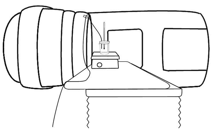
The hotplate (model PC-620D; Corning Incorporated, Corning, NY) included a magnetic stirring device which was used to facilitate a homogeneous temperature distribution of the water. The hotplate was placed on a lateral edge of a linac couch. The gantry head was rotated to 270 degrees to position the beam perpendicularly to the PSDs, and the couch was then moved laterally as close to the linac head as possible to maximize the signal from the PSDs (Figure 1).
Figure 1.
Experimental setup. The gantry was rotated to irradiate the plastic scintillation detector (PSD) perpendicularly and the couch was shifted laterally to maximize the signal from the PSD. A beaker of water was placed on a hotplate and the PSDs and a thermometer were held in place in the beaker by a Styrofoam cap. The optical fiber transmitting scintillation light was taped to the linac head to prevent motion in and out of the field, which would alter the Cerenkov contribution to the PSD signal.
Prior to obtaining each set of measurements, we filled the beaker with a combination of water and crushed ice. The cap with the PSDs and thermometer was then affixed to the top of the beaker, and the stirrer used to bring the water to thermal equilibrium. The beaker was filled completely with water, so that no air gap was present between the water and the bottom of the Styrofoam cap.
Each data point was acquired using the following protocol. First, the hotplate setting was adjusted to bring the water to the desired temperature. After the temperature was stabilized but before the irradiation, the water temperature was held constant for an additional 10 minutes to allow the PSDs to come to thermal equilibrium with the water. Although less time was likely required (the jacketing consisted of polyethylene, the scintillator of polystyrene, and the optical fiber of PMMA, each of which have a thermal conductivity similar to water), 10 minutes was chosen to ensure that the water temperature measured by the thermometer accurately reflected the PSD temperature. After this waiting time, the detectors were irradiated with 100 monitor units (MU) 3 times, for statistical purposes, and the output was captured with either the CCD or the spectrometer as described below.
It was not possible to stabilize the water temperature below room temperature; no means of cooling the setup without disturbing it were available. Therefore, for these measurements, we averaged the temperature measured before and after the irradiation (typically differing by 0.5°C) and assumed that this accurately reflected the PSD temperature.
II.C. Dose Measurements
Dose was measured for a pair of BCF-60 PSDs and a pair of BCF-12 PSDs to quantify the effect of temperature on measured dose. Measurements spanned a range from approximately 15°C to 40°C. Twenty-second CCD light-integrating acquisitions were used to measure the light output resulting from each 100 MU irradiation. An average background image was subtracted from the measurement images, from which dose values were subsequently obtained via analysis in ImageJ (Archambault et al 2008). The resulting dose values were normalized to the dose measured at room temperature, here defined as 22°C. If no measurement was made at 22°C, the value was obtained by interpolating between the 2 closest points.
II.D. Spectrometry
The effect of temperature on the intensity and spectral distribution of light of 4 different PSD configurations was quantified with a spectrometer. The following PSD configurations were used: a PSD built with BCF-60 scintillating fiber, a PSD built with BCF-12 scintillating fiber, a bare fiber (i.e. an optical fiber without a scintillating element or cyanoacrylate, thus capable only of generating Cerenkov light), and a PSD with an isolated cyanoacrylate optical coupling.
For measurements involving the bare fiber, only the submerged portion of the fiber that was subject to temperature changes was irradiated. This was done to ensure that any temperature dependence of the production and transmission of Cerenkov light would not be masked by Cerenkov generated elsewhere in the optical fiber.
To create an isolated optical coupling, the optical fiber of a BCF-60 PSD was cut 2 m below the scintillating element and reattached with cyanoacrylate. More cyanoacrylate than would typically be used in the fabrication of a PSD was employed to exaggerate any effects. Only this optical coupling was submerged in the water and subjected to temperature changes, while the scintillating element of the PSD was maintained at room temperature outside of the beaker and irradiated to generate a light signal that would be transmitted through the isolated coupling.
Twenty-second acquisitions were used for each 100-MU irradiation. An average background spectrum was subtracted from each measurement. To distinguish between the Cerenkov radiation and the scintillation contributions to the spectral measurements, a pure Cerenkov spectrum and a pure BCF-60 or BCF-12 scintillation spectrum were fitted to the total spectral output at room temperature for each configuration using the least squares method. The fitted Cerenkov spectrum was then subtracted from measurements at other temperatures, leaving only the scintillation spectra (the Cerenkov spectrum was demonstrated to be unchanged by changing temperature). The fitted room temperature Cerenkov spectrum needed to be subtracted at non-room temperatures because fitting the pure scintillation spectrum would not correctly determine the scintillation signal if the spectral distribution of the scintillator was temperature dependent. The pure Cerenkov spectrum was obtained from the bare fiber. The pure scintillation spectra were obtained at room temperature by irradiating the scintillators on a kV irradiation unit; the low-energy radiation of the kV irradiator produces a negligible amount of Cerenkov radiation (Therriault-Proulx et al 2012).
The spectra of each PSD configuration were analyzed to determine the wavelength at which the maximum intensity change occurred and the magnitude of that change, the change in the total intensity of the spectrum, and the change in the distribution of the spectrum. As a metric to quantify the change in the distribution, we calculated the ratio of the change in intensity of the portion of the spectrum that would be reflected by the dichroic mirror used in our CCD setup (520-550 nm) to the change in intensity of the portion that would be transmitted by it (the remaining visible spectrum).
II.E. Detector Stabilization
An additional experiment was performed to determine whether the temperature-dependent response of the PSDs at non-room-temperatures stabilized, and if so, how quickly. The water-filled beaker was heated to 29°C while a BCF-60 PSD was maintained at room temperature outside the beaker. After the water temperature stabilized at 29°C, the PSD was inserted through the Styrofoam cap and measurements of 100-MU irradiations were immediately commenced at a frequency of 1 per minute for 40 minutes. The first measurement was made approximately 50 seconds after introducing the PSD into the beaker. This delay was necessary to exit the room and close the vault door.
For comparison, these measurements were repeated in air (i.e., the beaker was not filled with water) and without the application of heat. This was done to eliminate the possibility that changes in output were due to other effects such as fatigue of the CCD camera.
III. Results
III.A. Dose Measurements
The measured dose for each pair of PSDs decreased with increasing temperature across the entire temperature range (Figure 2). The relationship between the BCF-60 PSD measured dose and temperature was predominantly linear, although a small nonlinear component was present. The measured dose for the BCF-60 PSD decreased by approximately 0.50% per °C increase relative to room temperature. The relationship between the BCF-12 PSD measured dose and temperature was linear, with the measured dose decreasing by 0.09% per °C increase.
Figure 2.
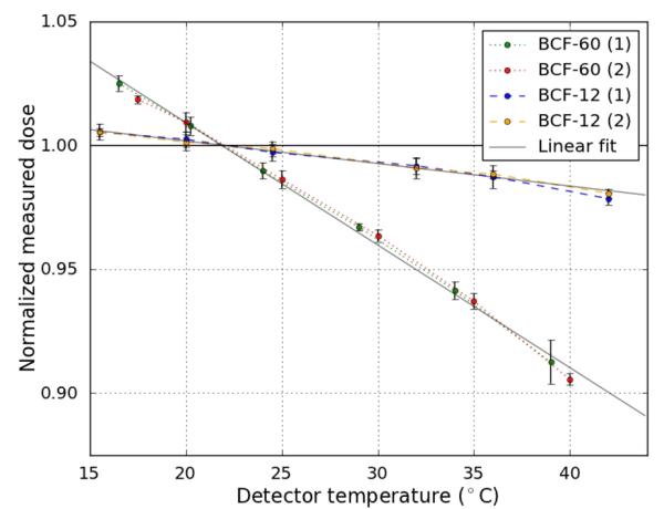
Dose measurements obtained under changing temperature conditions from 2 pairs of plastic scintillation detectors made with BCF-12 and BCF-60 scintillating fibers. A steady decrease in the measured dose was observed with increasing temperatures. Each point is the average of 3 measurements, and the error bars represent 2 standard deviations of those measurements. Linear fits show that BCF-60 exhibited slightly nonlinear temperature dependence, whereas the BCF-12 temperature dependence pattern was entirely linear.
III.B. Spectrometry
Spectrometry data for irradiation of the bare fiber revealed that neither the total intensity nor the distribution of the Cerenkov spectrum changed as a function of temperature (Figure 3).
Figure 3.
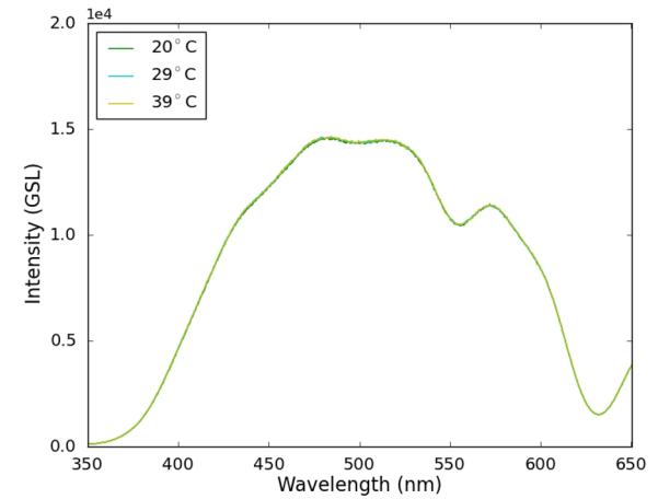
Cerenkov spectra. Only a staggered selection of data points is displayed because all spectra overlapped (the data points not shown in this chart adhered exactly to this phenomenon—see Figure 7). The shape and intensity of the Cerenkov spectrum did not change discernibly with rising temperatures.
However, considerable change in the intensity of the BCF-60 PSD output was observed for wavelengths between 500 nm and 600 nm, with no discernible change in output outside of that range (Figure 4). Between 500 nm and 600 nm, the maximum intensity loss occurred at 510 nm and was equal to 0.60% per °C relative to room temperature. The total light output of the BCF-60 PSD decreased at a rate of 0.32% per °C in a dominantly linear fashion, with a small nonlinear component. The portion of the spectrum that would be reflected by the dichroic filter decreased in intensity at a rate of 0.59% per °C increase, whereas the rest of the spectrum intensity decreased at a rate of only 0.43% per °C increase, a ratio of 1.37 (i.e., the reflected portion decreased in intensity 37% more rapidly than the rest of the spectrum).
Figure 4.
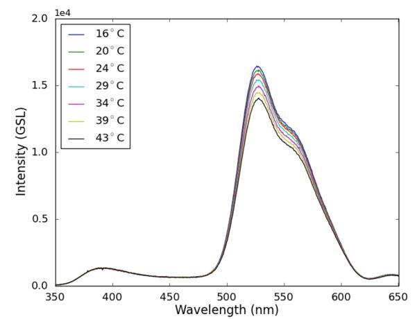
BCF-60 spectra. The spectrum intensity changed substantially between 500 nm and 600 nm and remained constant at other wavelengths.
A markedly less severe loss of intensity was observed in the spectra of the BCF-12 PSD, this time constrained to the regions between 400 nm and 500 nm (Figure 5). The maximum intensity loss occurred at approximately 410 nm: a 0.30% decrease per °C increase relative to room temperature. The total light output of the BCF-12 PSD decreased by 0.13% per °C in a linear fashion. The intensity of the portion of the spectrum corresponding to light reflected by the dichroic filter decreased at a rate of 0.02% per °C, whereas the intensity of the remaining spectrum decreased at a rate of 0.12% per °C, a ratio of 0.13.
Figure 5.
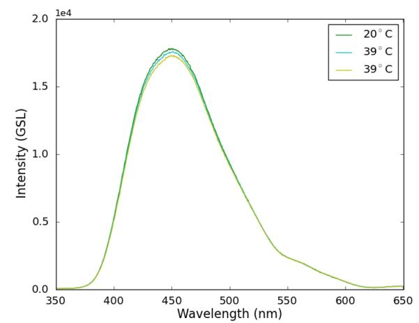
BCF-12 spectra. The spectrum intensity changed only slightly in the range of 400-500 nm. Owing to the close proximity of the spectral curves, only a staggered selection of data points is displayed to make the changes more visible. Other data points not shown here adhered well to the trend displayed above—see Figure 7.
For the cyanoacrylate coupling, a nonlinear decrease in transmitted light was observed with increasing temperature. Note that because the change was nonlinear, all values presented are the difference between the intensity at 38°C and 22°C. Intensity loss occurred primarily between 500 nm and 600 nm (Figure 6). The maximum intensity loss of 4.2% occurred at 650 nm. A 2.5% loss in total light intensity was observed. The intensity of the reflected portion of the spectrum decreased by 3.6%, whereas the intensity of the remaining spectrum decreased by only 2.5%, a ratio of 1.4.
Figure 6.
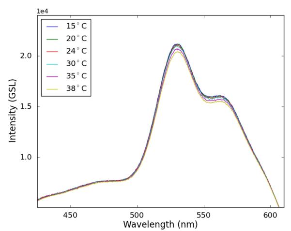
Isolated optical coupling. A small temperature-dependent decrease was observed in the light transmitted through the cyanoacrylate coupling. Note that the limits of the x and y axis here differ from those of other spectra figures to give a magnified view of this spectrum.
The total light output for each PSD configuration is plotted in Figure 7.
Figure 7.
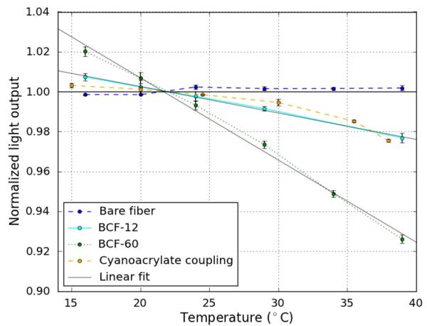
Total light output of each detector configuration as measured with a spectrometer. A more severe decrease in light output was observed for the BCF-60 PSD than for the BCF-12 PSD. Cerenkov light did not exhibit any temperature dependence. The cyanoacrylate coupling exhibited a temperature-dependent transmission. Each point is the average of 3 measurements, and the error bars represent 2 standard deviations of those measurements. Linear fits demonstrate that that the intensity change of the BCF-60 PSD had a small nonlinear component, whereas the temperature dependence pattern for the BCF-12 PSD was entirely linear.
III.C. Detector Stabilization
All measurements made with the PSD maintained at 29°C were within 0.50% of the average measured value. The measurements did increase very slightly over the course of the experiment; at the conclusion of the experiment it was noted that the water temperature had decreased by 1.5°C, which accounted for the increase in the measured values.
When the experiment was repeated in air, all measured values fell within approximately 0.50% of the average measured value, and no trend was observed. These results are displayed in Figure 8.
Figure 8.
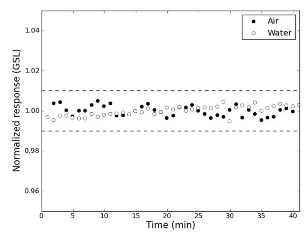
Stabilization. A room-temperature plastic scintillation detector immersed in 29°C water displayed a stable response over 40 minutes, from the first measurement at 50 seconds after immersion (dashed lines indicate ±1% from the average response). A slight upward trend owing to a small decrease in the temperature of the water over the 40 minutes was observed. Identical measurements in air confirmed that the plastic scintillation detector was stable under normal conditions.
IV. Discussion
Contrary to widely accepted knowledge, our results indicate that PSDs are not universally temperature independent. The temperature dependence of BCF-60 PSDs is on the order of 1% within a few degrees of room temperature and on the order of 10% at human body temperature. The effect of temperature on BCF-12 PSDs is much smaller but would still contribute a systematic error in measured dose at noncalibration temperatures if uncorrected. Clearly the effect of temperature must be accounted for in current PSDs and minimized in future PSDs if possible.
Measurements with pairs of PSDs revealed nearly identical temperature dependence for PSDs built with like scintillating-fibers. From these results, we conclude that the temperature dependence of one PSD is sufficient to characterize the dependence of a large number of PSDs if care is taken to standardize the construction.
Our spectrometry data revealed several points of interest. First, neither the intensity nor the spectral distribution of the Cerenkov light collected from the bare fiber was temperature dependent. From this, we conclude that Cerenkov production and attenuation of light in the range of wavelengths spanned by the Cerenkov light in the optical fiber are temperature independent. The Cerenkov spectrum spans both the BCF-12 and the BCF-60 spectrum, so the temperature-independent attenuation of the optical fiber holds for both types of scintillators. Accordingly, any temperature dependence of the PSDs must be caused by changes in the light emitted from the scintillator or the transmission of light through the optical coupling. Additionally, the temperature independence of Cerenkov radiation is important because the chromatic removal technique assumes that only the intensity and not the spectral distribution of Cerenkov light changes as irradiation conditions change.
Second, the distributions of the scintillators’ spectra were observed to change slightly in addition to the total intensity. This is problematic because the chromatic removal technique requires the spectral distribution of scintillation light to be constant to extract the dose from the total light output correctly. Our implementation of this technique using a dichroic mirror specifically requires that the ratio of the portion of scintillation light reflected to the portion transmitted is constant. As stated in the results, this was not the case. This means that not only will the dose measured be incorrect owing to the change in scintillation light produced per unit dose, but the error will also be compounded by the incorrect extraction of dose from the total light.
Lastly, our spectrometry data showed that the transmission of light through cyanoacrylate optical coupling exhibited a nonlinear temperature dependence. The temperature dependence was largely confined to the wavelengths between 500 nm and 600 nm, which is why the relationship between light output and temperature was partially nonlinear for BCF-60 PSDs (most of the BCF-60 emission spectra fell in this range) but not for BCF-12 . Because the quantity of cyanoacrylate used in our experiments was greater than that used in a typical PSD, it is not possible to determine how much of either the BCF-12 or BCF-60 temperature dependence stems from the use of a cyanoacrylate coupling. However, given the relatively small nonlinearity of the BCF-60 PSDs compared with the cyanoacrylate transmission, it is safe to say that the cyanoacrylate contributes only a small amount to the BCF-60 detectors’ temperature dependence.
The stability tests revealed that the PSD output stabilizes very rapidly when the temperature changes, reaching equilibrium in the first minute. This is likely due to the small size of the PSD, which has a diameter of only 2.3 mm, and the water-like thermal conductivity of the materials that make up the PSD. This is ideal for time-sensitive applications (e.g., integrating in-vivo dosimetry into the treatment workflow).
The disparity between the responses of BCF-60 and BCF-12 PSDs to temperature increases yields insight into an additional possible source of the temperature dependence. The key difference between these 2 scintillators is the wavelength shifting fluors used to convert the scintillation light, which is emitted primarily in the ultraviolet spectrum, to the visible spectrum. One fluor shifts ultraviolet light to blue light in BCF-12 and BCF-60 scintillators. In BCF-60 scintillators, a second fluor is responsible for shifting blue light to green light. This suggests that the wavelength shifting fluors may be partially responsible for the temperature dependence. Published data support this conclusion. Rozman and Kilin (1960) found that when a variety of fluors were incorporated into polystyrene scintillator, each combination exhibited temperature dependence and the dependence differed greatly depending on the fluor used. Surprisingly, Rozman and Kilin also observed temperature dependent emission of light in pure polystyrene.
Several methods to correct for the temperature dependence are possible. The simplest method is to determine the ratio of the measured dose at various temperatures to the measured dose at the temperature at which the detector was originally calibrated and use the inverse of these ratios as temperature-specific correction factors. However, this method does not account for the change in the spectral distribution. As stated previously, the changing distribution of the scintillation spectrum compromises the chromatic removal technique. Thus, this correction will only be exactly correct when irradiation conditions (e.g., field size, depth) are identical to the conditions used to determine the ratios. The change in distribution of the spectrum is small for both detectors, so this may introduce an acceptably small error into measurements. However, our study did not test this and cannot confirm that this is in fact the case; thus, further research is warranted.
A more effective but much more cumbersome solution would be to calibrate the detectors at a range of temperatures; then the resulting calibration coefficients would be based on the correct intensity and spectral distribution of light at each temperature. If the correct temperature-specific calibration was used, no further correction would be necessary.
Perhaps the best solution would be to construct a PSD that is minimally affected by temperature. To accomplish this, a scintillator with less intrinsic temperature dependence must be found. Rozman and Kilin’s (1960) finding of a broad range of temperature dependence patterns for different wavelength shifting fluors suggests that this is possible. The BC-400 scintillator may be a good candidate, because Beddar et al (1992a) found that it had no temperature dependence. Additionally, cyanoacrylate should not be used as optical coupling for scintillators emitting primarily in the green region of the visible spectrum. Other couplings need to be investigated before a recommendation can be made. Ayotte et al (2006) found that a detector with no optical coupling is feasible if well polished, outputting approximately the same amount of light that a coupled scintillator might. Unfortunately, this may result in a less robust detector because of the impermanent connection between the scintillator and the optical fiber. It does not appear to be necessary to replace the plastic optical fiber.
V. Conclusion
We have found that PSDs built with BCF-60 or BCF-12 scintillating fiber exhibit nonnegligible temperature dependence, with BCF-60 PSDs exhibiting greater temperature dependence than BCF-12 PSDs. For BCF-60 PSDs calibrated at room temperature, the dependence is on the order of 1% near room temperature and on the order of 10% at human body temperature. The exact mechanism of this temperature dependence is not known, but the wavelength shifting fluors and temperature-dependent transmission through cyanoacrylate appear to be at least partially responsible. We have suggested that a temperature-specific correction factors derived by characterizing the temperature dependence may be sufficient to account for this effect, although further research is required to validate this assertion. Alternatively, temperature-specific calibrations would account for the effect. In addition, carefully selecting the scintillator and optical coupling used in new PSDs may reduce the temperature dependence. Further research is needed to determine the optimal materials to use in a PSD to reduce or eliminate the temperature dependence.
References
- Archambault L, et al. Plastic scintillation dosimetry: Optimal selection of scintillating fibers and scintillators. Med. Phys. 2005a;32:2271–2278. doi: 10.1118/1.1943807. [DOI] [PubMed] [Google Scholar]
- Archambault L, et al. Measurement accuracy and Cerenkov removal for high performance, high spatial resolution scintillation dosimetry. Med. Phys. 2005b;33:128–135. doi: 10.1118/1.2138010. [DOI] [PubMed] [Google Scholar]
- Archambault L, et al. Transient noise characterization and filtration in CCD cameras exposed to stray radiation from a medical linear accelerator. Med. Phys. 2008;35:4342–4351. doi: 10.1118/1.2975147. [DOI] [PMC free article] [PubMed] [Google Scholar]
- Archambault L, et al. Toward a real-time in vivo dosimetry system using plastic scintillation detectors. Int. J. Radiat. Oncol. Biol. Phys. 2010;78:280–287. doi: 10.1016/j.ijrobp.2009.11.025. [DOI] [PMC free article] [PubMed] [Google Scholar]
- Ayotte G, et al. Surface preparation and coupling in plastic scintillator dosimetry. Med. Phys. 2006;33:3519–3525. doi: 10.1118/1.2256300. [DOI] [PubMed] [Google Scholar]
- Beddar AS, et al. Water-equivalent plastic scintillation detectors for high-energy beam dosimetry: I. Physical characteristics and theoretical considerations. Phys. Med. Biol. 1992a;37:1883–1900. doi: 10.1088/0031-9155/37/10/006. [DOI] [PubMed] [Google Scholar]
- Beddar AS, et al. Water-equivalent plastic scintillation detector for high energy beam dosimetry: II. Properties and measurements. Phys. Med. Biol. 1992b;37:1901–1913. doi: 10.1088/0031-9155/37/10/007. [DOI] [PubMed] [Google Scholar]
- Beddar AS, et al. Cerenkov light generated in optical fibres and other light pipes irradiated by electron beams. Phys. Med. Biol. 1992c;37:925–935. [Google Scholar]
- Beddar AS. On possible temperature dependence of plastic scintillator response. Med. Phys. 2012;39:6522. doi: 10.1118/1.4748508. [DOI] [PubMed] [Google Scholar]
- Fontebonne JM, et al. Scintillation Fiber Dosimeter for Radiation Therapy Accelerator. IEEE Trans. Nuc. Sci. 2002;49:2223–2227. [Google Scholar]
- Frelin AM, et al. Spectral discrimination of Čerenkov radiation in scintillating dosimeters. Med. Phys. 2005;32:3000–3006. doi: 10.1118/1.2008487. [DOI] [PubMed] [Google Scholar]
- Klein DM, et al. Measuring output factors of small fields formed by collimator jaws and multileaf collimator using plastic scintillation detectors. Med. Phys. 2010;37:5541–5549. doi: 10.1118/1.3488981. [DOI] [PMC free article] [PubMed] [Google Scholar]
- Klein DM, et al. In-phantom dose verification of prostate IMRT and VMAT deliveries using plastic scintillation detectors. Radiat. Meas. 2012 doi: 10.1016/j.radmeas.2012.08.005. In Press. [DOI] [PMC free article] [PubMed] [Google Scholar]
- Rozman IM, Kilin SF. Luminescence of Plastic Scintillators. Sov. Phys. Usp. 1960;2:856–873. [Google Scholar]
- Therriault-Proulx F, et al. Development of a novel multi-point plastic scintillation detector with a single optical transmission line for radiation dose measurement. Phys. Med. Biol. 2012;57:7147–7159. doi: 10.1088/0031-9155/57/21/7147. [DOI] [PMC free article] [PubMed] [Google Scholar]


