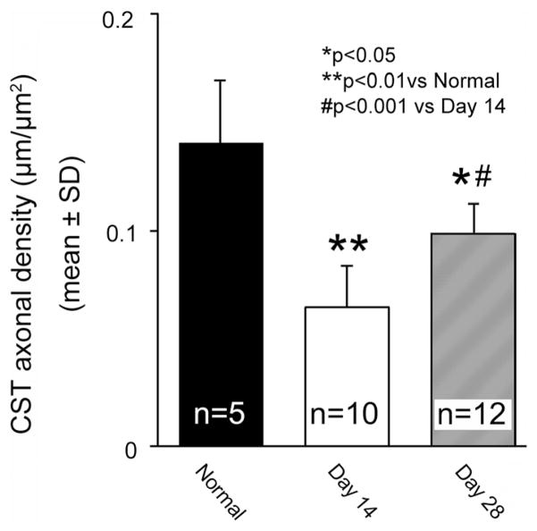
Figure 3.
Bar graph showing CST axonal density in the gray matter of the cervical cord. Note that the CST axonal density was decreased in the stroke-impaired gray matter at day 14 compared with normal mice, and significantly recovered at day 28 after MCAo-sham-BPT. BPT indicates bilateral pyramidotomy; CST, corticospinal tract; and MCAo, middle cerebral artery occlusion.

