

Abstract
To evaluate the results of facial nerve reanimation after facial nerve injury by means of hypoglossal to facial nerve anastomosis. Retrospective case review. Private neuro-otologic and cranial base quaternary referral center. Sixty patients underwent hypoglossal to facial nerve anastomosis for facial nerve reanimation between April 1987 and December 2010. Only forty patients completed a minimal follow up of 24 months at the time of evaluation and were included in the study population. Facial nerve paralysis was present for a mean duration of 11.3 months (range 2–42 months) and all the patients had a HB grade VI prior their surgery. Final facial nerve motor function. The most common cause of facial paralysis was vestibular Schwannoma surgery. All the patients achieved a postoperative HB grade III or IV after a mean follow-up time of 20 months. The facial movements were detected after a period that ranged from ranged from 5 to 9 months. Only 4 patients suffered from difficulties during eating and drinking and three of them had associated lower cranial nerve deficit. Despite the various techniques in facial reanimation following total facial nerve paralysis, the end to end of hypoglossal to facial nerve anastomosis remains one of the best treatments in cases of viable distal facial stump and nonatrophic musculature.
Keywords: Facial paralysis, Facial reanimation, Facial nerve anastomosis, Hypoglossal nerve
Introduction
Facial nerve (FN) paralysis has a major impact on patient’s quality of life, thus the main aim in temporal bone and cerebellopontine angle surgery is not only a complete eradication of the disease but also preservation of facial nerve function.
Despite the advances in intraoperative facial nerve monitoring and microsurgical techniques, injury of the nerve still occurred and required to be repaired as in resection of large vestibular Schwannoma and facial nerve tumors.
A tension-free primary end-to-end anastomosis of the facial nerve stumps is the method of choice for facial nerve reconstruction. In cases with a long nerve defect, a cable graft interposition or facial nerve rerouting are indicated; for a tensionless anastomosis the graft should be harvested 25% longer than the defect. [1].
When the central stump of the nerve is unavailable or too short to be sutured, restoring of the nerve function is accomplished using transposition of the hypoglossal nerve to the peripheral facial nerve stump.
This method is also advocated in cases of failure of cable graft and when functional recovery of the nerve did not occur despite its anatomical preservation.
Hypoglossal to facial nerve anastomosis (HFA) remains the procedure of choice in case of viable distal facial stump and non atrophic musculature. In the presence of atrophic or nonfunctional muscles following a long standing facial nerve paralysis, neuromuscular flap transfer or transposition, static techniques that include face lift, brow suspension and facial slings in addition to the lid gold weight implants and canthoplasty for eye management are the mainstay treatment to restore the function of facial muscle. [2].
This study reports the experience of the Gruppo Otologico with hypoglossal to facial nerve anastomosis.
Materials and Methods
Patient Population
Between April 1987 and December 2010, a total of 3,000 lateral skull base surgery operations were performed by the senior author (M.S.).
During this period sixty patients having a House-Brackmann grade VI facial nerve paralysis underwent hypoglossal to facial nerve anastomosis for restoring the function of facial musculature following FN paralysis.
After institutional review board approval, a retrospective chart review of the patients who completed 24 months of follow up were included in this study.
Surgical Technique
HFA can be performed under local anesthesia but we prefer to perform it under general anesthesia.
Through an extended Lahey incision, elevation of the anterior subplatysmal flap is done, the sternocleidomastoid muscle is retracted posteriorly and separated from the parotid gland; the facial nerve is identified at its exit from the stylomastoid foramen using the posterior belly of digastric muscle and the tragal pointer as landmarks. The nerve is dissected of the parotid gland until the level of the pes anserinus.
Then the hypoglossal nerve is identified deep to the posterior belly of the digastric muscle and lateral to the internal carotid artery just above the carotid bifurcation, it can be also identified by following up the ansa hypoglossi up until it emerges from the hypoglossal nerve (Fig. 1).
Fig. 1.
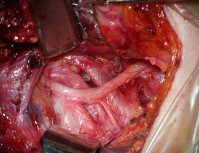
Hypoglossal with descendens hypoglossi nerve
The nerve is cut as far distally as possible and mobilized after transaction of the ansa hypoglossi. The facial nerve is transected at its exit from the stylomastoid foramen and reflected inferiorly; the ends of the two nerves are approximated without tension after cleaning of the epineurium and surrounding tissue to a limit of 5 mm from the ending cut. Over a large gelfoam piece, the ends of the nerves are anastomosed atraumatically using 3–4 per neural sutures of 8-0 monofilament Nylon; a piece of fascia is wrapped around the anastomosis and secured with fibrin glue (Fig. 2).
Fig. 2.

Distal end of the facial nerve sutured to the Hypoglossal Nerve using 8-0 monofilament Nylon
Digastric muscle could be transected to gain length for the hypoglossal nerve for a tensionless suturing but we prefer to preserve the muscle because it gives protection to the anastomosis. After haemostasis the closure is performed into two layers over a drain which is removed on the first postoperative day.
Evaluation Methods
All patients were subject to physiotherapy as soon as initial movements of the face appeared to enhance muscle activity.
The analysis of facial muscle activity post operatively was assessed using still photographs of the patients (at rest, smiling, eye closure, eyebrow raising and frowning).
Evaluation of the patients was performed using the House-Brackmann grading system, the IOWA facial recovery score, the Sunnybrook scale and the FDI (physical subscale).
Results
Forty out of sixty patients who completed a minimal follow up of 24 months at the time of evaluation constituted the study population. Two patients underwent lid weight implantation and tarsorrhaphy.
There were 22 females and 18 males; the mean age was 45.5 years (range 17–73 years).
The most common cause of facial paralysis was vestibular Schwannoma surgery where most of the patients had a tumour larger than 4 cm (Table 1).
Table 1.
Etiology of facial nerve paralysis
| Etiology | H-F anastomosis | Total number of cases operated |
|---|---|---|
| Vestibular Schwannoma | 34 | 2,232 |
| Meningioma | 3 | 180 |
| Petrous-bone cholesteatoma | 2 | 158 |
| Tympano-jugular paraganglioma | 1 | 199 |
| Total | 40 | 2,769 |
The mean interval time between the facial paralysis and hypoglossal-facial anastomosis was 11.3 months (range 2–42 months). All the patients achieved a postoperative HB grade III or IV after a mean follow-up time of 20 months (Table 2).
Table 2.
Time interval and results using the three methods
| Time interval (months) | H-B grades | IOWA score | S-B scale | |||||||||||
|---|---|---|---|---|---|---|---|---|---|---|---|---|---|---|
| I | II | III | IV | V | VI | A | B | C | D | E | F | 69–85 | <69 | |
| 0–6 | – | – | 11 | 6 | – | – | – | 14 | 3 | – | – | – | 15 | 2 |
| >6–12 | – | – | 5 | 1 | – | – | – | 3 | 2 | 1 | – | – | 5 | 1 |
| >12–24 | – | – | 9 | 5 | – | – | – | 12 | 1 | 1 | – | – | 11 | 3 |
| >24–48 | – | – | 1 | 2 | – | – | – | 2 | – | 1 | – | – | 2 | 1 |
| Total | – | – | 26 (65%) | 14 (35%) | – | – | – | 31 (77%) | 6 (15%) | 3 (7.5%) | – | – | 33 (82.5%) | 7 (17.5%) |
H-B House-Brackmann, IOWA IOWA facial recovery score; S-B Sunnybrook scale-composite score = voluntary movement-resting asymmetry-synkinesis (69–85/good, below 69/poor)
The facial movements were detected after a period that ranged from 5 to 9 months and appeared first in the midface in most of the cases.
After the hypoglossal-facial anastomosis, ten patients developed tongue atrophy, but only 4 of them suffered from difficulties during eating and drinking. Three of these four patients had associated LCN deficit.
Eight patients experienced mild facial synkinesis around the mouth. In 6 patients the synkinesis was not so bothersome to require any treatment and the other two patients were managed with botulin injections and avoiding extreme tongue movement. None of the patients suffered from hypertonia.
Discussion
Advances in intraoperative facial nerve monitoring help the surgeons to preserve the facial nerve during otologic and lateral skull base surgery. But facial nerve injury still occurs in cases where the FN is firmly attached to the tumor or when it lies between the surgeon and a bleeding that cannot be controlled without manipulation of the nerve.
It occurs also when the nerve acts as an obstacle impeding the surgical access and compromising the safety of the surgical procedure.
In our series 3.8% of patients who underwent resection of vestibular Schwannoma had interruption of the facial nerve during the procedure.
Although that the incidence of anatomic interruption of the FN during Vestibular Schwannoma surgery is low, this kind of surgery is considered as the most common cause of iatrogenic facial nerve paralysis in otologic and lateral skull base surgery.
It occurs usually with large tumors (>4 cm), irradiated tumors and revision surgeries when the facial nerve is highly adherent to the tumor [1–4].
When the proximal stump of the nerve at the brainstem couldn’t be identified hypoglossal to facial anastomosis is the procedure of choice for restoring of the facial nerve function [5].
The concept of facial function restoration using a crossover cranial motor nerve reinnervation was introduced by Drobnik in 1879 when he executed a facial to spinal accessory nerve anastomosis. In 1901 the German surgeon Werner Körte performed the first hypoglossal to facial nerve anastomosis in a patient having infectious petrositis when he transected the facial nerve at the styloid foramen and anastomosed the peripheral stump to the side of the hypoglossal nerve. Then in 1932 Ballance and Duel reported the first case of hypoglossal nerve transaction with direct end-to-end anastomosis to the facial nerve [6, 7]. Since then many reports of successful cases of end-to-end facial to hypoglossal anastomosis were published with good facial nerve outcomes [8–10].
The major disadvantage of this technique was the hemiglossal atrophy that resulted from sectioning of the hypoglossal nerve. To prevent the atrophy, different surgical modifications has been described in the literature like May techniques and longitudinal split of the hypoglossal nerve [11, 12].
Although the risk of tongue atrophy is less with these modifications, the facial nerve outcome is worse than the end to end anastomosis since the number of axons of the hypoglossal nerve incorporated in the reinnervation process is reduced.
This was supported by Yetiser et al. who conducted a metanalysis on the facial nerve outcome after HF anastomosis. The data in the literature was not enough to compare the outcomes of end-to-side versus end-to-end technique but facial reanimation in the latter technique occurs much earlier. Less tongue atrophy was noticed also using the end-to-side technique [13].
Other attempt to avoid tongue atrophy is to reinnervate the ipsilateral side of the tongue using an anastomosis of the distal trunk of the hypoglossal nerve to the ansa hypoglossi. This technique didn’t show any success in reducing the risk of tongue atrophy [14].
Thirty patients (75%) from our series had unilateral tongue atrophy with troubles during eating and swallowing for a transient period in 26 patients and permanently for the other 4 patients. Three of these 4 patients were complaining also from a superimposed lower cranial nerve palsy which aggravated their oropharyngeal disorder.
Hemiatrophy of the tongue doesn’t lead necessarily to a permanent deficit in tongue function. This is supported by the fact that patients following hemiglossectomy recuperate their normal tongue function without any problems in eating and swallowing. Clemis et al. [15] not only agreed with this theory but also believe that the hemiatrophy of the tongue is considered as a positive factor in enhancing the lingual function by reducing the mass of the nonfunctional side.
The lingual function might be affected shortly in the postoperative period but once the buccinator and orbicularis oris muscle recuperate their function, the oropharyngeal disorders will be relieved.
Although the HB scale is considered as a standard scale to assess the FN function and the results of facial reanimation procedures, we believe that other scales like IWAI and Sunny Brook should be also taken into consideration in conjunction to the HB scale. After HF anastomosis the recovery of the forehead muscles is so poor that compromises the HB scale. This is due to the fact that the temporalis branch of the FN takes off from the main trunk through a right angle and there are fewer nerve fibers that pass through this branch relatively to the other branches [16].
Conclusion
Despite the various techniques in facial reanimation following facial nerve paralysis, the end to end HFA remains for us the gold standard procedure with satisfying results in cases of viable distal facial stump and non-atrophic muscles.
Improvement in facial nerve function outweighed the minor problems encountered following this procedure.
References
- 1.Falcioni M, Taibah A, Russo A, Piccirillo E, Sanna M. Facial nerve grafting. Otol Neurotol. 2003;24(3):486–489. doi: 10.1097/00129492-200305000-00022. [DOI] [PubMed] [Google Scholar]
- 2.Rosenwasser RH, Liebman E, Jiménez DF, Buchheit WA, Andrews DW. Facial reanimation after facial nerve injury. Neurosurgery. 1991;29(4):568–574. doi: 10.1227/00006123-199110000-00014. [DOI] [PubMed] [Google Scholar]
- 3.Bacciu A, Falcioni M, Pasanisi E, Di Lella F, Lauda L, Flanagan S, Sanna M. Intracranial facial nerve grafting after removal of vestibular schwannoma. Am J Otolaryngol. 2009;30(2):83–88. doi: 10.1016/j.amjoto.2008.02.010. [DOI] [PubMed] [Google Scholar]
- 4.Godefroy WP, Malessy MJ, Tromp AA, van der Mey AG. Intratemporal facial nerve transfer with direct coaptation to the Hypoglossal. Otol Neurotol. 2007;28(4):546–550. doi: 10.1097/mao.0b013e31804301b8. [DOI] [PubMed] [Google Scholar]
- 5.Hammerschlag PE (1999) Facial reanimation with jump interpositional graft hypoglossal facial anastomosis and hypoglossal facial anastomosis: evolution in management of facial paralysis. Laryngoscope 109(2 Pt 2 Suppl 90):1–23 [DOI] [PubMed]
- 6.Ballance CA, Duel AB. The operative treatment of facial palsy by introduction of nerve grafts into the fallopian canal and by other intratemporal methods. Arch Otolaryngol. 1932;15:1–70. doi: 10.1001/archotol.1932.03570030008001. [DOI] [Google Scholar]
- 7.Waldman EH, Lustig LR. Sir Charles Alfred Ballance: contributions to otology and neurotology. Otol Neurotol. 2005;26:1073–1082. doi: 10.1097/01.mao.0000176176.79517.8b. [DOI] [PubMed] [Google Scholar]
- 8.Conley J, Baker D. Hypoglossal-facial nerve anastomosis for reinnervation of the paralysed face. Plast Reconstr Surg. 1979;63:3–72. doi: 10.1097/00006534-197901000-00011. [DOI] [PubMed] [Google Scholar]
- 9.Samii M, Matthies C. Indication, technique and results of facial nerve reconstruction. Acta Neurochir (Wien) 1994;130:125–139. doi: 10.1007/BF01405512. [DOI] [PubMed] [Google Scholar]
- 10.Luxford WM, Brackmann DE (1985) Facial nerve substitution: a review of 66 cases. Am J Otol (suppl):55–57 [PubMed]
- 11.May M, Sobol SM, Mester SJ. Hypoglossal-facial nerve interpositional-jump graft for facial reanimation without tongue atrophy. Otolaryngol Head Neck Surg. 1991;104:818–825. doi: 10.1177/019459989110400609. [DOI] [PubMed] [Google Scholar]
- 12.Atlas MD, Lowinger DS. A new technique for hypoglossal-facial nerve repair. Laryngoscope. 1997;107(7):984–991. doi: 10.1097/00005537-199707000-00028. [DOI] [PubMed] [Google Scholar]
- 13.Yetiser S, Karapinar U. Hypoglossal-facial nerve anastomosis: a meta-analytic study. Ann Otol Rhinol Laryngol. 2007;116(7):542–549. doi: 10.1177/000348940711600710. [DOI] [PubMed] [Google Scholar]
- 14.Chang CGS, Shen ALY. Hypoglossofacial anastomosis for facial palsy after resection of acoustic neuroma. Surg Neurol. 1984;21:282–286. doi: 10.1016/0090-3019(84)90203-9. [DOI] [PubMed] [Google Scholar]
- 15.Clemis JD, Gavron JP. Hypoglossal-facial nerve anastomosis: report on 36 cases with posterior fossa facial paralysis. In: Graham MD, House WF, editors. Disorders of the facial nerve. New York: Raven; 1982. pp. 499–505. [Google Scholar]
- 16.Pitty LF, Tator CH (1992) Hypoglossal-facial nerve anastomosis for facial nerve palsy following surgery for cerebello-pontine angle tumors. J. Neurosurg 77:724–731 [DOI] [PubMed]