Abstract
Despite recent advances in understanding mechanism of toxicity, the development of biomarkers (biochemicals that vary significantly with exposure to chemicals) for pesticides and environmental contaminants exposure is still a challenging task. Carbofuran is one of the most commonly used pesticides in agriculture and said to be most toxic carbamate pesticide. It is necessary to identify the biochemicals that can vary significantly after carbofuran exposure on earthworms which will help to assess the soil ecotoxicity. Initially, we have optimized the extraction conditions which are suitable for high-throughput gas chromatography mass spectrometry (GC-MS) based metabolomics for the tissue of earthworm, Metaphire posthuma. Upon evaluation of five different extraction solvent systems, 80% methanol was found to have good extraction efficiency based on the yields of metabolites, multivariate analysis, total number of peaks and reproducibility of metabolites. Later the toxicity evaluation was performed to characterize the tissue specific metabolomic perturbation of earthworm, Metaphire posthuma after exposure to carbofuran at three different concentration levels (0.15, 0.3 and 0.6 mg/kg of soil). Seventeen metabolites, contributing to the best classification performance of highest dose dependent carbofuran exposed earthworms from healthy controls were identified. This study suggests that GC-MS based metabolomic approach was precise and sensitive to measure the earthworm responses to carbofuran exposure in soil, and can be used as a promising tool for environmental eco-toxicological studies.
Introduction
Metabolomics, an omics science of systems biology, is the untargeted profiling of endogenous metabolites within a biological system under various physiological conditions and offers a unique opportunity to look at genotype-phenotype relationships as well as genotype-environmental relationships [1], [2]. Metabolomic profiling provides a powerful approach to identify and to quantitatively measure global changes in metabolites from biochemical pathways that are altered in response to disease, therapeutic intervention or toxicant. It has been widely employed in functional genomics, disease diagnosis, clinical [3], food and nutritional science [4], toxicology and pharmacology research [5]. Environmental metabolomics is an emerging field of science that provides wide opportunity to characterize the interactions of living organisms with the environment for assessing the organism functions at a molecular level, which will significantly contribute to the environment and human health [6], [7], [8], [9]. Recent metabolomic studies have identified novel biomarker patterns in terrestrial invertebrates [10], [11], [12], [13]. Earthworms, the soil engineers are a key terrestrial organism species and perform a number of essential functions like decomposition of organic matter, tillage and alteration of the soil and enhancement of microbial activity. Environmental pollutants, including organic chemicals and toxic metals may induce variety of adverse effects on ecosystems [14]. Indirectly, these effects of organic pollutants and metals are monitored by taking the effects on earthworms as an illustration [15]. Traditionally, a broad group of biomarkers such as cholinesterase (ChEs), DNA breakage, cytochorme P-450-dependent mono oxygenases, enzymes of oxidative stress have been used as biomarkers for heavy metal (Zn, Pb, Cd, Cu) and organic pollutant exposure by taking earthworm as a model organism for both laboratory and soil representative studies [16]. Over the past decade, varieties of endogenous metabolites have been identified as potential biomarkers of different environmental chemicals exposure to earthworms, for e.g. decrease in lactate and fatty acids for poly aromatic hydrocarbon (PAH) pyrene exposure [17], increase in alanine for pesticides DDT & endosulfan exposure [18], increase in histidine for copper exposure [19].
Metabolomics cover a vast range of metabolites and aim to determine the changes in low molecular weight organic metabolites in complex biological matrices. High-field proton nuclear magnetic resonance (1H NMR) spectroscopy and mass spectrometry (MS) based techniques are the major analytical tools used for metabolomic studies. NMR offers advantages of minimal sample preparation and quantitative measurement of metabolites. However, only most abundant peaks were observed due to its limited sensitivity with a detection threshold of 5 nmol and limited dynamic range. Extensive overlapping of signals in most of regions, especially carbohydrate and lipid regions of NMR spectrum makes difficult in metabolomic studies [20], [21]. Compared to NMR, MS methods are sensitive and can serve as a stand-alone method for identifying compounds from complex mixtures. The combination of mass spectrometry with gas chromatography has become a powerful metabolomic tool, provides high chromatographic resolution, reliable and reproducible mass fragmentation pattern of metabolites with large dynamic range, as well as the capability to identify unknown compounds for global metabolomic profiling of intracellular metabolites [22]. These advantages make gas chromatography-mass spectrometry (GC-MS) widely acceptable analytical tool for environmental metabolomic studies [23], [24], [25], [26], [27], [28].
Sample preparation strategy plays an important role in the metabolomic profiling. Metabolism quenching, tissue disruption and extraction were the key steps for metabolomic profiling. Various sample preparation protocols have been developed for species specific metabolome profile such as bacterial cells [29], different mammalian cells [30], [31], [32], microorganisms [33], Caenorhabditis elegans [34], ragworms [35], and different human matrices such as blood [36], urine [37], CSF [38], and human faecal water [39]. An optimal extraction method should obey the criterion, which includes extraction of large number of metabolites without affecting their stability, easily adaptable to analytical technique, and should be reliable and reproducible in order to identify metabolomic changes [40], [41]. Although previous studies for the optimization of tissue extraction protocols for non-targeted metabolomics of earthworm based on four solvent systems have been reported using different analytical platforms, but not been extensively studied using GC-MS based metabolomic platform [42] [43]. In the present communication, an attempt has been made to standardize the extraction conditions to explore the performance of non-targeted GC-MS based metabolomic approach for earthworm and applied to evaluate the toxicity of carbofuran in earthworm, Metaphire posthuma.
Carbofuran (2, 3-dihydro-2, 2-dimethyl-7-benzofuranyl-N-methyl carbamate) is a potent broad spectrum systemic insecticide, nematocide and acaricide commonly used for agriculture purposes [44], [45]. As a result of its wide spread use, it has been detected in surface and ground water [46]. European Union commission and United States EPA have considered that, it is very toxic to mammals, aquatic organisms and invertebrates [47]. It is having miticide activity, which acts by surface contact and through ingestion by interfering with the transmission of nerve impulses by inhibiting cholinesterase [48], [49]. It causes reversible acetyl cholinesterase carbamylation and allowing the accumulation of acetylcholine. Its application was found to reduce the total number of earthworms by 83% and the total biomass by 60% [50], [51].
Non-targeted GC-MS based metabolomic approaches can be subjected to supervised analysis methods in order to classify toxicity patterns or metabolic trajectories associated with carbofuran induced metabolomic changes. For this, initially we have optimized extraction solvent for global metabolomic profiling of earthworm Metaphire posthuma. Metaphire posthuma is a soil dwelling species and it is widely found in agriculture field. Due to this advantage we want to explore the metabolic responses to this species, which in turn can help to evaluate soil ecotoxicity. Previous studies have been conducted on other species at laboratory conditions [52], [53], [54], [55]. After that, we have investigated the metabolomic profiles for carbofuran induced toxicity evaluation, using non-targeted GC-MS based metabolomic approach combined with pattern recognition methods. Three different concentrations 0.6 mgkg−1, 0.3 mg kg−1 and 0.15 mg kg−1 corresponding to their LC50 of 1/20, 1/40, 1/80 are exposed by spiking in soil, in addition to an unspiked soil control [50], [51]. Metabolomic perturbations were identified in carbofuran exposed samples from healthy controls. Metabolomic approaches can be used in the investigation of the unique mode of action and ecotoxicological risk assessment of bioactive compounds [56], [57].
Materials and Methods
Chemicals and Reagents
All chemicals used were analytical grade unless otherwise stated. Methoxyamine hydrochloride, carbofuran and N-methyl-N-trimethylsilyl trifluoroacetamide (MSTFA) and all standards of amino acids, sugars and organic acids were procured from Sigma-Aldrich (St. Louis, MO, USA). Solvents like chloroform, acetonitrile, methanol (MeOH) and iso-propanol were obtained from Sigma-Aldrich (St. Louis, MO, USA). Pellet pestle with a cordless motor was procured from Sigma Aldrich (St. Louis, MO, USA). The ultra pure water was prepared by RiOsTM water purification system (Millipore, Billerica, MA, USA). IMECO ULTRA SONICS (Bombay, India) was used as sonicator. Heto GD-2 maxi dry plus was used as a lyophilizer.
Tissue disruption and extraction of metabolites
Earthworms were flash frozen in liquid nitrogen and stored at -80°C until further use. Earthworms were grounded to powder using a mortar and pestle in presence of liquid N2 in order to prepare a single homogenate sample. The homogenate was divided into three equal aliquots of 100 mg in a 2 ml of eppendorf tube. These tissue samples were extracted with different solvent systems such as methanol/chloroform/water (MCW; 2∶1∶1, v/v/v), methanol/water (MW; 4∶1, v/v), Acetonitrile/methanol/water (AMW; 2∶2∶1, v/v/v), pure methanol (MeOH), methanol/isopropanol/water (MIPW; 5∶2∶2, v/v/v) with a volume of 600 µl of each solvent system at 0°C. The resultant mixture was centrifuged at 10000 rpm for 3 min at 4°C. The procedure was repeated about three times.
Derivatization, GC-MS analysis and spectral acquisition
Tissue extracts were lyophilized and added 90 µL of O-methoxyamine hydrochloride (20 mg mL−1) solution in anhydrous pyridine to the dried extracts. The resultant mixture was mixed vigorously using cylcomixer for one min and incubated for 30 min at 60°C in a heating block. Subsequently, 200 µL of MSTFA with 1% TMCS as catalyst was added and the extracts were incubated at 60°C for further 60 min. Analyses were performed using Trace GC gas chromatograph coupled with Quantum XLS mass spectrometer (Thermo Scientific, FL, USA). The injector, ion source and transfer line temperatures were set at 250°C, 220°C and 290°C respectively. The initial oven temperature was held at 65°C for 2 min, increased to 230°C at a rate of 6°C/min and finally increased to 290°C at a rate of 10°C/min (held for 20 min). Helium was used as a carrier gas at a flow rate of 1.1 ml min−1. An aliquot of 1 µL of the extracts were injected into the TG-5 MS capillary column (30 m x 0.25 mm i.d. x 0.25 µm film thickness) consisting of a stationary phase of 5% phenyl 95% methyl polysiloxane in the split less mode. Detection was achieved using mass spectrometer in electron impact ionization at 70 eV. Full scan mass spectra was acquired in the mass range of 45–750 Da at 1 scans s−1 rate with an initial solvent delay of 6 min.
Data Pre-processing
All samples used for metabolomic profiling were analyzed as a single batch in a random order to minimize any analytical error, subjective interference and to keep the minimum retention shift. Chromatogram acquisition and data handling were carried out by using the GC-MS Xcalibur software (Thermo scientific, FL, USA). AMDIS software (Automated Mass Spectral Deconvolution and Identification System, version 2.0) was used to identify the metabolites in chromatographs. The mass spectra of all the detected compounds were compared with spectra in National Institute of Standards and Technology (NIST) library (version 2.0) or standards for confirmation. Baseline correction, noise reduction, smoothing and integration were performed using Thermo XCalibur software. Peaks resulting from the column bleeding and reagent peaks were excluded from the analysis. Blank experiments were also performed in order to minimize possible sources of contamination such as reagent impurities, contamination during sample preparation and any instrumental contamination. Intensity of target ion was used for peak integration due to its higher specificity, precision and identity [22], [58]. Only peaks shown higher reproducibility were taken for data analysis. Only those metabolites which have shown similarity index (SI) greater than 70% during spectral search using NIST library were taken into consideration for further analysis.
Statistical analysis
The GC/MS data matrix for metabolomic analysis was composed of the composition of metabolites (columns) and samples (rows). Data were mean-centred and unit variance scaled to remove the offsets and adjust the importance of low and high abundance metabolites to an equal level. The resulting scaled data were imported to Statistica (version 10, Statsoft, Tulsa, OK) and Metaboanalyst (version 2.0) [59], [60], for statistical analysis. Heat map combined with two-dimensional hierarchical cluster analysis (2D-HCA) for the visualization of the metabolomics data were created using the software PermutMatrix version 1.9.3 (http://www.lirmm.fr/~caraux/PermutMatrix/) [61]. Principal component analysis (PCA) was performed to explore the clustering behaviour of the metabolites/samples. To identify the differential metabolites that account for the separation between groups, Partial least square discriminate analysis (PLS-DA) was applied. PLS-DA model was validated using the leave one out cross validation method and the quality of model is assessed on R2 and Q2 scores [62], [63]. Excellent models are obtained when the cumulative values of R2Y and Q2Y are above 0.8 [64]. In addition to cross validation, model validation was also performed by 500 times permutation tests [65]. Variable importance in projection (VIP) scores was obtained from the PLS-DAs. VIP scores are weighed as sum of scores of the PLS loadings. VIP scores give the information of the relative contributions of individual metabolites to the variance between the control and exposed groups. The higher the VIP value, the greater is the individual contribution of that metabolite to group separation. Metabolites with variable importance in projection (VIP) values of greater than 1 were identified as potential marker metabolites [66], [67], [68]. Univariant analysis was performed to these metabolites by applying unpaired t-test with a significance value (p) less than 0.05. Classification performance of biomarkers was further evaluated with receiver operator characteristic curve (ROC) based on support vector machine (SVM). The area under the receiver operating characteristic curve (AUC) was used as a measure of classification performance. The AUC with value 0.9–1.0 considered as excellent classification performance [69]. ROC curve analysis was performed by using freely available web based software ROCCET (http://www.roccet.ca).
Soil spiking and earthworm exposure
The experimental procedure was adopted from previously reported procedure [70], [71], briefly, the procedure was as follows. The test worms (adult Metaphire posthuma) were collected from culture and placed in soil to acclimatize for seven days in a BOD incubator (Indian Equipment Corporation, Bombay, India) at 20±1°C. Before acclimatization soil composition was determined. The constituents of soil and pH were measured and found to be pH 7.1, Nitrogen 64 mg/kg, Phosphorus 8.3 mg/kg, Potassium 28 mg/kg, Magnesium 36 mg/kg, sand 87% [72]. Initially, four soil treatments were prepared including control. One kg of each soil was placed in three different trays and spiked with concentrations of 0.15, 0.3 and 0.6 mg kg−1 of carbofuran in equal volumes of acetone. Soil samples were then mixed well and vented for three hours to remove all the solvent. Then the soil samples were wetted with water to bring the soil to 50% of field capacity and then left to stabilize for 24 hours prior to addition of worms. Carbofuran spiked soil samples were transferred into l litre jars. An unspiked control treatment was prepared in the similar way. The adult earthworms with visible clitellum were collected from culture, weighed (average weight, 1.55±0.21 g). One kg of soil was used for six replicates per dose. Earth worms were transferred into individual jars and kept in dark during the exposure period as per Organisation for Economic Cooperation and Development (OECD) 1984 guidelines [71]. We did not observe the earthworms on the surface of the soil throughout the experimental period. After seven days of exposure, worms were weighed (average weight 1.20±0.16 g) and washed with distilled water to remove residual soil deposited on the surface of the earthworms. After washing with distilled water, earthworms were allowed to depurate for 15 hours on a filter paper and placed into a glass beaker to release their gut contents.
Results and Discussion
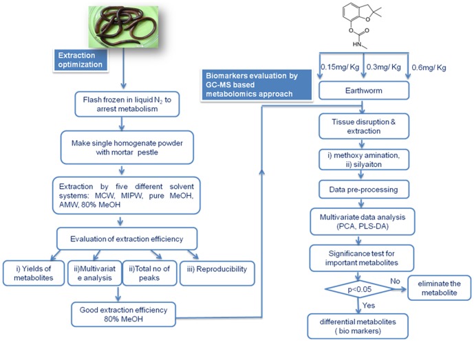
The workflow strategy was shown in fig 1 consisting of optimization of extraction conditions followed by biomarker evaluation of carbofuran exposure to earthworms.
Figure 1. Workflow for the optimization and evaluation of earthworm responses to sub-lethal toxicity of Carbofuran.
Optimization of extraction solvent system
Ideally, an extraction solvent should obey the following properties i) it has to cover a broad range of chemical properties of metabolites to enable extraction of all metabolites in high yields with good reproducibility [43], ii) solvent system should not affect the stability of metabolites extracted [42]. Since each extraction solvent has its own chemical and physical properties, it is not easy to generate such an extraction solvent for global metabolomic profiling of earthworms. To select an optimal extraction solvent system for the global metabolomic profiling of earthworm, Metaphire posthuma, we have evaluated five different solvent systems, MCW, 80% MeOH, MIPW, AMW and pure MeOH, which are known to have good extraction efficiency for various tissue and cell metabolome [30], [31], [32], [33], [34], [35]. Out of the five solvents tested, best extraction solvent was selected based on (i) peak intensity of structurally identified metabolites, (ii) multi variant analysis and (iii) distributions over individual metabolite features of coefficient of variation (reproducibility) [30], [34], [36], [73]. The extraction results in various compounds from the tissue of earthworm which includes, amino acids, organic acids, carbohydrates, fatty acids, phosphates, polyols and amines (table 1).
Table 1. Compounds structurally identified from earthworm, Metaphire.posthuma by GC-MS.
| Rt (min) | compound | fragmentations (m/z) | Idn | Rt (min) | compound | fragmentations (m/z) | Idn |
| Amino acids & their derivatives | Fatty acids | ||||||
| 9.16 | L-alanine | 116,73,147,190,59 | A | 21.41 | Tridecanoic acid | 117,73,271,129,145 | A |
| 9.48 | L-glycine | 102,73,147,75,204 | A | 21.54 | Tetradecanoic acid | 285,117,75,129,145 | A |
| 11.53 | L-valine | 144,73,218,145,100 | A | 19.03 | Tri decanoic acid methyl ester | 74,87,55,143,185 | A |
| 12.23 | L-leucine | 158,73,147,102,59 | A | 19.58 | Dodecanoicacid | 75,117,257,132,145 | A |
| 12.34 | L-isoleucine | 158,73,218,147,232 | A | 26.98 | Hexadecanoic acid | 117,313,73,129,145 | A |
| 14.70 | L-serine | 204,73,218,147,100 | A | 27.71 | 9,12-octa deca dienoic acid (Z,Z) methyl ester | 67,81,95,55,109 | B |
| 15.29 | L-threonine | 73,117,218,147,101 | A | 27.80 | 9-Octadecenoic acid (Z)methyl ester | 55,74,69,83,97 | B |
| 17.90 | L-methionine | 176,128,73,61,147 | A | 27.90 | 11-octadecenoic acid (E) methyl ester | 55,69,74,83,97 | B |
| 17.75 | L-aspartic acid | 73,232,100,147,218 | A | 28.24 | Octadecanoic acid methyl ester | 74,87,143,298,255 | B |
| 19.87 | L-glutamic acid | 246,73,128,147,156 | A | 28.46 | Heptadecanoic acid | 73,117,327,132,145 | A |
| 17.82 | L-lysine | 84,73,156,102,128 | A | 28.98 | 6-Hexadecenoic acid-7-methyl ester (Z) | 138,55,69,97,83 | B |
| 18.47 | L-phenylalanine | 218,192,73,91,147 | A | 29.45 | 9,12-octa deca dienoic acid (Z,Z) | 75,81,67,129,337 | B |
| 22.23 | L-ornithine | 174,73,142,186,348 | A | 29.61 | 11-Cis octadecenoic acid | 339,73,117,129,55 | B |
| 24.43 | L-tyrosine | 218,73,280,100,147 | A | 29.94 | Octadecanoic acid | 117,341,73,132,145 | B |
| 23.94 | N-α-acetyl-L- lysine | 174,73,156,86,59 | B | 30.16 | 8,11,14-Eicosa trienoic acid mehtyl ester (Z,Z,Z) | 79,67,93,55,150 | A |
| 18.09 | pyroglutamic acid | 156,73,147,230,258 | B | 30.24 | 5,8,11,14-Eicosa tetraenoic acid methyl ester(all Z) | 79,91,67,105,55 | B |
| Organic acids | 30.37 | 5,8,11,14,17-Eicosa pentaenoic acid methyl ester(all Z) | 79,91,67,105,119 | B | |||
| 8.09 | pyruvic acid | 73,174,45,89,59 | A | 30.62 | Cis-13-Eicosenoic acid methyl ester | 55,69,74,83,97,297 | A |
| 13.59 | scuccinic acid | 147,73,247,129,45 | A | 31.47 | Arachidonic acid | 73,91,67,117,55 | A |
| 14.29 | 2-butenedioic acid | 245,147,73,45,83 | B | 31.55 | Cis-5,8,11,14,17-Eicosapenta enoic acid | 79,73,91,117,67 | B |
| 16.43 | malonic acid | 73,147,305,45,69 | A | 31.70 | α-Linoleic acid | 73,79,67,95,55 | A |
| 17.44 | malic acid | 73,147,233,245,133 | A | 32.45 | Cis-7,10,13,16-Docosa tetra enoic acid methyl ester | 79,91,67,105,55 | B |
| 18.07 | 2-ketoisovaleric acid | 73,147,157,232,260 | B | 33.45 | 2-mono palmitin | 129,218,73,147,103 | B |
| 19.08 | glutaric acid | 73,147,198,156,288 | B | 33.76 | Eicosanoic acid glycerate ester | 73,57,147,43,129 | B |
| 11.01 | 2-butenoic acid | 147,73,231,45,66 | B | 33.67 | Hexadecanoic acid glycerate ester | 371,460,239,73,147 | B |
| polyols (poly hydric alcohols) | |||||||
| 8.26 | lactic acid | 147,117,73,191,133 | A | 12.78 | glycerol | 73,147,205,117,103 | A |
| 19.05 | 2-hydroxy glutaric acid | 73,129,147,247,349 | B | 27.19 | Inositol | 73,318,147,217,305 | A |
| 26.72 | d-(+)gluconicacid | 73,319,147,129,220 | B | 28.15 | myo Inositol | 73,217,147,305,191 | A |
| Carbohydrates | 21.22 | threitol | 73,147,103,205,217 | B | |||
| 24.31 | fructose | 73,103,217,307,147 | A | 25.65 | mannitol | 73,319,147,205,217 | A |
| 25.30 | glucose | 73,319,205,147,218 | A | 31.38 | 1-O-pentadecylglycerol | 205,147,73,117,131 | B |
| 25.16 | mannose | 73,147,319,205,160 | A | 32.78 | 1-O-hexadecylglycerol | 205,147,117,73,133 | B |
| 25.47 | Galactose | 73,205,319,147,217 | A | Phosphates | |||
| 34.22 | turanose | 73,361,147,217,103 | B | 12.98 | phosphoric acid | 299,73,133,314,211 | B |
| 35.02 | lactose | 204,73,191,217,361 | A | 10.78 | methyl phosphate | 241,73,133,256,211 | B |
| 35.51 | maltose | 204,73,191,361,217 | A | 22.80 | 1-glycero phosphate | 357,73,299,147,103 | B |
| 34.79 | sucrose | 361,73,217,147,271 | A | 22.96 | O-phospho ethanolamine | 73,299,188,174,315 | B |
| 35.69 | melibiose | 204,73,191,217,129 | B | galactose-6-phosphate | 73,299,387,315,147 | B | |
| 26.47 | talose | 204,73,191,217,147 | B | amines | |||
| 24.85 | methyl mannopyranoside | B | 21.95 | cadaverine | 174,73,86,59,100 | B | |
| Others | 9.59 | ethanolamine | 174,73,147,100,86 | B | |||
| 39.31 | cholesterol | 129,329,368,73,353 | A | 34.57 | N-acetyl glucosamine | 73,147,103,205,129 | B |
| 36.50 | cholesta-3,5-diene | 368,147,105,91,145 | B | 14.67 | 2-amino-3-phenyl propane | 188,73,100,147,114 | B |
| 25.91 | Indole acetic acid | 202,319,73,188,154 | B |
Rt = retention time, Idn = identification, A = These compounds are confirmed by authentic standards, B = These compounds are identified by NIST library Mass Spectral fragmentation pattern.
For an unbiased analysis of metabolome, it is important to extract all the existing metabolites in high yields, including those metabolites present in relatively low concentrations. So as a first criterion, peak intensities of representative intracellular metabolites from various chemical groups, including amino acids, organic acids, carbohydrates, fatty acids, polyhydric compounds, phosphates and amines were compared by using five different solvent extraction systems. Fig S1 shows the extraction efficiency of these solvents in terms of total intensity over a wide range of chemical classes. Fig S1A shows that highest yields for amino acids were obtained using AMW as extraction solvent, which is similar to the earlier reported study on earthworm species Lumbricus rubellus. Highest extraction efficiencies of carbohydrates were obtained with MIPW and 80% MeOH as compared to pure methanol (Fig S1B). It may be due to high polar nature of sugars (poly hydroxyl compounds) and their solubility in solvents with high polarity, like MIPW and 80% MeOH as compared to less polar solvent MeOH. In case of fatty acids, pure MeOH has shown the highest extraction efficiency followed by 80% MeOH as depicted in Fig S1C. Organic acids, phosphates and polyols exhibited highest peak intensities when 80% MeOH was used as an extraction solvent depicted in Fig S1D-F.
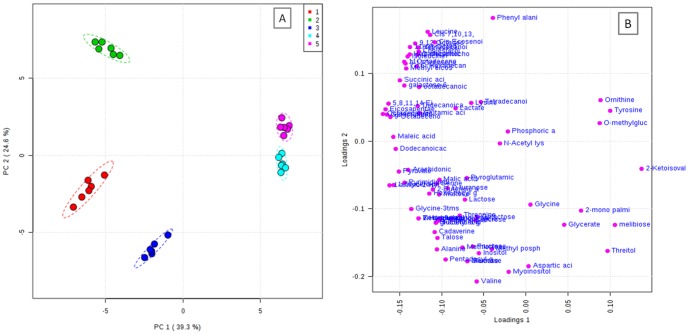
Multivariate analysis was used to provide visualization of the overall affects of different solvent systems on the metabolomic profiles of earthworms [34], [35], [36]. PCA model has been constructed to obtain the overview of comparison of metabolic profiles in different solvent systems. The PCA model with R2X = 0.639, Q2X = 0.507 has been obtained based on cumulative values up to PC 2 shown in Fig 2. From this model, it was evident that, there were clear differences between the solvent systems in the extraction profile of metabolites. Score plot reflects the contribution of each principal component of all the solvent systems used for extraction and loading plot reflects the importance of the weight of metabolites. Based on the score plot, extraction of metabolites with 80% MeOH, MIPW and pure MeOH were shown completely different pattern of extraction than from the extraction with AMW, MCW. On the basis of differential distribution of metabolites on the bi plot, most of the metabolites significantly extracted into solvent systems like 80% MeOH, MIPW and pure MeOH, which were clustered to one side of PC1 (Fig S2), which is similar to the trend observed in metabolite peak intensities as shown in Fig. S1.
Figure 2. PCA of optimization of extraction solvent system for global metabolite profiling of earthworm Metaphire posthuma A) scores plot, explaining the extraction efficiency of different solvent systems 1. 80% MeOH, 2. Pure MeOH, 3. MIPW, 4. MCW, 5. AMW. B) Loadings plot.
Overall 325 peaks were detected by GC-MS in that 84–91 peaks were identified and other 136–233 peaks were remain unidentified in all five extraction solvent systems. Maximum numbers of peaks were detected by 80% MeOH and MIPW with 325 and 317 peaks respectively. In the same way maximum number of identified peaks was detected in 80% MeOH and MIPW respectively. The total number of identified and unidentified peaks was depicted in table S1. Fewer numbers of peaks were detected and identified in Pure MeOH and MCW.
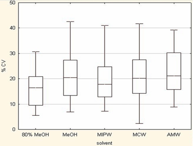
Reproducibility is another criterion for the selection of optimal extraction solvent and it is an important analytical parameter to assess the efficiency of extraction solvent. The box plot in Fig. 3 shows the distribution of coefficient of variation of identified metabolites for different extraction solvent systems. The median %CV values of identified metabolites for 80% MeOH, pure MeOH, MIPW, MCW and AMW were found to be 16.45, 20.39, 17.76, 20.12 and 21.14 respectively. Highest reproducibility was shown by 80% MeOH and after that MIPW. Previous methods showed a median CV values in the range of 15–30% across all analytical platforms. The present results coincide with the previously reported extraction protocols [28].
Figure 3. Coefficient of variation of metabolite features for different solvent systems extracted from earthworm Metaphire posthuma.
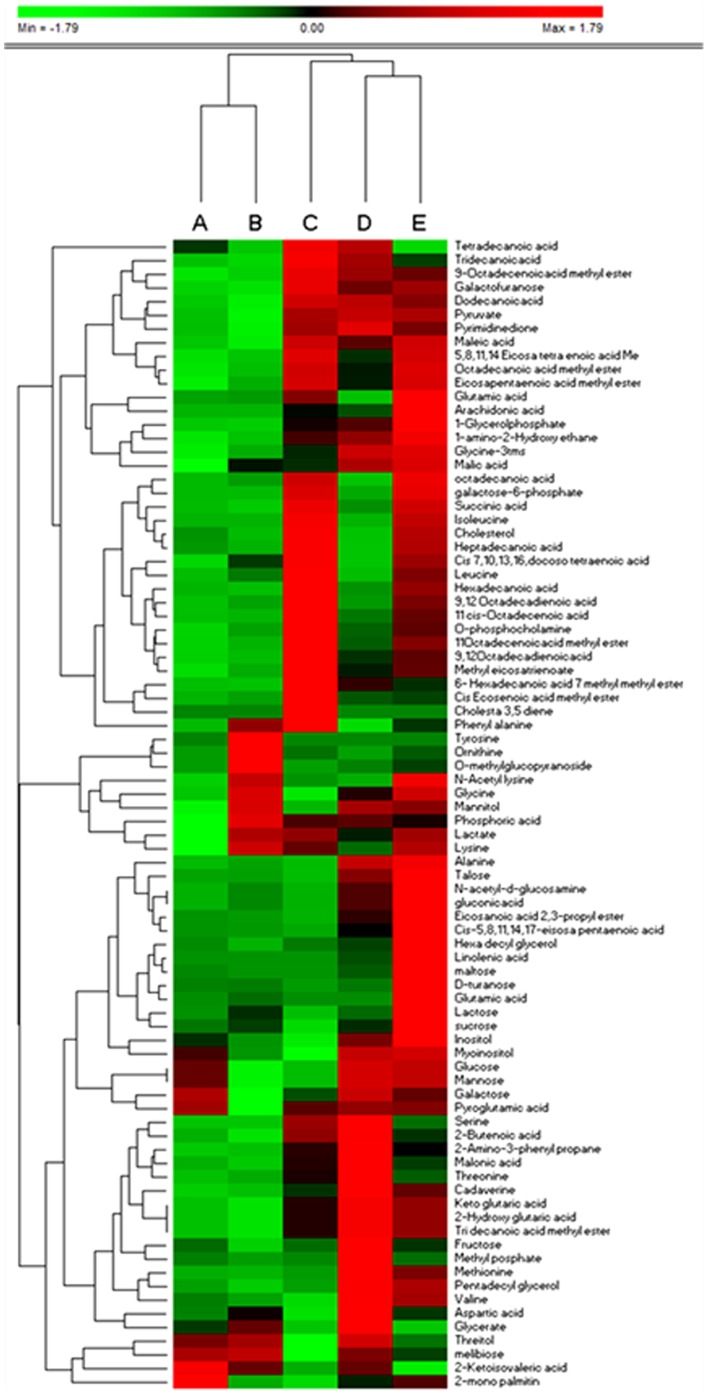
Hierarchical clustering was performed to identify significant differences in the extraction of metabolites using different solvent systems. The Hierarchical clustering analysis for the identified metabolites was depicted in Fig 4. The metabolites were placed in rows, and the sum of the peak intensities were normalized by employing unit variance. Ward's algorithm was used for this Hierarchical clustering. It clearly clustered the solvent systems, 80% MeOH, pure methanol and MIPW as one group, MCW and AMW as the other group with 80% MeOH producing the highest yield with respect to recovery. Very less recovery was observed with MCW. The results of Hierarchical clustering analysis were very well correlated with that of the multivariate analysis in terms of clustering and extraction efficiency of 80% methanol. The GC-MS chromatograph for the optimized extracted solvents for M.posthuma was shown in Fig S3. Overall, 80% methanol was found to be an optimal extraction solvent system based up on the criteria like highest peak intensity, multivariate analysis, reproducibility and hierarchical clustering with good extraction efficiency.
Figure 4. Clustered heat map of intracellular metabolites extracted using A) MCW B) AMW C) Pure MeOH D) MIPW E) 80% MeOH.
Evaluation of carbofuran toxicity on earthworms by metabolomic approach
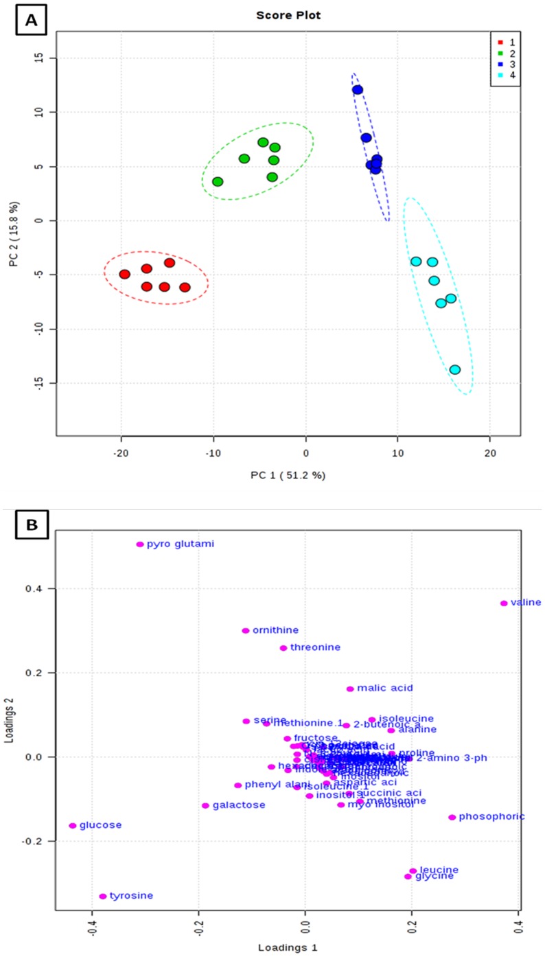
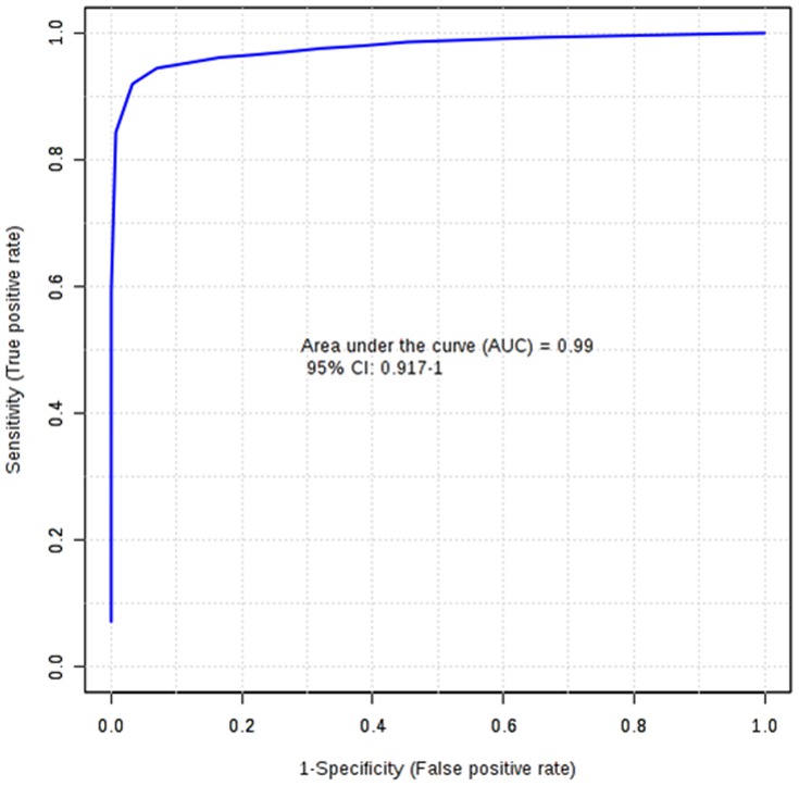
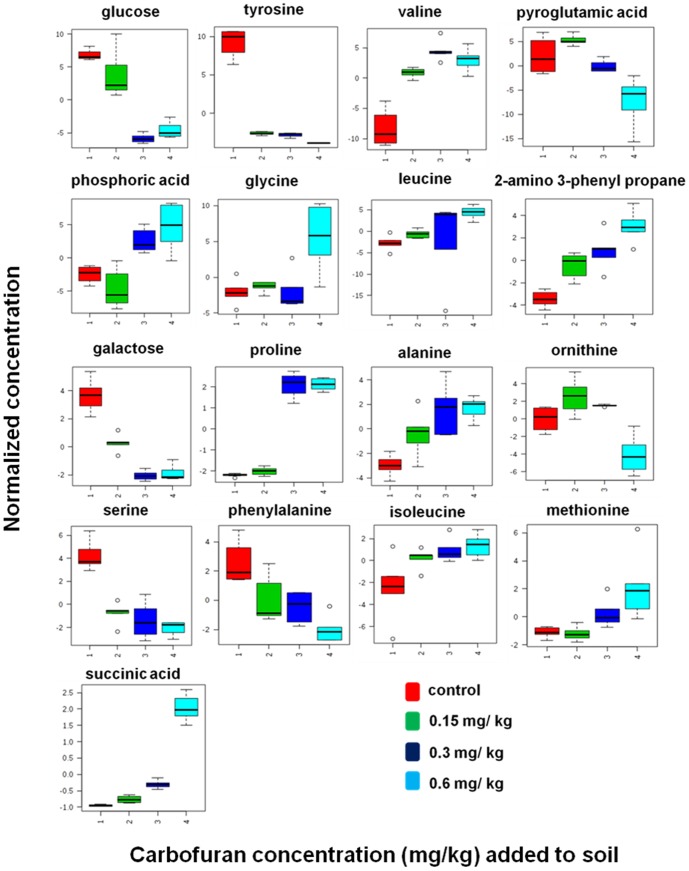
The potential application of the developed extraction method was evaluated to identify the metabolic perturbations of earthworms exposed to highly toxic carbamate pesticide, Carbofuran at three different concentrations (0.15, 0.3 and 0.6 mg kg−1). Total area normalization was performed. Initially an unsupervised PCA was applied to identify the clustering behaviour. PCA score plot has displayed the classification trend between control and carbofuran exposed samples. PCA model explained 67.0% of the total data variance with principal component 1 (PC1) explained 51.2% and principal component 2 (PC2) explained 15.8% of the total variance, respectively shown in figure 5. The PCA model with R2X = 0.614, Q2X = 0.364 has been obtained based on cumulative values up to PC 2. PLS-DA was performed to find a small number of linear combinations of the original variables (called latent variables), that was predictive for the class membership and that described most of the variability of the GC-MS metabolic profiles of control and exposed samples. Four different clusters were identified in PLS-DA score plot as depicted in Fig S4. Clear separation was identified between controls, and carbofuran exposed samples at baseline in the first PLS-DA component, suggesting that metabolomic perturbations were evident in the exposed samples. Characteristic fit criteria such as R2Xcum, R2Ycum and Q2Ycum were examined in order to validate the PLS-DA model. R2X and R2Y represent the fraction of the variance of X matrix and Y matrix, respectively and Q2Y represents the predictive accuracy of the built model. The cumulative values of PLS-DA model with R2Xcum = 0.500, R2Ycum = 0.974, Q2Ycum = 0.968 shows good fit of the model. The supervised PLS-DA model was further validated with 500 times permutation tests (Fig S5). For this both separation distance (B/W) and prediction accuracy test statistics were performed. Permutation test statistics clearly indicated that the original class assignment is much higher compared to the B/W ratios based on permutation class assignments. The differences between the classes are statistically significant p<0.002. The magnitude of this response was higher at a concentration of 0.6 mg kg−1 than lower induced concentration of 0.15 mg kg−1. The PLS-DA model of earthworm samples was employed to explore the intrinsic differences in metabolomic profiles of control and exposed samples. Variables with VIP greater than 1 were considered to be influential for the discrimination of samples in the score plots obtained after PLS-DA experiment. Uni-variant t-test was performed to find out the p-value along with their fold change between the exposed and control samples of earthworms. Seventeen metabolites were identified as potential biomarkers among all the differential metabolites according to the VIP values (greater than 1) and p values less than 0.05. These metabolites belong to the classes of amino acids, carbohydrates and fatty acids. The marker metabolites along with their VIP scores and p-values were shown in table 2. Model sensitivity and specificity were summarised using ROC curves for the models distinguishing carbofuran exposed from healthy controls (AUROC = 0.99) Fig 6. Results were indicative of strong predictive power of the metabolite markers between Carbofuran exposed and healthy control samples. From the loading plots, the metabolites like glucose and galactose (sugars); tyrosine, valine, glycine, leucine, proline, alanine, ornithine, serine, phenyl alanine, isoleucine mehionine (amino acids) and succinic acid, pyroglutamic acid, phosphoric acid, 2-amino-3-phenyl propane (other metabolites) were showed significant fluctuations in response to carbofuran exposure. PC1, which was shown to explain the variation due to exposure concentration, was influenced more by amino acids and sugars. The relative concentration changes in discernible metabolite intensities were examined to determine the fluctuations in the metabolites of earthworms after their exposure to carbofuran as depicted in fig 7. Sugars like glucose and galactose were shown significant decrease in their concentration levels in carobofuran exposed earthworms in comparison to controls. This decrease was suggested to be as a result of increase in the energy requirements of earthworms exposed to carbofuran. The decrease was more in the concentrations of 0.3 mg/kg and 0.6 mg/kg in comparison to 0.15 mg/kg. This observation may also be a result of energy metabolism being disturbed by the toxicity of carbofuran at high exposure concentrations.
Figure 5. Principle component analysis (PCA) A) scores and B) loadings plots for 1.control earthworms and earthworms exposed to soil spiked with 2. Concentration of 0.1 mg/Kg, 3. Concentration of 0.3 mg/Kg, and 4. Concentration of 0.6 mg/Kg of carbofuran.
Table 2. Key putatively identified metabolic perturbations from GC MS based metabolomic analyses of earthworms after exposed to carbofuran.
| S.no | Marker metabolite | p-value | VIP | fold change |
| 1 | Glucose | 2.1518E-10 | 3.20 | decreased |
| 2 | Tyrosine | 1.4389E-16 | 2.89 | decreased |
| 3 | Valine | 8.8602E-10 | 2.77 | increased |
| 4 | Pyroglutamic acid | 1.0541 E-5 | 2.39 | decreased |
| 5 | Phosphoric acid | 4.0752E-6 | 2.10 | increased |
| 6 | Glycine | 1.3342E-4 | 1.62 | increased |
| 7 | Leucine | 2.0883E-8 | 1.54 | increased |
| 8 | 2-amino,3-phenyl propane | 1.4289E-7 | 1.51 | increased |
| 9 | Galactose | 1.127E-11 | 1.39 | decreased |
| 10 | Proline | 3.8104E-17 | 1.29 | increased |
| 11 | Alanine | 3.5999E-5 | 1.26 | increased |
| 12 | Ornithine | 2.4149E-6 | 1.21 | decreased |
| 13 | Serine | 1.1611E-7 | 1.19 | decreased |
| 14 | Phenyl alanine | 3.4904E-4 | 1.16 | decreased |
| 15 | Isoleucine | 1.6252E-5 | 1.12 | increased |
| 16 | Methionine | 1.5858E-4 | 1.06 | increased |
| 17 | Succinic acid | 4.7329E-16 | 1.04 | increased |
Figure 6. Predictive accuracy of the model discriminating carbofuran exposed and healthy control earthworms summarised using ROC curve analysis.
Area under the curve = 0.99.
Figure 7. Earthworm metabolite responses to carbofuran exposure 1) control group 2) concentration of 0.15 mg kg−1 exposed group 3) concentration of 0.3 mg kg−1 exposed group 4) concentration of 0.6 mg kg−1 exposed group.
Increase in levels of alanine is an universal indicator for stress in various organisms exposed to different external contaminants [74]. In the present study, alanine was significantly increased in earthworms exposed to carbofuran. The increased response of alanine after carbofuran exposure was observed with increase in the dose of carbofuran from 0.15 mg/kg to 0.6 mg/kg indicates that alanine may be an indicator for carbofuran exposure. Succinate, a Krebs cycle intermediate showed a significant increase in carbofuran exposed earthworms. It may be due to possible disruption in the succinate dehydrogenase enzyme function [75]. Succinate dehydrogenase is the only enzyme of the Krebs cycle that is bound to inner mitochondrial membrane [75]. Five amino acids such as tyrosine, pyroglutamic acid, ornithine, serine and phenyl alanine were down regulated in carbofuran exposed earthworms in comparison to control. Seven amino acids valine, glycine, leucine, proline, alanine, isoleucine, methionine were up-regulated in carbofuran exposed earthworms in comparison to control. The present study indicated that amino acid and carbohydrate metabolism were mainly disturbed in carbofuran exposed earthworms. The present results were well in agreement with earlier reports on fish exposure to carbofuran, where the disturbances were shown to occur in protein and carbohydrate metabolism [76].
Conclusion
Environmental metabolomics is a rapidly developing and emerging sub-discipline of metabolomics and has the potential to relate between earthworm toxicity and bioavailability of soil contaminants. It necessitates the need for simple and robust methods with precise and accurate extraction of metabolites from earthworms. As the sample preparation is crucial for the metabolomic profiling of earthworms, extraction conditions for non targeted metabolomic approach was first optimized. Then, we applied the nontargeted metabolomic approach to identify the biomarkers of carbofuran induced toxicity in earthworm, Metaphire posthuma with the aid of multivariate analysis. Earthworms exhibited significant perturbations in their metabolomic profiles after their exposure to carbofuran. This study suggests that, the high sensitivity, specificity and availability of spectral libraries of mass spectrometer combined with gas chromatography having good resolution ability, makes the gas chromatography-mass spectrometry an excellent tool for metabolomics application to identify the biomarkers for environmental toxicants and contaminants exposure.
Supporting Information
Yields of the identified metabolites. A) Amino acids B) Carbohydrates C) Fatty acids D) Organic acids E) Phosphates F) Polyols.
(TIF)
PCA bi plot for solvent systems (80% MeOH, MIPW, Pure MeOH, AMW, MCW) and extracted metabolites. Bi plot clearly indicate most of the metabolites clustered together with 80% Methanol, 100% MeOH, MIPW.
(TIF)
GC-MS chromatogram for extracted metabolites using different solvent systems include, 80% MeOH, MIPW, Pure MeOH, AMW, MCW.
(TIF)
PLS-DA scores plot for 1) control earthworms and earthworms exposed to soils spiked with 2) 0.15 mg/kg 3) 0.3 mg/kg 4) 0.6 mg/kg.
(TIF)
Validation results. A) screen shot showing a PLS-DA cross validation. B) Permutation analysis of PLS-DA models derived from carbofuran exposed and healthy controls. Statistical validation of the PLS-DA by permutation analysis using 500 different model permutations. The goodness of fit and predictive capability of the original class assignments is much higher compared to ratios based on the permutation class assignments.
(TIF)
Total number of peaks in earthworm Metaphire posthuma .
(DOC)
Acknowledgments
The authors are thankful to Dr. K.C. Gupta, Director, CSIR-IITR for scientific discussions and providing the necessary infrastructural facilities to carry out this work. R Ch is thankful to Council of Scientific and Industrial Research for providing the fellowship.
Funding Statement
The authors greatly acknowledge CSIR, New Delhi for their financial support through INDEPTH and EMPOWER scheme (OLP-0004) to carry out this research work. The funders had no role in study design, data collection and analysis, decision to publish, or preparation of the manuscript.
References
- 1. Fiehn O (2002) Metabolomics - the link between genotypes and phenotypes. Plant Mol Biol 48: 155–171. [PubMed] [Google Scholar]
- 2. Lewis NE, Nagarajan H, Palsson BO (2012) Constraining the metabolic genotype- phenotype relationship using a phylogeny of Insilco methods. Nat Rev Microbiology 10: 291–305. [DOI] [PMC free article] [PubMed] [Google Scholar]
- 3. Spratlin JL, Serkova NJ, Eckhardt SG (2009) Clinical applications of Metabolomics in Oncology: a review Clin Cancer Res. 15: 431–440. [DOI] [PMC free article] [PubMed] [Google Scholar]
- 4. Gibney MJ, Walsh M, Brennan L, Roche HM, German B, et al. (2005) Metabolomics in human nutrition: opportunities and challenges. Am J. Clin Nutr 82: 497–503. [DOI] [PubMed] [Google Scholar]
- 5. Johnson CH, Patterson AD, Idle JR, Gonzalez FJ (2012) Xenobiotic Metabolomics: major impact on the Metabolome. Annu Rev Pharmacol and Toxicol 52: 37–56. [DOI] [PMC free article] [PubMed] [Google Scholar]
- 6. Bundy PJG, Davey MP, Viant MR (2009) Environmental metabolomics: a critical review and future perspectives. Metabolomics 5: 3–21. [Google Scholar]
- 7. Miller MJ (2007) Environmental Metabolomics: a SWOT analysis (Strengths, Weaknesses, Opportunities and Threats). J. Proteome Res 6: 540–545. [DOI] [PubMed] [Google Scholar]
- 8. Viant MR (2009) Applications of metabolomics to the environmental sciences. Metabolomics 5: 1–2. [Google Scholar]
- 9. Viant MR (2008) Recent developments in environmental metabolomics. Mol. BioSyst 4: 980–986. [DOI] [PubMed] [Google Scholar]
- 10. Hughes SL, Bundy JG, Want EJ, Kille P, Stürzenbaum SR (2009) The metabolomic responses of Caenorhabditis elegans to cadmium are largely independent of metallothionein status, but dominated by changes in cystathionine and phytochelatins. J. Proteome Res 8: 3512–3519. [DOI] [PubMed] [Google Scholar]
- 11. Southam AD, Lange A, Hines A, Hill EM, Iguchi YKT, et al. (2011) Metabolomics reveals target and off-target toxicities of a model organophosphate pesticide to Roach (Rutilus rutilus): implications for biomonitoring. Environ. Sci. Technol 45: 3759–3767. [DOI] [PMC free article] [PubMed] [Google Scholar]
- 12. Poynton HC, Taylor NS, Hicks J, Colson K, Chan S, et al. (2011) Metabolomics of microleter hemolymph samples enables an improved understanding of the combined metabolic and transcriptional responses of Dephnia magna to cadmium. Environ. Sci. Technol 45: 3710–3717. [DOI] [PubMed] [Google Scholar]
- 13. Jones OAH, Swain SC, Svendsen C, Griffin JL, Sturzenbaum SR, et al. (2012) Potential new method of mixture effects testing using metabolomics and Caenorhabditis elegans . J. Proteome Res 11: 1446–1453. [DOI] [PubMed] [Google Scholar]
- 14. Peijnenburg WJGM, Vijver MG, Devillers J, (ed) ( Earthworms and Their Use in Eco(toxico)logical Modeling. Ecotoxicology Modeling, Emerging Topics in Ecotoxicology: Principles, Approaches and Perspectives 2: 177–204. [Google Scholar]
- 15. Simpson MJ, McKelvie JR (2009) Environmental metabolomics: new insights into earthworm ecotoxicity and contaminant bioavailability in soil. Anal Bioanal Chem 394: 137–149. [DOI] [PubMed] [Google Scholar]
- 16. Sanchez-Hernandez JC (2006) Earthworm biomarkers in ecological risk assessment. Rev Environ Contam Toxicol 188: 85–126. [DOI] [PubMed] [Google Scholar]
- 17. Jones OAH, Spurgeon DJ, Svendsen C, Griffin JL (2007) A metabolomics based approach to assessing the toxicity of the poly aromatic hydrocarbon pyrene to earthworm Lumbricus rubellus . Chemosphere 71: 601–609. [DOI] [PubMed] [Google Scholar]
- 18. McKelvie JR, Yuk J, Xu Y, Simpson AJ, Simpson MJ (2009) 1H NMR and GC/MS metabolomics of earthworm responses to sub-lethal DDT and endosulfan exposure. Metabolomics 5: 84–94. [Google Scholar]
- 19. Gibb JOT, Svendsen C, Weeks JM, Nicholson JK (1997) 1H NMR spectroscopic investigations of tissue metabolite biomarker response to Cu II exposure in terrestrial invertebrates: identification of free histidine as a novel biomarker of exposure to copper in earthworms. Biomarkers 2: 295–302. [DOI] [PubMed] [Google Scholar]
- 20. Krishnan P, Kruger NJ, Ratcliffe RG (2005) Metabolite fingerprinting and profiling in plants using NMR. J Exp Bot 56: 255–265. [DOI] [PubMed] [Google Scholar]
- 21. Lankadurai BP, Wolfe DM, Whitfield ML, Aslund MLW, Simpson AJ, et al. (2013) 1H NMR metabolomic analysis of polar and non polar earthworm metabolites after sub-lethal exposure to phenanthrene. Metabolomics 9: 44–56. [Google Scholar]
- 22. Koek MM, Jellema RH, Greef JVD, Tas AC, Hankemeier T (2011) Quantitative metabolomics based on gas chromatography mass spectrometry: status and perspectives. Metabolomics 7: 307–328. [DOI] [PMC free article] [PubMed] [Google Scholar]
- 23. Michaud MR, Denlinger DL (2007) Shifts in the carbohydrate, polyol, and amino acid pools during rapid cold-hardening and diapauses-associated cold-hardening in flesh flies (Sarcophaga crassipalpis): a metabolmic comparison. J Comp Physiol B 177: 753–763. [DOI] [PubMed] [Google Scholar]
- 24. Podrabsky JE, Lopez JP, Fan TW, Higashi R, Somero GN (2007) Extreme anoxia tolerance in embryos of the annual killfish Austrofundulus limnaeus: insights from metabolmics analysis. J Exp Biol 210: 2253–2266. [DOI] [PubMed] [Google Scholar]
- 25.Currie F, Broadhurst D, Dunn W, Goodacre R (2007) Proceedings of the Metabolomics Society's 3rd Annual International Conference, Manchester, UK.
- 26. Ralston-Hooper K, Hopf A, Oh C, Zhang X, Adamec J, et al. (2008) Development of GCxGC/TOF-MS metabolomics for use in ecotoxicological studies with invertebrates. Aquat Toxicol 88: 48–52. [DOI] [PubMed] [Google Scholar]
- 27. Li X, Xu Z, Lu X, Yang X, Yin P, et al. (2009) Comprehensive two-dimensional gas chromatography/time-of-flight mass spectrometry for metabonomics: Biomarker discovery for diabetes mellitus. Anal Chim Acta 633: 257–262. [DOI] [PubMed] [Google Scholar]
- 28. Meng J, Zhang X, Wu H, Bu J, Shi C, et al. (2012) Morphine-induced conditioned place preference in mice: metabolomic profiling of brain tissue to find molecular switch of drug abuse by gas chromatography/mass spectrometry. Anal. Chim. Acta 2012 710: 125–130. [DOI] [PubMed] [Google Scholar]
- 29. Shin MH, Lee D, Liu KH, Fiehn OH, Kim KH (2010) Evaluation of sampling and extraction methodologies for the global metabolomic profiling of Saccharophagus degradans. . Anal. Chem 82: 6660–6666. [DOI] [PubMed] [Google Scholar]
- 30. Lorenz MA, Burant CF, Kennedy RT (2011) Reducing time and increasing sensitivity in sample preparation for adherent mammalian cell metabolomics. Anal. Chem 83: 3406–3414. [DOI] [PMC free article] [PubMed] [Google Scholar]
- 31. Wu H, Southam AD, Hines A, Viant MR (2008) High-throughput tissue extraction protocol for NMR and MS based metabolomics. Anal Biochem 372: 204–212. [DOI] [PubMed] [Google Scholar]
- 32. Parab GS, Rao R, Lakshminarayanan S, Bing YV, Moochhala SM, et al. (2009) Data driven optimization of metabolomics methods using rat liver samples. Anal. Chem 81: 1315–1323. [DOI] [PubMed] [Google Scholar]
- 33. Ewald JC, Heux SP, Zamboni N (2009) High-throughput quantitative metabolomics: workflow for cultivation, quenching and analysis of yeast in a multiwall format. Anal. Chem 81: 3623–3629. [DOI] [PubMed] [Google Scholar]
- 34. Geier FM, Want EJ, Leroi AM, Bundy JG (2011) Cross-platform comparison of Caenorhabditis elegans tissue extraction strategies for comprehensive metabolome. Anal. Chem 83: 3730–3736. [DOI] [PubMed] [Google Scholar]
- 35. Alvarez MC, Donarski JA, Elliott M, Charlton AJ (2010) Evaluation of extraction methods for use with NMR-based metabolomics in the marine polychaete ragworm, Hediste diversicolor. . Metabolomics 6: 541–549. [Google Scholar]
- 36. Jiye A, Trygg J, Gullberg J, Johansson AI, Jonsson P, et al. (2005) Extraction and GC/MS analysis of the human blood plasma metabolome. Anal Chem 77: 8086–8094. [DOI] [PubMed] [Google Scholar]
- 37. Rocha SM, Caldeira M, Carrola J, Santos M, Cruz N, et al. (2012) Exploring the human urine metabolomic potentialities by comprehensive two-dimensional gas chromatography coupled to time of flight mass spectrometry. J Chromatogr A 1252: 155–163. [DOI] [PubMed] [Google Scholar]
- 38. Wishart DS, Lewis MJ, Morrissey JA, Flegal MD, Jeroncic K, et al. (2008) The human cerebrospinal flluid metabolome. J. Chromatogr. B 871: 164–173. [DOI] [PubMed] [Google Scholar]
- 39. Gao X, Estelle PG, Sebedio JL (2010) Development of a quantitative metabolomic approach to study clinical human fecal wáter metabolome based on trimethyl silylation derivatization and GC/MS analysis. Anal. Chem 82: 6447–6456. [DOI] [PubMed] [Google Scholar]
- 40. Sellick CA, Knight D, Croxford AS, Maqsood AR, Stephens GM, et al. (2010) Evaluation of extraction process for intracellular metabolite profiling of mammalian cells: matching extraction approaches to cell type and metabolite targets. Metabolomics 6: 427–438. [Google Scholar]
- 41. Winder CL, Dunn WB, Schuler S, Broadhurst D, Jarvis R, et al. (2008) Global metabolomic profiling of Escherichia coli cultures: an evaluation of methods for quenching and extraction of intracellular metabolites. Anal Chem 80: 2939–2948. [DOI] [PubMed] [Google Scholar]
- 42. Brown SAE, Simpson AJ, Simpson MJ (2008) Evaluation of sample preparation methods for nuclear magnetic resonance metabolomic profiling studies with Eisenia fetida . Environ Toxicol Chem 27: 828–836. [DOI] [PubMed] [Google Scholar]
- 43. Liebeke M, Bundy JG (2012) Tissue disruption and extraction methods for metabolomic profiling of an invertebrate sentinel species. Metabolomics 8: 819–830. [Google Scholar]
- 44.Rubin AL, Evert S (2006) Risk characterization document for Carbofuran. California environmental protection agency. (www.cdpr.ca.gov/docs/risk/rcd/carbofuran/pdf)
- 45. Gupta RC (1994) Carbofuran toxicity. J Toxicol Environ Health 43: 383–418. [DOI] [PubMed] [Google Scholar]
- 46. Campbell S, David MD, Woodward LA, Li QX (2004) Persistence of carbofuran in marine sand and water. Chemosphere 54: 1155–1161. [DOI] [PubMed] [Google Scholar]
- 47.European Commision health and consumer protection Deirectorate general. Unit E 3. Chemicals, contaminants, pesticides. Review report for carbofuran; USEPA. Carbofuran I.R.E.D facts, 2012 Available: http://www.epa.gov/oppsrrd1/REDs/factsheets/carbofuran_ired_fs.htm.
- 48. Hernandez MD, Perez LM, Soler F, Gravato C, Guilhermino L (2011) Effect of carbofuran on sea bass (Dicentrarchus labrax L): study of biomarker behaviour alterations. Eotoxicol Environ Saf 74: 1905–1912. [DOI] [PubMed] [Google Scholar]
- 49. Milatovic D, Gupta RC, Dekundy A, Montine TJ, Dettbarn WD (2005) Carbofuran-induced oxidative stress in slow and fast skeletal muscles: prevention by memantine and atropine. Toxicology 208: 13–24. [DOI] [PubMed] [Google Scholar]
- 50. Stenersen J, Gilman A, Vardanis A (1973) Carbofuran: its toxicity to and metabolism by earthworm (Lumbricus terrestris). J Agric Food Chem 21: 166–171. [DOI] [PubMed] [Google Scholar]
- 51. Gilman AP, Vardanis A (1974) Carbofuran. Comparative toxicity and metabolism in the worms Lumbricus teerrestris L. and Eisenia foetida S. J Agric Food Chem 22: 625–628. [DOI] [PubMed] [Google Scholar]
- 52. Sundararaman V, Gupta SK (1992) An ultrastructural study of the clitellar epithelium of the earthworm Metaphire posthuma. Tissue Cell 24: 745–750. [DOI] [PubMed] [Google Scholar]
- 53. Tripathi G, BhardwarJ P (2005) Biodiversity of earthworm resources of arid environment. J Environ Biol 26: 61–71. [PubMed] [Google Scholar]
- 54. Liang SH, Jenq YP, Chiu YW, Chen JH, Shieh BS, et al. (2009) Cloning, expression, and characterization of cadmium-induced metallothionein-2 from the earthworm Metaphire posthuma and Polypheretima elongate. Comp Biochem Physiol C Toxicol Pharmacol 149: 349–357. [DOI] [PubMed] [Google Scholar]
- 55. Liang SH, Chen MH, Chen CC, Chen CS, Chen JH, et al. (2009) Impact of cadmium on the bacterial communities in the gut of Metaphire posthuma. J Hazard Mater 172: 1212–7. [DOI] [PubMed] [Google Scholar]
- 56. Aliferis KA, Chrysayi TM (2011) Metabolomics in pesticide research and development: review and future perspectives. Metabolomics 7: 35–53. [Google Scholar]
- 57. Whitfield Åslund ML, McShane H, Simpson MJ, Simpson AJ, Whalen JK, et al. (2012) Earthworm sublethal responses to titanium dioxide nonmaterial in soil detected by 1H NMR metabolomics. Environ Sci Technol 46: 1111–1118. [DOI] [PubMed] [Google Scholar]
- 58. Koek MM, Muilwijk B, Vanderwerf MJ, Hankemeier T (2006) Microbial metabolomics with Gas chromatography Mass spectrometry. Anal. Chem 78: 1272–1281. [DOI] [PubMed] [Google Scholar]
- 59. Xia J, Psychogios N, Young N, Wishart DS (2009) Metaboanalyst: a web server for metabolomic data analysis and interpretation. Nucl. Acids Res 37: W652–660. [DOI] [PMC free article] [PubMed] [Google Scholar]
- 60.Xia J, Mandal R, Sinelnikov I, Broadhurst D, Wishart DS (2012) Metaboanalyst 2.0. a comprehensive servor for metabolomic data analysis. Nucl. Acids Res 40: : W 127–133. [DOI] [PMC free article] [PubMed] [Google Scholar]
- 61. Caraux G, Pinloche S (2005) Permut Matrix: a graphical environment to arrange gene expression profiles in optimal linear order. Bioinformatics 21: 1280–1281. [DOI] [PubMed] [Google Scholar]
- 62.Varmuza K, Filzmoser P (2009) Introduction to multivariate statistical analysis in chemometrics; CRC Press: Boca Raton, FL
- 63. Rubingh CM, Bijlsma S, Derks EPPA, Bobeldijk I, Verheij ER, et al. (2006) Assessing the performance of statistical validation tools for megavariate metabolomics data. Metabolomics 2: 53–61. [DOI] [PMC free article] [PubMed] [Google Scholar]
- 64. Lundstedt T, Seifert E, Abramo L, Thelin B, Nystrom A, et al. (1998) Experimental design and optimization. Chemometr. Intell Lab 42: 3–40. [Google Scholar]
- 65. Westerhuis JA, Hoefsloot HCJ, Smit S, Vis DJ, Smilde AK, et al. (2008) Assessment of PLS-DA cross validation. Metabolomics. 4: 81–89. [Google Scholar]
- 66.Hastie T, Tibshirani R, Friedman JH (2001) The elements of statistical learning: Data mining, inference and prediction: with 200 full-color illustrations. New York, Springer.
- 67. Hu ZP, Browne ER, Liu T, Angel TE, Ho PC, et al. (2012) Metabolomic profiling of TASTPM transgenic Alzheimer's disease mouse model. J.Proteome Res 11: 5903–5913. [DOI] [PubMed] [Google Scholar]
- 68. Xuan J, Pan G, Qiu Y, Yang L, Su M, et al. (2011) Metabolomic profiling to identify potential serum biomarkers for schizophrenia and risperidone action. J.Proteome Res 10: 5433–5443. [DOI] [PubMed] [Google Scholar]
- 69. Xia J, Broadhurst DI, Wilson M, Wishart DS (2013) Translational biomarker discovery in clinical metabolomics: an introductory tutorial. Metabolomics 9: 280–299. [DOI] [PMC free article] [PubMed] [Google Scholar]
- 70. Brown PJ, Long SM, Sprugeon DJ, Svendsen C, Hankard PK (2004) Toxicological and biochemical responses of the earthworm Lumbricus rubellus to pyrene, a non carcinogenic aromatic hydrocarbon. Chemosphere 57: 1675–1681. [DOI] [PubMed] [Google Scholar]
- 71.Organisation for Economic Cooperation and Development (OECD) (1984) Guidelines for testing chemicals 207. Earthworm acute toxicity tests. OECD.
- 72. Singh R, Mishra V, Mudiam MKR, Chauhan LKS, Singh RP (2012) Degradation of γ- HCH spiked soil using stabilized Pd/Fe0 bimetallic nanoparticles: Pathway, kinetics and effect of reaction conditions. Journal of Hazardous Materials 237: 355–364. [DOI] [PubMed] [Google Scholar]
- 73. Bruce SJ, Tavazzi I, Parisod V, Rezzi S, Kochhar S, et al. (2009) Investigation of human blood plasma sample preparation for performing metabolomics using ultrahigh performance liquid chromatography/mass spectrometry. Anal Chem 81: 3285–3296. [DOI] [PubMed] [Google Scholar]
- 74. Ben-Izhak Monselise E, Parola AH, Kost D (2003) Low frequency electromagnetic fields induce a stress effect up on higher plants, as evident by the universal stress signal, alanine. Biochemical and Biophysical Research communications 2: 427–434. [DOI] [PubMed] [Google Scholar]
- 75.Horton HR, Moran LA, Scrimgeour KG, Perry MD, Rawn JD (2006) Principles of biochemistry (4th ed). Upper Saddle river, NJ:Pearson Prentice Hall.
- 76. Begum G (2004) Carbofuran insecticide induced biochemical alterations in liver and muscle tissues of the fish Clarias batrachus (linn) and recovery response. Aquatic toxicology 66: 83–92. [DOI] [PubMed] [Google Scholar]
Associated Data
This section collects any data citations, data availability statements, or supplementary materials included in this article.
Supplementary Materials
Yields of the identified metabolites. A) Amino acids B) Carbohydrates C) Fatty acids D) Organic acids E) Phosphates F) Polyols.
(TIF)
PCA bi plot for solvent systems (80% MeOH, MIPW, Pure MeOH, AMW, MCW) and extracted metabolites. Bi plot clearly indicate most of the metabolites clustered together with 80% Methanol, 100% MeOH, MIPW.
(TIF)
GC-MS chromatogram for extracted metabolites using different solvent systems include, 80% MeOH, MIPW, Pure MeOH, AMW, MCW.
(TIF)
PLS-DA scores plot for 1) control earthworms and earthworms exposed to soils spiked with 2) 0.15 mg/kg 3) 0.3 mg/kg 4) 0.6 mg/kg.
(TIF)
Validation results. A) screen shot showing a PLS-DA cross validation. B) Permutation analysis of PLS-DA models derived from carbofuran exposed and healthy controls. Statistical validation of the PLS-DA by permutation analysis using 500 different model permutations. The goodness of fit and predictive capability of the original class assignments is much higher compared to ratios based on the permutation class assignments.
(TIF)
Total number of peaks in earthworm Metaphire posthuma .
(DOC)


