Summary
Despite accumulating evidence suggesting local self-maintenance of tissue macrophages in the steady state, the dogma remains that tissue macrophages derive from monocytes. Using parabiosis and fate mapping approaches, we confirmed that monocytes do not show significant contribution to tissue macrophages in the steady state. Similarly, we found that after depletion of lung macrophages, the majority of repopulation occurred by stochastic cellular proliferation in situ in an macrophage-colony stimulating factor (M-CSF) and granulocyte macrophage (GM)-CSF-dependent manner but independently of interleukin-4 (IL-4). We also found that after bone marrow transplantation, host macrophages retained the capacity to expand when the development of donor macrophages was compromised. Expansion of host macrophages was functional and prevented the development of alveolar proteinosis in mice transplanted with GM-CSF receptor-deficient progenitors. Collectively, these results indicate that tissue resident macrophages and circulating monocytes should be classified as mononuclear phagocyte lineages that are independently maintained in the steady state.
Introduction
A foundational dogma in immunology is the derivation of macrophages from monocytes, bolstering the conceptual framework that monocytes and macrophages can be grouped since they arise from the same developmental spectrum. However, this dogma was established with the mononuclear phagocyte system (MPS) theory (van Furth and Cohn, 1968) prior to the advent of flow cytometry and other more precise techniques to help discriminate the individual components of the MPS. Macrophages broadly consist of two classes: tissue resident macrophages and infiltrating macrophages. Examples of tissue resident macrophages include liver Kupffer cells, microglia, peritoneal, lung, splenic red pulp, and bone marrow (BM) macrophages, which by definition, reside in and perform homeostatic functions in their respective tissues in the steady state (Murray and Wynn, 2011). On the other hand, infiltrating macrophages are only found after an inciting pathology. It has become clear that classical monocytes (also known as "inflammatory" or Ly6Chigh monocytes) are the source of infiltrating macrophages found in pathological settings, such as cancer (Qian et al., 2011), atherosclerosis (Ingersoll et al., 2011) and metabolic disease (Chawla et al., 2011). However, monocyte contribution to tissue resident macrophages is still controversial. In vivo labeling studies with radioisotopes together with radiation chimera experiments in the 1960s made immunologists confident that BM-derived cells, likely monocytes, replenish the tissue resident macrophage compartment (van Furth and Cohn, 1968; Virolainen, 1968). These studies were supported by early parabiosis studies that reported monocyte contribution to peritoneal macrophages and Kupffer cells (Parwaresch and Wacker, 1984; Wacker et al., 1986) and indeed, adoptively transferred Gr1low monocytes were shown to contribute to lung macrophage replacement after depletion (Landsman et al., 2007).
However, many observations conflict with the supposed monocyte origin of tissue resident macrophages. For example, the first macrophages, called primitive macrophages, appear embryonically prior to the development of monocytes (Takahashi et al., 1989). In addition, monocytopenic animals have normal tissue macrophage density (Kuziel et al., 1997; Takahashi, 2001). Moreover, the cytokine interleukin-4 (IL-4) has recently been shown to promote local expansion of pleural tissue macrophages in response to parasitic infection (Jenkins et al., 2011). We have demonstrated that central nervous system (CNS)-resident macrophages derive from yolk sac macrophages and are maintained independently of monocytes (Ginhoux et al., 2010), which is in line with a recent report that a minor fraction of tissue resident macrophages also derive from yolk sac macrophages and are maintained independently of monocytes in the steady state (Schulz et al., 2012).
In this study, we sought to re-examine this controversy in the literature. Using a combination of monocyte fate mapping, parabiosis, depletion, radiation chimera, and adoptive transfer strategies, we show that in contrast to the current dogma, tissue resident macrophages repopulate locally throughout adult life in a predominantly independent fashion from circulating monocytes in the steady state and after cell turnover.
Results
Fate mapping studies indicate lack of monocyte contribution to tissue resident macrophages
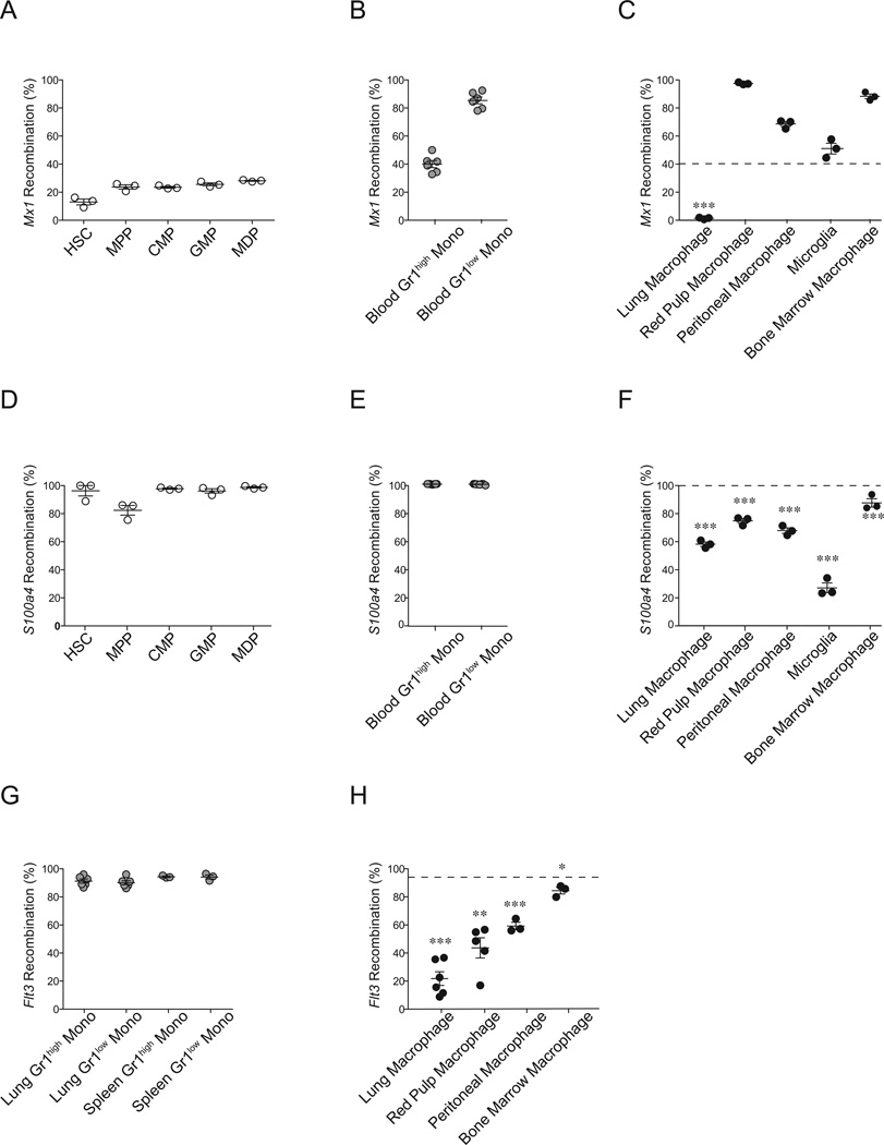
To study the independence of tissue macrophages from circulating monocytes, we probed microarray data from the Immunological Genome Project to find fate mapping candidate genes to trace monocyte progeny. Candidate genes were selected if they were absent in BM progenitors, expressed in Gr-1high and Gr-1low monocytes and absent in certain tissue resident macrophages. Monocytes and tissue macrophages were defined based on their localization and phenotypic markers described in Figure S1. We found two such candidates in Mx1 and S100a4 (Figure S1F and S1G). To fate map the progeny of monocytes, we crossed Mx1-cre (Kuhn et al., 1995) and S100a4-cre (Bhowmick et al., 2004) mice with Rosa26Tomato reporter animals (Madisen et al., 2010). In Mx1-crexR26Tomato animals, there was an increase of tdTomato signal in successive stages of myeloid differentiation and high expression in monocytes (39.7 ± 2.5 % in Gr1high and 85.0 ± 2.4 % in Gr1low) (Figure 1A-B and S1I). Although a relatively high tdTomato signal can lead to multiple non-definitive interpretations (as for peritoneal, red pulp, and BM macrophages in this model), tdTomato positive macrophages (1.2 ± 0.35 %), clearly indicating the lack of contribution of monocytes to lung macrophages (Figure 1B-C and S1J).
Figure 1. Fate mapping reveals that tissue resident macrophages are maintained independently of adult hematopoiesis.
(A-F) The percentage of tdTomato+ cells among purified hematopoietic progenitors (A, D, open circles), monocytes (B, E, gray circles), and tissue resident macrophages (C, F, black filled circles) in Mx1cre×R26Tomato (A-C) and S100a4cre×R26Tomato (D-F) are shown. Representative data from two separate experiments are shown. (G, H) The percentage of GFP+ cells among monocytes (G, gray circles), and tissue resident macrophages (H, black circles) in Flk2cre×R26Tomato/GFP mice are shown. Data were pooled from two independent experiments. Dashed lines in C, F, H indicate % recombination in blood (C, F) or spleen (H) Gr1high monocytes. LT-HSC: long-term hematopoietic stem cells; MPP: multipotent hematopoietic progenitors; CMP: common myeloid progenitors; GMP: granulocyte macrophage progenitor; MDP: macrophage dendritic cell progenitor. *: P < 0.05; **: P < 0.01; ***: P < 0.001 compared to Gr1high monocytes. See also Figure S1.
In the S100a4cre×R26Tomato fate mapping model, we observed unexpectedly high expression of tdTomato in hematopoietic stem cells (HSC) and other progenitors, despite the negligible expression of S100a4 transcripts in these populations, suggesting cre recombination in more primitive hematopoietic cells that had given rise to the adult HSC early in development (Figure 1D and S1G). Consistently, we also observed that more than 99% of monocytes expressed tdTomato (Figure 1E and S1J). However, macrophages from the lung, splenic red pulp, peritoneum, BM, and microglia (Figure 1F and S1J) had substantially lower tdTomato compared to both hematopoietic progenitors and monocytes (Figure 1D and 1E), suggesting an origin of tissue macrophages independent not only from monocytes, but also from adult hematopoietic progenitors.
We confirmed this possibility using, Flk2cre×R26Tomato/GFP mice, which constitutively express tdTomato in all cells, except in the progeny of adult hematopoietic progenitors, which expressed GFP due to Flk2-cre-mediated recombination (Boyer et al., 2011). Although the GFP signal was ~90% in hematopoietic progenitors, circulating leukocytes (data not shown), and lung and spleen monocytes (Figure 1G and S1K), GFP expression was very low in tissue macrophages (Figure 1H and S1K). This was not due to tissue macrophage-specific shutdown of the Rosa26 locus, as all the GFP− tissue macrophages in the Flk2cre×R26Tomato/GFP mice were tdTomato+, indicating unabated activity of the Rosa26 locus (Figure S1K). Altogether, these three fate mapping models strongly suggest that tissue macrophages develop independently of significant contribution of both blood monocytes and adult hematopoietic progenitors, consistent with recent findings (Schulz et al., 2012).
Parabiosis indicates lack of monocyte contribution to tissue resident macrophages
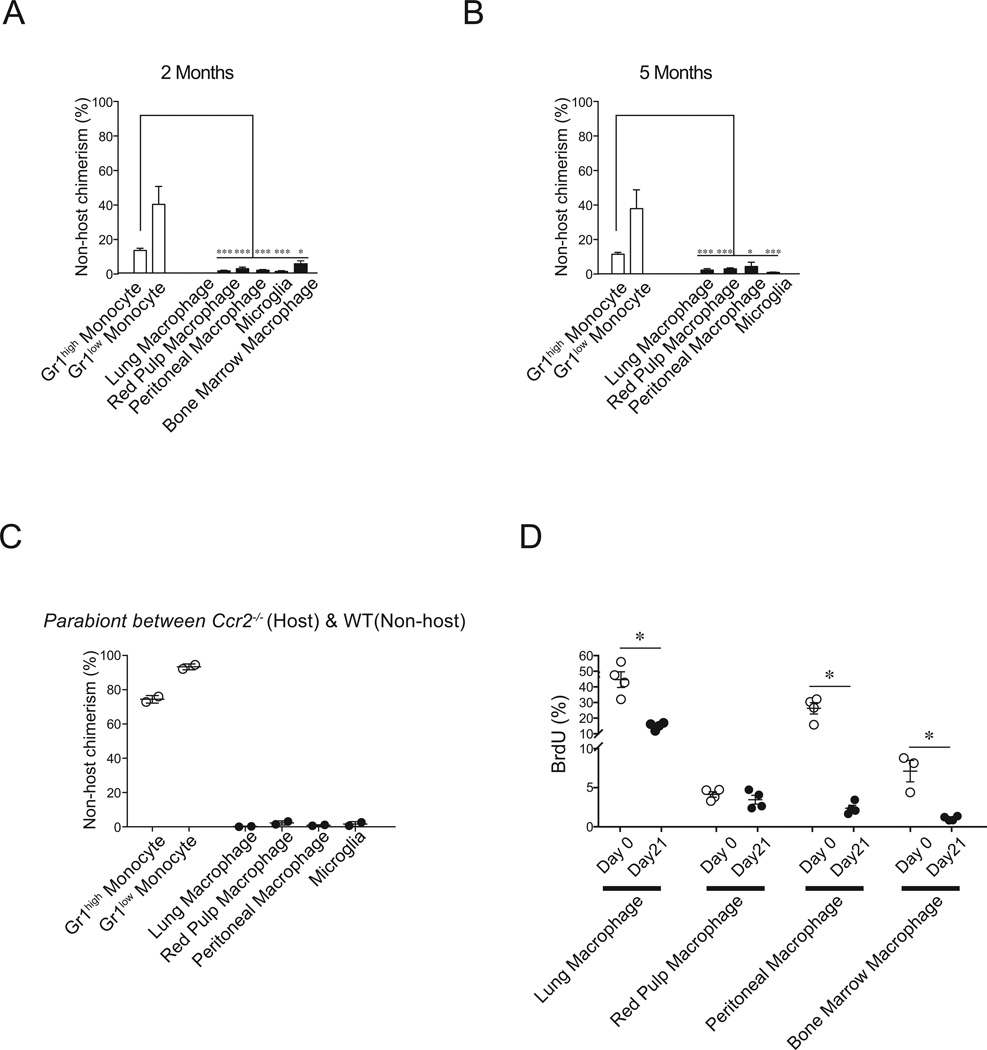
To further establish the relationship between monocytes and tissue macrophages, we parabiotically joined CD45.2+ and congenic CD45.1+ mice and calculated the percentages of non-host-derived cells. After 2 or 5 months of parabiosis, circulating Gr1high monocytes showed ~15% non-host chimerism, while Gr1low monocytes had ~40% non-host chimerism (Figure 2A and 2B), consistent with prior work (Jakubzick et al., 2008). If either monocyte population were differentiating into tissue macrophages in the steady state, a level of non-host chimerism comparable to monocyte chimerism would be expected for tissue macrophages (as gated in Figure S1 A-E). Consistent with their local self-maintenance and their derivation from embryonic macrophages – and not monocytes (Ajami et al., 2007; Ginhoux et al., 2010), microglia demonstrated <1% non-host chimerism. However, we also found that lung, peritoneal, splenic, red pulp and BM macrophages also demonstrated minimal reduced chimerism after two and five months of parabiosis (Figure 2A and 2B).
Figure 2. Parabiosis studies indicate that tissue resident macrophages are maintained independently of monocytes.
(A, B) C57BL/6 CD45.2+ and CD45.1+ parabionts were surgically connected for 2 months (A) or 5 months (B). Bars show the percentage non-host cells among peripheral blood monocytes (white bars) and tissue resident macrophages (black bars) in host parabionts (n=4–6). Representative data from two separate experiments are shown as mean +/− SEM. *: P < 0.05; **: P < 0.01; ***: P < 0.001 compared to Gr1high monocytes. (C) Parabiotic pairs were generated between wild-type CD45.1+ mice and Ccr2−/− CD45.2+ mice. The percentage of non-host cells in the CD45.2+ mouse among peripheral blood monocytes (open circles) and tissue macrophages (filled circles) were analyzed 2 months after surgery. (D) C57BL/6 mice were pulsed with BrdU for 3 weeks (1mg i.p. daily) and BrdU incorporation in tissue macrophages was assessed 1 day (Day 0, open circles) and 22 days (Day 21, filled circles) after the last pulse. *: P < 0.05. See also Figure S2.
To further accentuate the discordance between monocyte and tissue macrophage chimerism, we generated parabiotic mice between wild-type CD45.1+ and either Ccr2+/− or Ccr2−/− CD45.2+ mice. Although Ccr2+/−CD45.2+ parabionts showed similar non-host chimerism to wild-type parabionts (compare Figure 2A-B and S2A), Ccr2−/−CD45.2+ parabionts demonstrated ~70% and ~90% non-host chimerism among Gr1high and Gr1low monocytes, respectively (Figure 2C) due to defective monocyte emigration from the BM into the circulation of Ccr2−/− animals (Serbina and Pamer, 2006). Nonetheless, non-host chimerism of lung, red pulp, peritoneal and BM macrophages remained negligible after two months of parabiosis (Figure 2C). Importantly, this discordance in monocyte versus macrophage chimerism was still observed after seven months and one year of parabiosis (Figure S2B). To assess whether absence of monocyte contribution to macrophage homeostasis could be due to their prolonged half-life in tissues, we used bromodeoxyuridine (BrdU) labeling to measure macrophage turnover in situ. We observed substantial tissue macrophage turnover over the course of 21 days with the exception of spleen red pulp macrophages (Figure 2D), suggesting that the lack of non-host chimerism up to one year after parabiosis does not result from a long lifespan of tissue macrophages or a lack of opportunity for monocyte replacement.
Tissue resident macrophage repopulate through local proliferation after non-genotoxic ablation
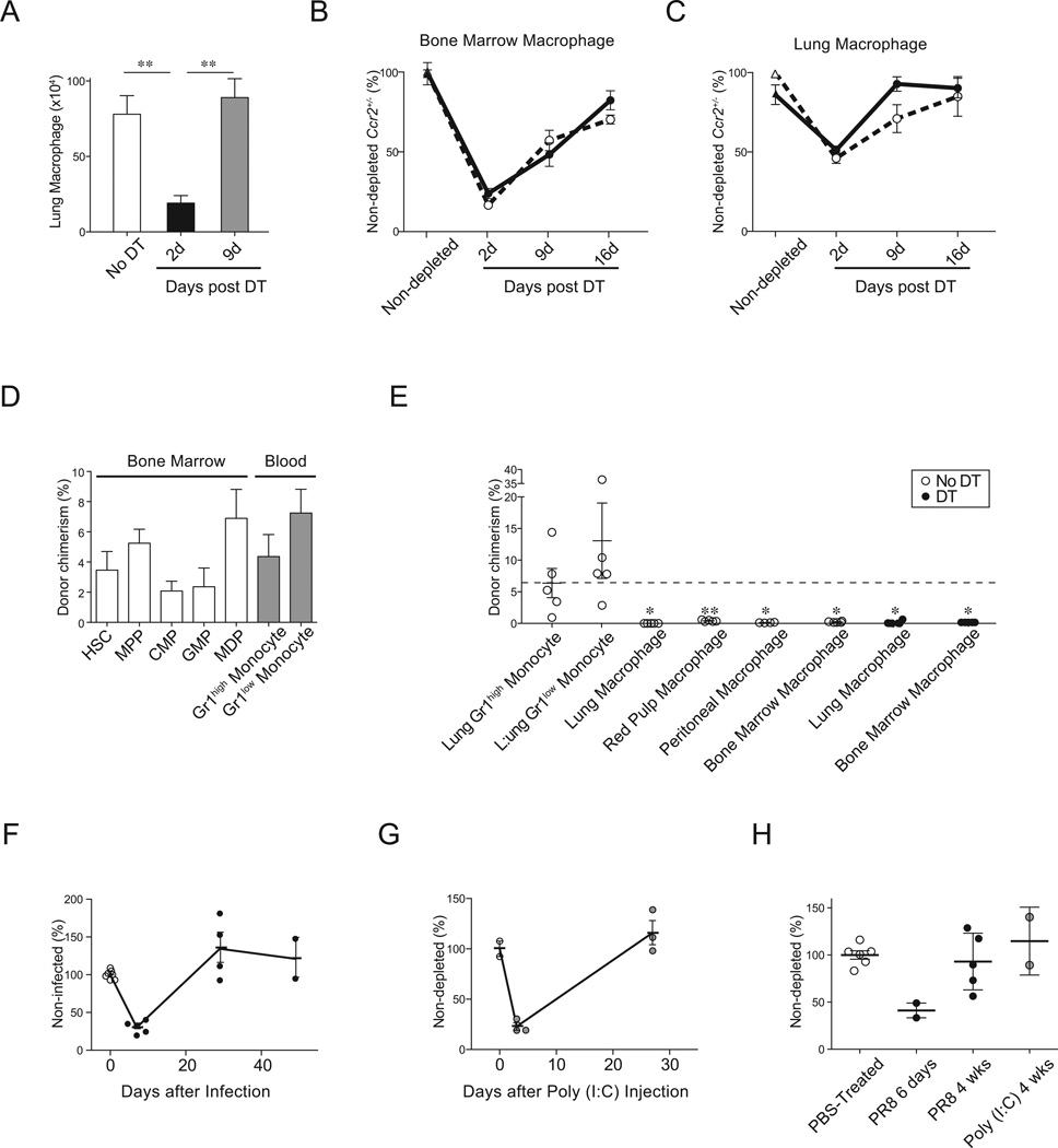
The preceding data suggest that tissue macrophages locally self-maintain in the steady state with minimal contribution from monocytes. We next asked whether this paradigm persisted after enforced cell turnover. To this end, we utilized CD169-DTR animals, which express the diphtheria toxin receptor (DTR) downstream of the Siglec1 (also called CD169 or Sialoadhesin) locus (Miyake et al., 2007). Administration of diphtheria toxin (DT) into CD169-DTR animals allows for specific depletion of certain tissue resident macrophages without ablating monocytes or dendritic cells (Asano et al., 2011; Chow et al., 2011; Miyake et al., 2007). Short-term DT administration into CD169-DTR animals resulted in efficient reduction of BM and lung macrophages (Figure 3A); however, peritoneal macrophages or red pulp macrophages are not reduced (data not shown), precluding analyses of these macrophages in the CD169-DTR depletion model.
Figure 3. Tissue resident macrophages repopulate locally independently of CCR2+ progenitors.
(A) CD169DTR/+ mice were injected i.p. with 10 µg/kg DT (n=5, filled bars) or control diluent (n=3, open bars) on day 1 and 4 and the absolute numbers of lung macrophages were enumerated 2 days and 9 days after the last injection. Representative data from three separate experiments are shown as mean +/− SEM. **: P < 0.01. (B, C) CD169DTR/+×Ccr2+/− (open circles, n=5/time point), and CD169DTR/+×Ccr2−/− mice (filled circles, n=6/time point) were injected with DT on day 0 and the number of BM macrophages (B) and lung macrophages (C) were analyzed at 2, 9, and 16 days after DT administration. CD169+/+×Ccr2+/− (open triangle) and CD169+/+×Ccr2−/− (filled triangle) mice treated with DT were used as non-depleted controls. The absolute cell numbers are represented as relative values with the mean absolute number of lung macrophages in non-depleted CD169+/+×Ccr2+/− mice set at 100%. Data were pooled from two independent experiments and shown as mean +/− SEM. (D-E) CD169DTR/+ (CD45.2+, n=5) mice were infused with 7.5 × 10⁷ congenic CD45.1+ BM cells, followed by DT (filled circles) or PBS (open circles) administration on days 4 and 7 after BM cell transfer. The percentage of donor-derived CD45.1+ cells among (D) BM progenitor cells (white bars) and blood monocytes (Mono, gray bars) and (E) lung monocytes and tissue macrophages were analyzed sixteen days after the transfer. Representative data from three independent experiments are shown as mean +/− SEM. *: P < 0.05; **: P < 0.01 compared to Gr1high monocytes. (F-H) S100a4Cre×R26Tomato mice were infected intranasally with 100 pfu PR8 influenza virus (F and H filled circles) or instilled intranasally with 50µg Poly (I:C) (G and H gray circles) or control PBS (F-H, open circles). Kinetics of lung macrophages number at different times after PR8 infection (F) or Poly (I:C) injection (G) and numbers of tdTomato− lung macrophages at six days and four weeks following PR8 infection or Poly (I:C) injection are shown (H). Data are represented as mean +/− SEM. See also Figure S3.
We generated CD169DTR/+Ccr2−/− mice to determine whether CCR2-dependent migration of candidate progenitor cells, including monocytes and hematopoietic progenitors, contribute to tissue macrophage recovery after turnover (Boring et al., 1997). Consistent with prior work (Kuziel et al., 1997; Serbina and Pamer, 2006), Ccr2−/− mice indeed had lower circulating monocyte numbers (Figure S3A) and also lower steady-state monocyte numbers in the lung and spleen (Figure S3B and data not shown) compared to Ccr2+/− littermates, but comparable numbers of BM and lung tissue macrophages (Figure 3B-C). Consistent with the lack of CCR2-dependent contribution to steady-state tissue macrophages, we also found that both the extent of the depletion and the kinetics of the recovery were equivalent in DT-infused CD169DTR/+Ccr2+/− and CD169DTR+/− Ccr2−/− mice (Figure 3B-C), indicating that the recovery of the tissue resident macrophage compartment is independent of CCR2-dependent cells such as monocytes (Serbina and Pamer, 2006) and hematopoietic progenitors (Si et al., 2010).
To further examine the contribution of circulating hematopoietic cells to tissue macrophage recovery after non-genotoxic ablation, we infused 7.5×10⁷ BM cells isolated from CD45.1+ congenic mice into CD169DTR/+ CD45.2+ recipient mice without prior conditioning. With this adoptive transfer approach, we obtained a low but stable engraftment of donor BM progenitors and consequently stable donor monocyte chimerism in the recipient blood (4.4 ± 1.5 % for Gr1high monocytes and 7.2 ± 1.6% for Gr1low monocytes) (Figure 3D). Consistent with the parabiosis studies (Figure 2), we observed that the percentage of CD45.1+ donor-derived cells among lung, red pulp, peritoneum, and BM macrophages in recipient mice, without DT injection was negligible (0.002 % ± 0.002%, 0.34 % ± 0.06 %, 0.05 % ± 0.02 %, and 0.07 % ± 0.05%, respectively) (Figure 3E). Tissue macrophage depletion and their subsequent recovery 9 days later did not increase the donor tissue macrophage chimerism (Figure 3E), indicating that circulating hematopoietic progenitors or monocytes did not contribute to the recovery of the tissue macrophage pool.
The recent observation that macrophages are depleted after Toxoplasma infection, led us to examine the contribution of monocytes to tissue macrophages in an infectious setting (Goldszmid et al., 2012). We intranasally infected S100a4cre×R26Tomato with the PR8 strain of influenza virus, which resulted in dramatic lung macrophage cytoablation 6 days post infection, substantial infiltration of Gr1high monocytes, and recovery of macrophage numbers by four weeks post infection (Figure 3F, S3C-D). Consistent with Figure 1, 35–40% of lung macrophages were tdTomato−. If adult hematopoietic progenitors or monocytes, which are >99% tdTomato+, were replacing lung macrophages, one would expect that the absolute number of tdTomato− macrophages would not recover after influenza infection and would instead be replaced by tdTomato+ progenitors. On the contrary, we observed that the number of tdTomato− macrophages returned to uninfected baseline numbers four weeks after infection, indicating recovery from local repopulation of tdTomato− macrophages (Figure 3H). Similarly, when we intranasally instilled S100a4cre×R26Tomato mice with 50µg of Poly (I:C) to induce macrophage cytoablation and monocyte infiltration (Figure 3G, S3E-F), tdTomato− macrophages returned to normal numbers four weeks after instillation (Figure 3H).
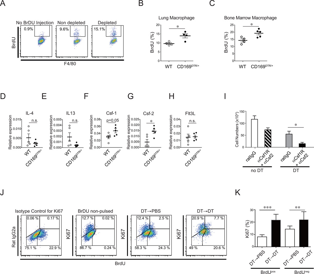
Since we observed no replacement of tissue macrophages by circulating progenitor candidates, we hypothesized that tissue macrophage replenishment occurred via local proliferation. Assessing proliferation with a short pulse of BrdU, we observed that macrophage depletion in CD169-DTR mice resulted in a significant increase in proliferation of lung and BM macrophages (Figure 4A-C). In CD11c-DTR transgenic mice, DT administration depleted red pulp but not lung macrophages (Figure S4A) as previously reported (Meredith et al., 2012). Accordingly, we found increased macrophage proliferation in the red pulp but not in lung tissues (Figure S4B). Using systemic administration of clodronate liposomes, we also observed a depletion of lung, red pulp and BM macrophages that was associated with enhanced subsequent local proliferation (Figure S4C-E). Collectively, using three distinct conditional depletion models, our results reveal that macrophages enhance proliferation in order to repopulate locally after cell turnover.
Figure 4. Repopulation of lung tissue resident macrophages is dependent on local cytokine production.
(A, B) Wild-type or CD169DTR/+ mice were administered 10µg/kg DT on days −5 and −2 and injected i.p. with 1mg BrdU /mouse on day −1 prior to analysis. Flow cytometric plots depict percentage of BrdU positive cells among total lung macrophages in non-BrdU injected mice (left panel), WT mice treated with DT (center panel), or CD169DTR/+ mice treated with DT (right panel). (B, C) Quantitation of the percentage of BrdU+ cells among lung macrophages (B) and BM macrophages (C) in WT (open circles) and CD169DTR/+ (filled circles) mice treated with DT. (D-H) mRNA was purified from lung homogenates and expression of IL-4 (D), IL-13 (E), CSF-1 (F), CSF-2 (G), and Flt3L (H) were quantified with real-time PCR relative to the expression of GAPDH. (I) CD169DTR/+ mice were injected with PBS or DT on days −12 and −9 prior to analysis followed by i.n. injection of 600µg of anti-Csf-1R Ab (clone: AFS98) on days −8, −6, −4, and −1 and 100µg of anti-Csf-2 neutralizing Ab (clone: MP122E9) on days −7, −5, −2 prior to the analysis. The numbers of lung tissue resident macrophages in mice that received rat IgG alone (open bar, n=16), anti-Csf-1R mAb and Csf-2 mAb blockade without DT (hatched bar, n=5), DT + control rat IgG (gray filled bar, n=9), and DT in addition to anti-Csf-1R mAb and Csf-2 mAb blockade (black filled bar, n=10) are shown. Data were pooled from three independent experiments and shown as mean +/− SEM. (J, K) CD169DTR/+ mice were treated with 10 εg/kg DT on day −16, pulsed with 1mg/day BrdU on days −15 through −5, and re-injected with DT (black bars, n=8) or PBS (white bars, n=8) on day −2 prior to analysis. Mice were sacrificed on day 0 and BrdU incorporation and Ki67 expression were analyzed using flow cytometry. Representative FACS plots of Ki67 and BrdU staining in lung macrophages (J) are shown. Ki67 expression (cycling cells) among BrdU positive fraction and BrdU negative fraction of lung macrophages were quantified (K). Data were pooled from two independent experiments and shown as mean +/− SEM. *: P < 0.05; **: P < 0.01; ***: P < 0.001. See also Figure S4.
Although IL4 has been shown to be the critical cytokine that promotes local expansion of the tissue macrophage pool in parasitic infection (Jenkins et al., 2011), the expression of IL4 and IL13, which also utilizes the IL4 receptor, did not increase in the lungs of macrophage-depleted mice (Figure 4D and 4E). On the other hand, we observed an increased in the expression of Csf-1 (also called M-Csf) and Csf-2 (also called GM-Csf), but not fms-like tyrosine kinase 3 ligand (Flt3L) in the lung tissue of macrophage-depleted mice (Figure 4F-H). To determine whether recovery of the lung tissue macrophage compartment after ablation was dependent on Csf-1 and Csf-2 cytokines, we administered anti-Csf-1R and anti-Csf-2 blocking Abs after macrophage depletion. The recovery of tissue macrophages was impaired in the cytokine-blocked mice compared to rat IgG-treated control mice (Figure 4I). Anti-Csf-1R and anti-Csf-2 blocking Abs also reduced tissue macrophages in non-depleted mice. These data indicate that local repopulation of macrophages in both the steady state and upon enforced depletion is dependent on Csf-1R and/or Csf-2 signaling.
Next, we examined whether the ability of macrophages to repopulate was restricted to a particular progenitor population or whether all macrophages could self renew in a stochastic manner. Thus, we depleted lung macrophages in the CD169-DTR model and subsequently pulsed daily with BrdU injections for 10 days to label proliferating cells. Two days after completion of the 10-day pulse, the CD169-DTR mice were injected with DT once again. Two days later, mice were sacrificed and we used an antibody against the cell cycle protein Ki-67 to distinguish between cycling macrophages that had proliferated after the initial DT injection (Ki-67+BrdU+) from the newly proliferating population (Ki-67+BrdU−). Ki-67 was equally increased among BrdU+ and BrdU− macrophages, indicating that repopulation capacity was a stochastic event and not restricted to a subset of macrophages. (Figure 4J and 4K). In contrast, DT injection failed to induce B cell proliferation, establishing that induction of cellular proliferation upon DT injection in CD169-DTR mice was specific to macrophages (Figure S4F and S4G). Altogether, these data suggest that differentiated macrophages proliferate locally and stochastically during cell turnover.
Recovery of tissue resident macrophages after genotoxic insult occurs via donor circulating progenitors
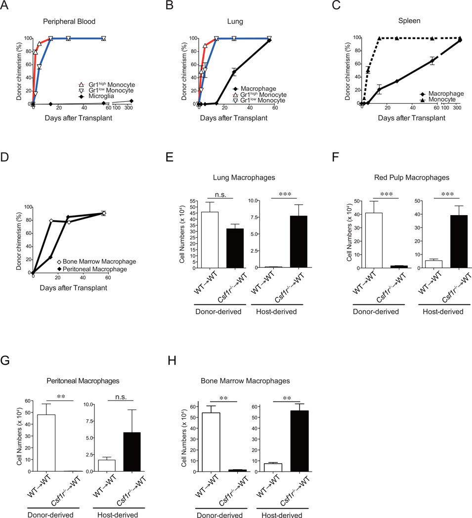
With the exception of microglia (Ginhoux et al., 2010) (Figure 5A), after lethal irradiation and bone marrow transplantation, tissue macrophages derive from donor origin (Haniffa et al., 2009; Virolainen, 1968), indicating that unlike the CD169-DTR depletion and inflammatory models above, lethal irradiation represents an insult that facilitates impairment of local repopulation capacity and thus donor engraftment. In this context in which macrophages do arise from donor cells, we sought to determine whether the kinetics of tissue macrophage recovery was consistent with derivation from monocytes. We found that within 2 weeks of bone marrow transplantation, all Gr1high and Gr1low monocytes in the peripheral blood, lung, and spleen were of donor origin (Figure 5A-C). Although, tissue resident macrophages in the lung, splenic red pulp, peritoneum and BM were eventually replaced by donor cells (Figure 5B-5D), donor repopulation of macrophages was markedly delayed compared to that of monocytes, suggesting that monocytes may not contribute to the recovery of tissue macrophages after genotoxic insult.
Figure 5. X-ray irradiation does not abrogate host tissue macrophage repopulation potential.
(A-D) C57BL/6 CD45.2+ mice were lethally irradiated and transplanted with 5 × 10⁶ congenic BM CD45.1+ cells. The percentage of donor (CD45.1+) cells among circulating monocytes and CNS microglia (A), monocytes and/or macrophages in the lung (B), spleen (C), BM (D), and peritoneum (D) are shown (n=3–4/time points). (E-H) C57BL/6 CD45.1+ mice were lethally irradiated and transplanted with 1 × 10⁶ fetal liver cells isolated from C57BL/6 CD45.2+ Csf-1r−/− (black bars, n=7) mice or control littermate (white bars, n=7). The absolute numbers of host and donor tissue resident macrophages in the lung (E), spleen (F), peritoneum (G), and BM (H) were enumerated 4 months after transplant. Data were pooled from two independent experiments and shown as mean +/− SEM. **: P < 0.01; ***: P < 0.001. See also Figure S5.
Host tissue resident macrophages retain the capability to self-maintain even after genotoxic insult
The inability of host tissue macrophages to regenerate tissue macrophage chimerism after bone marrow transplantation could potentially be due to irreversible radiation-induced destruction of tissue macrophages or incomplete radiation-induced suppression of tissue macrophage self-renewal. In order to distinguish between these two possibilities, we evaluated a system in which, donor repopulation of the tissue macrophage compartment was specifically impaired. To achieve this, we established a transplantation system in which donor-derived cells were deficient in Csf-1R by infusing 1×10⁶ wild-type or Csf1r−/− fetal liver cells into lethally-irradiated adult animals. Four months after transplantation, a time point at which monocytes were completely of donor origin both in [WT into WT] chimera and [Csf-1r−/− into WT] chimeras (Figure S5A), we enumerated donor-derived and host-derived tissue macrophages. In the lung of the recipients of Csf-1r−/− fetal liver, there was a slight non-significant reduction in the number of donor-derived tissue macrophages compared to recipients of WT fetal liver cells (Figure 5E and S5B). Nonetheless, this slight reduction in donor-derived macrophages was associated with a substantial expansion of the host macrophages (Figure 5E), suggesting that host tissue macrophages retained the capacity to self-renew in the context of reduced competition from donor-derived cells. This impaired donor contribution and consequent host tissue macrophage repopulation was even more profound in the spleen and BM, as the entire deficit in donor-derived splenic red pulp and BM macrophages was recovered by the expansion of the host tissue macrophage compartment (Figure 5F, 5H, S5C, and S5E). In the peritoneum, there was also a substantial reduction in donor-derived macrophages in recipients of Csf1r−/− fetal liver cells, and there was a small, although not statistically significant, expansion of host peritoneal macrophages (Figure 5G and S5D). Together, these results highlight that the recovery of the tissue resident macrophage compartment in the spleen, peritoneum, and BM is dependent on Csf-1R signaling whereas it is partially dependent on Csf-1R in the lung. Furthermore, at least in the lung, spleen, and BM, host tissue resident macrophages can expand in the setting of reduced donor competition.
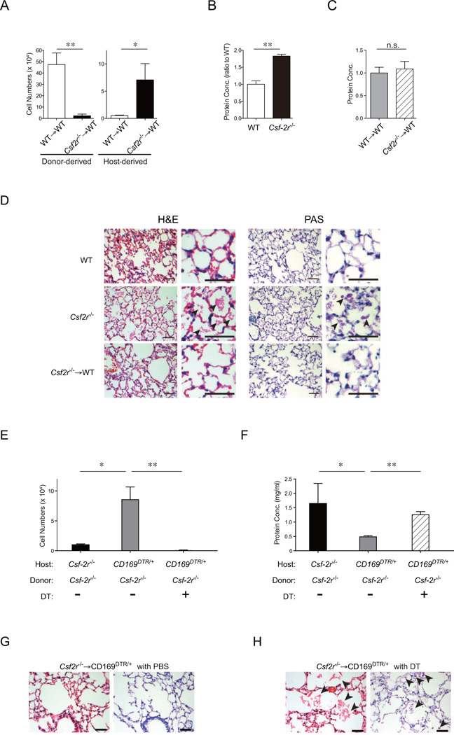
Since Csf-2 is critical in lung macrophage homeostasis (Trapnell and Whitsett, 2002) (Figure S6A and S6B), we sought to examine whether impaired donor Csf-2-dependent donor repopulation could also result in the development of host lung macrophage expansion after lethal irradiation. Two months after bone marrow transplantation of wild-type or Csf-2 receptor-deficient (Csf-2r−/−) CD45.2+ cells into lethally irradiated CD45.1+ mice, all circulating monocytes were completely donor-derived (Figure S6C). Although recipients of wild-type BM achieved donor-derived recovery of the lung macrophage compartment, we observed that donor-derived recovery of lung tissue macrophages was substantially reduced in Csf-2r−/− BM chimeric mice (Figure 6A and Figure S6D). Importantly, the severe impairment of donor-derived macrophage development resulted in a compensatory expansion of the host tissue macrophage pool over time (Figure 6A and S6E). Mice deficient in Csf-2 (Stanley et al., 1994) and Csf-2R (Robb et al., 1995) manifest a severe lung inflammatory disease characteristic of clinical alveolar proteinosis due to impaired surfactant clearance by defective pulmonary macrophages. Defects in Csf-2 and Csf-2R (Trapnell et al., 2003) have been described in patients with this disorder. We evaluated whether the host macrophages that recovered from genotoxic insult were functionally competent by examining their ability to clear surfactant and prevent the development of alveolar proteinosis in these chimeras. Naïve Csf-2r−/− mice had significantly elevated alveolar protein concentration compared to wild-type mice (Figure 6B). Furthermore, the lung histology showed conspicuous granular eosinophilic material within the alveolar spaces visualized with H&E staining, and this material was positive for periodic acid-shiff (PAS), indicative of clinical alveolar proteinosis (Figure 6D). In contrast, [Csf-2r−/− → WT] chimeras had an alveolar protein concentration comparable to that of [WT→WT] chimeras (Figure 6C). Blinded examination by two independent lung pathological examiners did not detect any pathology in either [Csf-2r−/−→WT] or [WT→WT] chimeras (Figure 6D and S6F). To further assess the functional ability of self-renewing host macrophages to clear alveolar proteins, CD169DTR/+ mice were reconstituted with Csf-2r−/− BM cells. BM chimeric mice were injected twice weekly with either DT or PBS for up to 8 weeks post transplant to deplete host CD169+ lung tissue macrophages. We found that host macrophage inability to repopulate in DT treated [Csf-2r−/−→CD169DTR/+] chimeric mice was associated with elevated bronchoalveolar lavage fluid (BALF) protein concentrations and the development of alveolar proteinosis (Figure 6E-F and 6H), whereas PBS treated [Csf-2r−/−→CD169DTR/+] chimeric mice were protected from lung disease (Figure 6E-G). Hence, host-derived lung tissue macrophages can resist radiation injury, and functionally repopulate when donor repopulation is impaired, and this expansion of host-derived wild-type macrophages is sufficient to prevent the development of alveolar proteinosis.
Figure 6. Lung macrophages repopulate locally after lethal irradiation and promote lung tissue integrity.
(A-D) CD45.1+ mice were lethally irradiated and transplanted with 5 × 10⁶ BM cells isolated from CD45.2+ Csf-2r−/− (black bars, n=6) mice or control CD45.2+ littermate (white bars, n=5). (A) The absolute numbers of donor- and host-derived lung macrophages in right lung lobes were enumerated 2 months after bone marrow transplantation. (B, C) Protein concentrations in the bronchial alveolar lavage fluid (BALF) were quantified in wild-type mice (white bar), Csf-2r−/− mice (black bar), and wild-type mice transplanted with wild-type (gray bar) or Csf-2r−/− (hatched bar) BM (n=3/group) two months after bone marrow transplantation. Data were pooled from two independent experiments and shown as mean +/− SEM. (D) Representative sections of left lung lobes stained with H&E (left) and PAS (right) obtained from steady-state wild-type (WT) mice (top), steady-state Csf-2r−/− mice (center), and WT mice transplanted with Csf-2r−/− (bottom) BM. Arrow heads: PAS+ eosinophilic material within the alveolar spaces. Scale bar: 25µm. (E-H) Csf-2r−/− mice and CD169DTR/+ mice were lethally irradiated and reconstituted with 5 × 10⁶ BM cells isolated from C57BL/6 WT or Csf-2r−/− mice. A group of CD169DTR/+Csf-2r+/+ recipients were injected with DT (10µg/kg, twice weekly) starting from day +3 post-transplant. The absolute numbers of macrophages in the right lung lobes (E) and protein concentrations in BALF (F) in [Csf-2r−/− into Csf-2r−/−] (black bars, n=3) and [Csf-2r−/− into CD169DTR/+] chimera treated with PBS (gray bars, n=6) or DT (hatched bars, n=6) were enumerated two months after transplant. Data were pooled from two independent experiments and shown as mean +/− SEM. Paraffin sections of the left lung lobes of recipients of [Csf-2r−/− into CD169DTR/+] chimeras treated with PBS (G) or DT (H) at 2 months post transplant were stained with H&E (left panel) and PAS stain (right panel). Sections isolated from [Csf-2r−/− into CD169DTR/+] chimera treated with DT (H) demonstrate granular eosinophilic material positive for PAS within the alveolar spaces (arrow heads) whereas sections from [Csf-2r−/− into CD169DTR/+] chimera treated with diluent (G) demonstrate normal alveolar structure. Scale bars: 25 µm. See also Figure S6. *: P < 0.05; **: P < 0.01.
Discussion
Recent findings have called into question the dogma that monocytes constitutively give rise to tissue resident macrophages (Ajami et al., 2007; Ginhoux et al., 2010; Jenkins et al., 2011; Schulz et al., 2012). In order to address this controversy, we took advantage of animal models that have allowed us to comprehensively re-examine the origin of tissue resident macrophages. Utilizing three distinct fate mapping models, parabiosis studies, and adoptive transfer of total BM cells, we use discordant chimerism between monocytes and tissue macrophages to demonstrate that monocytes are not the progenitors of lung, splenic red pulp, peritoneal, and BM tissue macrophages in the steady state. Even when host tissue macrophages were eliminated in the lung and BM and forced to turnover, repopulation of macrophages still occurred independently of circulating monocytes. Our finding that these tissue macrophages can extensively self-maintain independently of the adult HSC in the steady state complements the recent report that the yolk sac-derived fraction of splenic, hepatic, and pancreatic macrophages can also self-maintain (Schulz et al., 2012) and goes further in describing that this self-maintenance capacity is not restricted to the steady state, but is also preserved after cell turnover. These results establish that similar to Langerhans cells and microglia, several tissue macrophages self-renew in situ and can repopulate locally after tissue injuries (Ajami et al., 2007; Merad et al., 2002))).
If circulating monocytes and hematopoietic progenitors are not contributing, then how are tissue macrophages maintained in the steady state and after turnover? In line with previous reports of tissue macrophage proliferation in the steady state (Daems and de Bakker, 1982; Jenkins et al., 2011), we found that non-genotoxic depletion of lung tissue macrophages was followed by local proliferation and enhanced local cytokine production of Csf-1 and Csf-2. Consistent with the importance of these two cytokines with lung macrophage repopulation after cell turnover, concomitant blockade of Csf-1R and Csf-2 signaling compromised lung tissue macrophage repopulation after non-genotoxic depletion. Thus, we conclude that tissue macrophages are capable of local repopulation in the steady state and after cell turnover at least in some tissues. Local proliferation potential was stochastic and not restricted to a subset of tissue macrophages.
Importantly, we also found that host tissue macrophages can repopulate locally after macrophage cytoablation caused by infection with influenza or instillation of Poly (I:C), which complement prior results showing that pleural tissue resident macrophages can proliferate locally in mice in response to parasitic infection (Jenkins et al., 2011). Unfortunately, we were unable to trace the monocyte progeny in inflamed tissues and it is unclear at this point whether monocyte-derived macrophages transiently infiltrate inflamed tissues or remain locally once the inflammation resolves.
In humans, there have been numerous reports of congenital causes of monocytopenia that were not associated with a reduction in tissue macrophages. In reticular dysgenesis, a rare inherited immunodeficiency characterized by the lack of blood monocytes, neutrophils, and lymphocytes, macrophages are present in normal numbers in the dermis and in the atrophic lymphoid tissues of these patients (Emile et al., 2000). Moreover, there was a report of 18 patients with an autosomal dominant susceptibility to opportunistic infections and monocytopenia, who had normal numbers of dermal macrophages (Vinh et al., 2010). In addition, in four patients with a syndrome of monocytopenia and deficiency of dendritic, B and NK cells, dermal macrophages were found to be present (Bigley et al., 2011). Similarly, a patient with IRF8 null mutation was recently found to have a block in monocyte and DC differentiation and increased susceptibility to mycobacterium. However, despite profound peripheral monocytopenia, macrophages were present in the lymph nodes and in the BM of the IRF8 mutated patient (Hambleton et al., 2011). The normalcy of macrophages in these monocytopenic patients further suggests that tissue macrophages can develop in the absence of monocytes in humans.
It has been known for decades that tissue macrophages are replaced, albeit slowly, by BM-derived cells after bone marrow transplantation (Thomas et al., 1976; Virolainen, 1968), and in fact, these results were used as the early arguments for circulating contribution to tissue macrophages. However, our data indicates that there is extensive host-derived tissue macrophage recovery when donor-derived repopulation is impaired, as in the chimeras generated from Csf1r−/− fetal liver or Csf2r−/− BM. These data are consistent with the observation that fractionation of a lethal irradiation dose allows substantial recovery of host-derived lung macrophages (Tarling et al., 1987). Importantly, this expansion of host-derived wild-type macrophages is function as they prevented the development of alveolar proteinosis observed in transplant recipients of Csf2r−/− BM. These observations in the myeloablative setting dovetail nicely with our findings that tissue resident macrophages self maintain locally throughout adult life in the steady state and after non-genotoxic tissue macrophage turnover. Besides the well-described effect that Csf-2 has on macrophage phagocytosis (Trapnell et al., 2003), its control of macrophage numbers is likely another mechanism by which defects in Csf-2 and Csf-2R result in clinical alveolar proteinosis (Sakagami et al., 2009; Uchida et al., 2007). The capacity of host macrophages to repopulate after radiation injuries may have consequences in the bone marrow transplantation setting in which remaining host antigen presenting cells have been shown to contribute to graft versus host disease outcome (Hashimoto and Merad, 2011).
Since we now have the tools to distinguish monocytes and infiltrating macrophages from resident macrophages, we advocate for more stringent distinctions to be made for these two populations. We believe that this will be important in understanding tissue resident macrophage homeostasis in humans subjected to various opportunistic (e.g. pathogenic infections) or iatrogenic (e.g. myeloablative procedures) insults, but more importantly this will be critical to understand the contribution of tissue resident macrophages and BM-derived macrophages to tissue immunity, homeostasis and repair.
Experimental Procedures
Mice
The mouse strains used are described in the Supplemental Experimental Procedures.
Flow cytometry
The detailed procedures, antibodies used and gating schemes are described in the Supplemental Experimental Procedures.
Parabiosis
Parabiotic mice were generated as reported (Ginhoux et al., 2009) using age- and weight-matched CD45.2+ (C57BL/6 or Ccr2−/−) and CD45.1+ (C57BL/6) mice between 6 and 8 weeks old. Consistent with prior publications (Ginhoux et al., 2009; Liu et al., 2007), circulating B cells and neutrophils achieved efficient (~50%) non-host chimerism at 2 and 5 months.
Macrophage depletion
The detailed protocols are described in the Supplemental Experimental Procedures.
Microbial-induced lung inflammation
Mice were infected intranasally with 100 pfu of Influenza strain A/Puerto Rico/8/34 (PR8) diluted in 25 µl PBS as previously described (Helft et al., 2012). Other Mice were intranasally instilled with 50 µg of high molecular weight polyinosinic-polycytidylic acid (Poly (I:C)) that was purchased from InvivoGen (San Diego, CA) and reconstituted in normal saline (1 mg/ml).
RNA Isolation, reverse transcription and quantitative real-time PCR (Q-PCR)
The detailed protocols are described in the Supplemental Experimental Procedures.
Concomitant Csf1-R and Csf-2 blockade
Blocking antibody to Csf-1R (clone AFS98) was produced from a hybridoma in-house. Blocking antibody to Csf-2 (clone 22E9) was purchased from R&D. 600µg anti-Csf-1R was infused intranasally on days −8, −6, −4, and −1 and 100µg anti-Csf-2 was infused intranasally on days −7, −5, −2 prior to harvest.
Bone marrow adoptive transfer
In order to achieve donor circulating chimerism without parabiosis or conditioning, we infused 7.5×10⁷ total BM cells from CD45.1+ mice into CD169DTR/+ (CD45.2+) animals.
BM and fetal liver transplantation
CD45.1+ mice were irradiated (1,200 cGy, two split doses, 3h apart) in a Cesium Mark 1 irradiator (JL Shepperd & associates) and then infused with 5×10⁶ BM cells from wild-type or Csf-2r−/− CD45.2+ cells or 1×10⁶ fetal liver cells from wild-type or Csf1r−/− animals. Mice were harvested and analyzed as detailed in the manuscript text and figure legends.
Statistical analyses
The unpaired Student’s t test was used in all analyses, data in bar graphs are represented as mean ± SEM, and statistical significance was expressed as follows: *, P < 0.05; **, P < 0.01; ***, P < 0.001; n.s., not significant
Supplementary Material
Acknowledgments
MM is supported by NIH grants CA154947A, AI10008, AI089987. P.S.F. is supported by NIH grants HL116340, DK056638, HL097700, and HL069438. A.C. is supported by predoctoral fellowship from NHLBI (5F30HL099028). S.W.B. is supported by predoctoral NIH Training Grant 2T32GM008646 to UC Santa Cruz; E.C.F. is supported by a California Institute for Regenerative Medicine (CIRM) New Faculty Award.
Footnotes
Publisher's Disclaimer: This is a PDF file of an unedited manuscript that has been accepted for publication. As a service to our customers we are providing this early version of the manuscript. The manuscript will undergo copyediting, typesetting, and review of the resulting proof before it is published in its final citable form. Please note that during the production process errors may be discovered which could affect the content, and all legal disclaimers that apply to the journal pertain.
References
- Ajami B, Bennett JL, Krieger C, Tetzlaff W, Rossi FM. Local self -renewal can sustain CNS microglia maintenance and function throughout adult life. Nat. Neurosci. 2007;10:1538–1543. doi: 10.1038/nn2014. [DOI] [PubMed] [Google Scholar]
- Asano K, Nabeyama A, Miyake Y, Qiu CH, Kurita A, Tomura M, Kanagawa O, Fujii S, Tanaka M. CD169-Positive Macrophages Dominate Antitumor Immunity by Crosspresenting Dead Cell-Associated Antigens. Immunity. 2011;34:85–95. doi: 10.1016/j.immuni.2010.12.011. [DOI] [PubMed] [Google Scholar]
- Bhowmick NA, Chytil A, Plieth D, Gorska AE, Dumont N, Shappell S, Washington MK, Neilson EG, Moses HL. TGF-beta signaling in fibroblasts modulates the oncogenic potential of adjacent epithelia. Science. 2004;303:848–851. doi: 10.1126/science.1090922. [DOI] [PubMed] [Google Scholar]
- Bigley V, Haniffa M, Doulatov S, Wang XN, Dickinson R, McGovern N, Jardine L, Pagan S, Dimmick I, Chua I, et al. The human syndrome of dendritic cell, monocyte, B and NK lymphoid deficiency. J. Exp. Med. 2011;208:227–234. doi: 10.1084/jem.20101459. [DOI] [PMC free article] [PubMed] [Google Scholar]
- Boring L, Gosling J, Chensue SW, Kunkel SL, Farese RV, Jr, Broxmeyer HE, Charo IF. Impaired monocyte migration and reduced type 1 (Th1) cytokine responses in C-C chemokine receptor 2 knockout mice. J. Clin. Invest. 1997;100:2552–2561. doi: 10.1172/JCI119798. [DOI] [PMC free article] [PubMed] [Google Scholar]
- Boyer SW, Schroeder AV, Smith-Berdan S, Forsberg EC. All hematopoietic cells develop from hematopoietic stem cells through Flk2/Flt3-positive progenitor cells. Cell Stem Cell. 2011;9:64–73. doi: 10.1016/j.stem.2011.04.021. [DOI] [PMC free article] [PubMed] [Google Scholar]
- Chawla A, Nguyen KD, Goh YP. Macrophage-mediated inflammation in metabolic disease. Nat. Rev. Immunol. 2011;11:738–749. doi: 10.1038/nri3071. [DOI] [PMC free article] [PubMed] [Google Scholar]
- Chow A, Lucas D, Hidalgo A, Mendez-Ferrer S, Hashimoto D, Scheiermann C, Battista M, Leboeuf M, Prophete C, van Rooijen N, et al. Bone marrow CD169+ macrophages promote the retention of hematopoietic stem and progenitor cells in the mesenchymal stem cell niche. J. Exp. Med. 2011;208:261–271. doi: 10.1084/jem.20101688. [DOI] [PMC free article] [PubMed] [Google Scholar]
- Daems WT, de Bakker JM. Do resident macrophages proliferate? Immunobiology. 1982;161:204–211. doi: 10.1016/S0171-2985(82)80075-2. [DOI] [PubMed] [Google Scholar]
- Emile JF, Geissmann F, Martin OC, Radford-Weiss I, Lepelletier Y, Heymer B, Espanol T, de Santes KB, Bertrand Y, Brousse N, et al. Langerhans cell deficiency in reticular dysgenesis. Blood. 2000;96:58–62. [PubMed] [Google Scholar]
- Ginhoux F, Greter M, Leboeuf M, Nandi S, See P, Gokhan S, Mehler MF, Conway SJ, Ng LG, Stanley ER, et al. Fate mapping analysis reveals that adult microglia derive from primitive macrophages. Science. 2010;330:841–845. doi: 10.1126/science.1194637. [DOI] [PMC free article] [PubMed] [Google Scholar]
- Goldszmid RS, Caspar P, Rivollier A, White S, Dzutsev A, Hieny S, Kelsall B, Trinchieri G, Sher A. NK cell-derived interferon-gamma orchestrates cellular dynamics and the differentiation of monocytes into dendritic cells at the site of infection. Immunity. 2012;36:1047–1059. doi: 10.1016/j.immuni.2012.03.026. [DOI] [PMC free article] [PubMed] [Google Scholar]
- Hambleton S, Salem S, Bustamante J, Bigley V, Boisson-Dupuis S, Azevedo J, Fortin A, Haniffa M, Ceron-Gutierrez L, Bacon CM, et al. IRF8 mutations and human dendritic-cell immunodeficiency. N. Engl. J. Med. 2011;365:127–138. doi: 10.1056/NEJMoa1100066. [DOI] [PMC free article] [PubMed] [Google Scholar]
- Haniffa M, Ginhoux F, Wang XN, Bigley V, Abel M, Dimmick I, Bullock S, Grisotto M, Booth T, Taub P, et al. Differential rates of replacement of human dermal dendritic cells and macrophages during hematopoietic stem cell transplantation. J. Exp. Med. 2009;206:371–385. doi: 10.1084/jem.20081633. [DOI] [PMC free article] [PubMed] [Google Scholar]
- Hashimoto D, Merad M. Harnessing dendritic cells to improve allogeneic hematopoietic cell transplantation outcome. Semin. Immunol. 2011;23:50–57. doi: 10.1016/j.smim.2011.01.005. [DOI] [PubMed] [Google Scholar]
- Ingersoll MA, Platt AM, Potteaux S, Randolph GJ. Monocyte trafficking in acute and chronic inflammation. Trends Immunol. 2011;32:470–477. doi: 10.1016/j.it.2011.05.001. [DOI] [PMC free article] [PubMed] [Google Scholar]
- Jakubzick C, Tacke F, Ginhoux F, Wagers AJ, van Rooijen N, Mack M, Merad M, Randolph GJ. Blood monocyte subsets differentially give rise to CD103+ and CD103- pulmonary dendritic cell populations. J. Immunol. 2008;180:3019–3027. doi: 10.4049/jimmunol.180.5.3019. [DOI] [PubMed] [Google Scholar]
- Jenkins SJ, Ruckerl D, Cook PC, Jones LH, Finkelman FD, van Rooijen N, MacDonald AS, Allen JE. Local macrophage proliferation, rather than recruitment from the blood, is a signature of TH2 inflammation. Science. 2011;332:1284–1288. doi: 10.1126/science.1204351. [DOI] [PMC free article] [PubMed] [Google Scholar]
- Kuhn R, Schwenk F, Aguet M, Rajewsky K. Inducible gene targeting in mice. Science. 1995;269:1427–1429. doi: 10.1126/science.7660125. [DOI] [PubMed] [Google Scholar]
- Kuziel WA, Morgan SJ, Dawson TC, Griffin S, Smithies O, Ley K, Maeda N. Severe reduction in leukocyte adhesion and monocyte extravasation in mice deficient in CC chemokine receptor 2. Proc. Natl. Acad. Sci. U. S. A. 1997;94:12053–12058. doi: 10.1073/pnas.94.22.12053. [DOI] [PMC free article] [PubMed] [Google Scholar]
- Landsman L, Varol C, Jung S. Distinct differentiation potential of blood monocyte subsets in the lung. J. Immunol. 2007;178:2000–2007. doi: 10.4049/jimmunol.178.4.2000. [DOI] [PubMed] [Google Scholar]
- Madisen L, Zwingman TA, Sunkin SM, Oh SW, Zariwala HA, Gu H, Ng LL, Palmiter RD, Hawrylycz MJ, Jones AR, et al. A robust and high-throughput Cre reporting and characterization system for the whole mouse brain. Nat. Neurosci. 2010;13:133–140. doi: 10.1038/nn.2467. [DOI] [PMC free article] [PubMed] [Google Scholar]
- Merad M, Manz MG, Karsunky H, Wagers A, Peters W, Charo I, Weissman IL, Cyster JG, Engleman EG. Langerhans cells renew in the skin throughout life under steady-state conditions. Nat. Immunol. 2002;3:1135–1141. doi: 10.1038/ni852. [DOI] [PMC free article] [PubMed] [Google Scholar]
- Meredith MM, Liu K, Darrasse-Jeze G, Kamphorst AO, Schreiber HA, Guermonprez P, Idoyaga J, Cheong C, Yao KH, Niec RE, Nussenzweig MC. Expression of the zinc finger transcription factor zDC (Zbtb46, Btbd4) defines the classical dendritic cell lineage. J. Exp. Med. 2012;209:1153–1165. doi: 10.1084/jem.20112675. [DOI] [PMC free article] [PubMed] [Google Scholar]
- Miyake Y, Asano K, Kaise H, Uemura M, Nakayama M, Tanaka M. Critical role of macrophages in the marginal zone in the suppression of immune responses to apoptotic cell-associated antigens. J. Clin. Invest. 2007;117:2268–2278. doi: 10.1172/JCI31990. [DOI] [PMC free article] [PubMed] [Google Scholar]
- Murray PJ, Wynn TA. Protective and pathogenic functions of macrophage subsets. Nat. Rev. Immunol. 2011;11:723–737. doi: 10.1038/nri3073. [DOI] [PMC free article] [PubMed] [Google Scholar]
- Parwaresch MR, Wacker HH. Origin and kinetics of resident tissue macrophages. Parabiosis studies with radiolabelled leucocytes. Cell Tissue Kinet. 1984;17:25–39. doi: 10.1111/j.1365-2184.1984.tb00565.x. [DOI] [PubMed] [Google Scholar]
- Qian BZ, Li J, Zhang H, Kitamura T, Zhang J, Campion LR, Kaiser EA, Snyder LA, Pollard JW. CCL2 recruits inflammatory monocytes to facilitate breast-tumour metastasis. Nature. 2011;475:222–225. doi: 10.1038/nature10138. [DOI] [PMC free article] [PubMed] [Google Scholar]
- Robb L, Drinkwater CC, Metcalf D, Li R, Kontgen F, Nicola NA, Begley CG. Hematopoietic and lung abnormalities in mice with a null mutation of the common beta subunit of the receptors for granulocyte-macrophage colony-stimulating factor and interleukins 3 and 5. Proc. Natl. Acad. Sci. U. S. A. 1995;92:9565–9569. doi: 10.1073/pnas.92.21.9565. [DOI] [PMC free article] [PubMed] [Google Scholar]
- Sakagami T, Uchida K, Suzuki T, Carey BC, Wood RE, Wert SE, Whitsett JA, Trapnell BC, Luisetti M. Human GM-CSF autoantibodies and reproduction of pulmonary alveolar proteinosis. N. Engl. J. Med. 2009;361:2679–2681. doi: 10.1056/NEJMc0904077. [DOI] [PMC free article] [PubMed] [Google Scholar]
- Schulz C, Gomez Perdiguero E, Chorro L, Szabo-Rogers H, Cagnard N, Kierdorf K, Prinz M, Wu B, Jacobsen SE, Pollard JW, et al. A lineage of myeloid cells independent of Myb and hematopoietic stem cells. Science. 2012;336:86–90. doi: 10.1126/science.1219179. [DOI] [PubMed] [Google Scholar]
- Serbina NV, Pamer EG. Monocyte emigration from bone marrow during bacterial infection requires signals mediated by chemokine receptor CCR2. Nat. Immunol. 2006;7:311–317. doi: 10.1038/ni1309. [DOI] [PubMed] [Google Scholar]
- Si Y, Tsou CL, Croft K, Charo IF. CCR2 mediates hematopoietic stem and progenitor cell trafficking to sites of inflammation in mice. J. Clin. Invest. 2010;120:1192–1203. doi: 10.1172/JCI40310. [DOI] [PMC free article] [PubMed] [Google Scholar]
- Stanley E, Lieschke GJ, Grail D, Metcalf D, Hodgson G, Gall JA, Maher DW, Cebon J, Sinickas V, Dunn AR. Granulocyte/macrophage colony-stimulating factor-deficient mice show no major perturbation of hematopoiesis but develop a characteristic pulmonary pathology. Proc. Natl. Acad. Sci. U. S. A. 1994;91:5592–5596. doi: 10.1073/pnas.91.12.5592. [DOI] [PMC free article] [PubMed] [Google Scholar]
- Takahashi K. Development and Differentiation of Macrophages and Related Cells: Historical Review and Current Concepts. J Clin Exp Hematop. 2001;41:1–33. [Google Scholar]
- Takahashi K, Yamamura F, Naito M. Differentiation, maturation, and proliferation of macrophages in the mouse yolk sac: a light-microscopic, enzyme-cytochemical, immunohistochemical, and ultrastructural study. J. Leukoc. Biol. 1989;45:87–96. doi: 10.1002/jlb.45.2.87. [DOI] [PubMed] [Google Scholar]
- Tarling JD, Lin HS, Hsu S. Self-renewal of pulmonary alveolar macrophages: evidence from radiation chimera studies. J. Leukoc. Biol. 1987;42:443–446. [PubMed] [Google Scholar]
- Thomas ED, Ramberg RE, Sale GE, Sparkes RS, Golde DW. Direct evidence for a bone marrow origin of the alveolar macrophage in man. Science. 1976;192:1016–1018. doi: 10.1126/science.775638. [DOI] [PubMed] [Google Scholar]
- Trapnell BC, Whitsett JA. Gm-CSF regulates pulmonary surfactant homeostasis and alveolar macrophage-mediated innate host defense. Annu. Rev. Physiol. 2002;64:775–802. doi: 10.1146/annurev.physiol.64.090601.113847. [DOI] [PubMed] [Google Scholar]
- Trapnell BC, Whitsett JA, Nakata K. Pulmonary alveolar proteinosis. N. Engl. J. Med. 2003;349:2527–2539. doi: 10.1056/NEJMra023226. [DOI] [PubMed] [Google Scholar]
- Uchida K, Beck DC, Yamamoto T, Berclaz PY, Abe S, Staudt MK, Carey BC, Filippi MD, Wert SE, Denson LA, et al. GM-CSF autoantibodies and neutrophil dysfunction in pulmonary alveolar proteinosis. N. Engl. J. Med. 2007;356:567–579. doi: 10.1056/NEJMoa062505. [DOI] [PubMed] [Google Scholar]
- van Furth R, Cohn ZA. The origin and kinetics of mononuclear phagocytes. J. Exp. Med. 1968;128:415–435. doi: 10.1084/jem.128.3.415. [DOI] [PMC free article] [PubMed] [Google Scholar]
- Vinh DC, Patel SY, Uzel G, Anderson VL, Freeman AF, Olivier KN, Spalding C, Hughes S, Pittaluga S, Raffeld M, et al. Autosomal dominant and sporadic monocytopenia with susceptibility to mycobacteria, fungi, papillomaviruses, and myelodysplasia. Blood. 2010;115:1519–1529. doi: 10.1182/blood-2009-03-208629. [DOI] [PMC free article] [PubMed] [Google Scholar]
- Virolainen M. Hematopoietic origin of macrophages as studied by chromosome markers in mice. J. Exp. Med. 1968;127:943–952. doi: 10.1084/jem.127.5.943. [DOI] [PMC free article] [PubMed] [Google Scholar]
- Wacker HH, Radzun HJ, Parwaresch MR. Kinetics of Kupffer cells as shown by parabiosis and combined autoradiographic/immunohistochemical analysis. Virchows ArchBCell Pathol. Incl. Mol. Pathol. 1986;51:71–78. doi: 10.1007/BF02899017. [DOI] [PubMed] [Google Scholar]
Associated Data
This section collects any data citations, data availability statements, or supplementary materials included in this article.


