Abstract
Context:
Retinal nerve fiber layer (RNFL) thinning has been demonstrated in cases of optic neuritis (ON) and multiple sclerosis (MS) in Caucasian eyes, but no definite RNFL loss pattern or association with visual functions is known in Indian eyes.
Aim:
To evaluate RNFL thickness in cases of ON and MS, and to correlate it with visual function changes in Indian patients.
Settings and Design:
Cross-sectional case-control study at a tertiary level institution.
Materials and Methods:
Cases consisted of patients of (i) typical ON without a recent episode (n = 30:39 ON eyes and 21 fellow eyes), (ii) MS without ON (n = 15;30 eyes) while the controls were age-matched (n = 15; 30 eyes). RNFL thickness was measured using the Stratus 3°CT. The visual functions tested included the best-corrected visual acuity (BCVA), contrast sensitivity, stereopsis, visual evoked responses, and visual fields.
Statistical analysis used:
Intergroup analysis was done using ANOVA and Pearson's correlation coefficient used for associations.
Results:
RNFL thickness was reduced significantly in the ON and MS patients compared to the controls (P-0.001). Maximum loss is in the temporal quadrant. Lower visual function scores are associated with reduced average overall RNFL thickness. In ON group, RNFL thinning is associated with severe visual field defects while contrast sensitivity has strongest correlation with RNFL in the MS group.
Conclusions:
RNFL thickness is reduced in ON and MS cases in a pattern similar to Caucasians and is associated with the magnitude of impairment of other visual parameters. Contrast sensitivity and stereoacuity are useful tests to identify subclinical optic nerve involvement in multiple sclerosis.
Keywords: Multiple sclerosis, optic neuritis, optical coherence tomography, retinal nerve fiber layer, visual functions
Multiple Sclerosis (MS) is a demyelinating relapsing remitting inflammation and although sparing of axons is typical, indirect evidence suggests that axonal loss is the major cause of irreversible neurological disability.[1] Axonal loss has also been documented in optic neuritis (ON) by demonstrating Retinal Nerve Fiber layer (RNFL) thinning on Optical Coherence Tomography (OCT).[2] OCT changes have been previously studied in cases of MS, and RNFL loss has been documented in Caucasian eyes.[3,4,5,6,7,8] However, literature is lacking in studies, which have correlated these RNFL changes to visual function changes, and no study has comprehensively examined this aspect in the setting of multiple sclerosis or optic neuritis. Also, no study has examined the pattern and nature of loss of RNFL in Indian eyes and its association with visual functions. Therefore, in this study, we intend to evaluate the RNFL changes in cases of MS and ON in India and correlate these with visual function changes.
Materials and Methods
A cross-sectional study was conducted at a tertiary care ophthalmology and neurology setup in India after prior approval from the institutional review board. The sampling frame consisted of patients recruited from the outpatient department who met the study criteria.
The subjects were divided into three groups with the following respective inclusion criteria. The first group consisted of 30 consecutive patients who had past history of typical optic neuritis, but had not had an attack in the preceding one month. The second group consisted of 15 patients with diagnosed MS on the basis of the revised McDonald's criteria, who were not having optic nerve involvement.[9] The third group consisted of 15 disease-free controls, who didn't have any history of ocular or neurological disease. Patients with co-morbid ocular conditions (not related to MS) likely to affect visual function parameters, and patients where a reliable OCT could not be performed by virtue of being uncooperative or unwilling were excluded from the study.
All enrolled patients underwent a detailed history, ocular and neurological examination and investigations to guage visual functions. Best-corrected visual acuity was recorded on the ETDRS chart under standard illumination. Contrast sensitivity was measured by the Pelli-Robson chart; stereoacuity by the TNO stereotest, visual evoked response (Pattern\Flash) was performed using the Nicolet Gansfield stimulator, and visual fields were recorded by the Humphrey automated perimetry using HVF 24-2 SITA standard protocol. The RNFL thickness was measured using the optic nerve cube program on the Stratus 3°CT in the four quadrants i.e. superior, nasal, inferior, and temporal. OCT scans with signal strength above 7 were acceptable for analysis else they were repeated.
Analysis was done using SPSS version 15 (SPSS Inc., Chicago, IL, USA) and Stata 8.0 (StataCorp LP, College Station, TX, USA) using appropriate tests. Parametric and Non-Parametric tests (T-test and Mann Whitney U) were used for intergroup comparison while the Pearson's correlation coefficient was derived for examining intergroup associations. Multivariate analysis was done wherever deemed appropriate.
Results
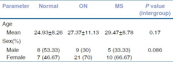
Thirty cases of ON, 15 cases of MS (30 eyes), and 15 controls (30 eyes) were enrolled in the study. Nine of the 30 cases of ON had bilateral symptoms, resulting in 39 ON eyes and 21 fellow eyes. The demographic profile of both the ON and MS group show a female predominance and is depicted in Table 1. The groups are well-matched with no statistically significant difference between the groups regarding demographics.
Table 1.
Depicting demographic profile of the subgroups

In the optic neuritis group, the mean time interval between an acute attack and the conduct of the study was 6.2 ± 3.3 months.
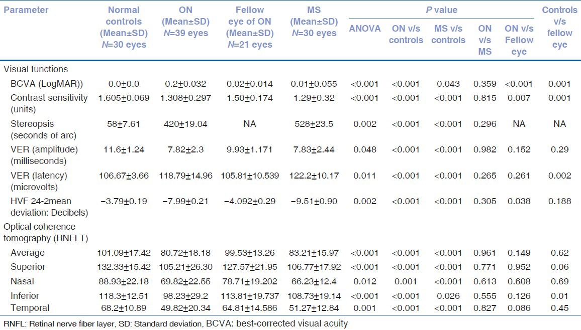
The visual functions, RNFL thickness measurements, and comparison of the four groups, namely optic neuritis eyes, eyes of patients with multiple sclerosis, fellow eyes of optic neuritis patients, and normal controls are depicted in Table 2. The optic neuritis group has significantly worse visual acuity, contrast sensitivity, and a more severe visual field affliction than the fellow eyes or normal controls while the VER amplitude was lower and the VER latency delayed in comparison to the normal controls. There is no statistically significant difference between the optic neuritis and multiple sclerosis groups for any of these parameters even though none of the MS patients had history of optic neuritis. The multiple sclerosis group has a significantly worse contrast sensitivity, stereoacuity, lower VER amplitude and delayed VER latency and more severe visual field affliction than the normal controls. An intergroup analysis for each of the visual function parameters is depicted in Table 2.
Table 2.
Depicting visual function changes and RNFL changes in various subgroups along with significance of the differences

The mean RNFL thickness, both the average and the quadrant-wise thickness are depicted in Table 2. The average and quadrant-wise RNFL thickness of the optic neuritis subgroup is significantly thinner than the normal controls. Conversely, there is no significant difference between the RNFL thickness of the optic neuritis group from the fellow eyes and the multiple sclerosis groups. The RNFL of the multiple sclerosis groups is different from the normal controls being thinner in each of the quadrants and the average mean. An intergroup analysis is depicted in Table 2.
Associations were tested between the RNFL changes and visual functions for the subgroups using the Pearson's correlation coefficient [Tables 3 and 4]. In the optic neuritis group [Table 3], the average RNFL thickness has significant correlations with VER (amplitude), VER (latency) and also HVF-24-2 visual fields, although of a mild to moderate strength. In a multivariate analysis, only the association with HVF-24-2 visual fields was significant (P = 0.049). When a quadrant-wise analysis was done, the RNFL thickness of the superior quadrant has significant correlation with VER (latency) with a thinner RNFL mirroring a delayed latency. No other visual parameter correlated significantly. The RNFL thickness of the inferior and temporal quadrants have significant correlation with VER (amplitude) and HVF-24-2 visual fields each, of which, only the association with visual fields remains significant in a multivariate analysis for each of these two quadrants (P = 0.038 each). The RNFL thickness of the nasal quadrant did not have significant correlation with any of the visual parameters in our study. RNFL loss was not found to be predictive of visual acuity or higher visual functions such as contrast sensitivity or stereopsis.
Table 3.
Depicting the association between OCT RNFL with visual function parameters in eyes with optic neuritis (pearson's correlation coefficient

Table 4.
Depicting the association between OCT RNFL with visual function parameters in eyes of patients with multiple sclerosis (pearson's correlation coefficient)

The multiple sclerosis group has more and stronger correlations between the RNFL and visual function parameters. [Table 4] The average RNFL thickness had significant and strong correlation with contrast sensitivity, stereoacuity, VER (amplitude), VER (latency), and HVF-24-2 visual fields. In a multivariate setting, the contrast sensitivity and VER latency continued to have significant correlations [P = 0.01 and P = 0.029, respectively]. The RNFL thickness of the superior and inferior quadrants mirrors the correlations of the average RNFL. For both the superior and inferior quadrant, only the association with contrast sensitivity remains significant during a multivariate analysis [P = 0.01 and P = 0.035, respectively]. The RNFL thickness of the temporal quadrant had significant correlation with s tereoacuity, VER latency, and HVF-24-2 visual fields, of which, only the association with stereoacuity remains significant in a multivariate analysis [P = 0.017]. The RNFL thickness of the nasal quadrant has significant correlation with VER latency and HVF-24-2 visual fields, of which VER latency is significantly correlated in a multivariate setting [P = 0.02]. There is no significant association with visual acuity for either of the quadrants or the average RNFL thickness.
Discussion
The demographic profile of our study population is in accordance to that noted in previous OCT studies in literature.[6,7,8]
The optic neuritis group has shown poorer vision than either the fellow eye or controls indicating that even though visual acuity is expected to recover in cases of optic neuritis, there is a residual loss of vision which is significant in the Indian eyes. The difference in visual acuity between the MS groups and the normal control group was not statistically significant due to the fact that the patients of multiple sclerosis at enrollment had no history of ocular involvement. It is also pertinent to note that despite vision being normal in the multiple sclerosis group in comparison to the optic neuritis group, this does not rule out subclinical involvement, which is demonstrated by the other visual parameters showing subnormal values. Thus, while visual acuity can be used to distinguish optic neuritis from the normal, it is not a relevant parameter to differentiate multiple sclerosis from the normal. Our study detected a significant difference between contrast sensitivity values of the ON/MS and control groups, which is in accordance to large cross-sectional and longitudinal studies.[10,11] Therefore, contrast sensitivity could be used to identify subclinical optic nerve involvement in cases of multiple sclerosis while in cases of optic neuritis, it would be important to evaluate contrast sensitivity along with visual acuity for follow up after recovery.[12]
The stereoacuity in case of optic neuritis and multiple sclerosis is also found to be significantly lower than that of controls. While vision is subnormal in optic neuritis and may have an effect on stereoacuity, subnormal stereoacuity in cases of multiple sclerosis having normal vision may indicate subclinical optic nerve involvement in such cases. This aspect has not been examined in previous literature.
The visual evoked response (VER) in the optic neuritis patients and in multiple sclerosis patients showed decreased amplitude as well as increased latency in comparison to the controls. The increase in latency seen in demyelinating diseases is due to the impaired saltatory conduction and the consequent reduction in the speed of transmission of nerve impulses. The decreased amplitude points to the actual decrease in number of axons carrying the impulses, possibly secondary to axonal loss in optic neuritis and multiple sclerosis patients.
The visual fields in case of optic neuritis and multiple sclerosis also showed a higher mean deviation as compared to controls. The visual field correlated strongest with RNFL loss in optic neuritis patients and is indicative of a post-neuritic sequelae, possibly due to a permanent damage to the axons reflected in the thinned RNFL.
The RNFL thickness was measured using the Stratus 3 OCT, which has been shown to give a reproducible, quantitative, and objective measurement of the RNFL thickness.[5,13,14] The RNFL thickness measured by OCT also showed a significant thinning in the average value and each of the four quadrants in both optic neuritis and multiple sclerosis patients as compared to normal controls. The validity of the RNFL readings can be confirmed by the fact that the mean of the average RNFLT in the controls was comparable to that found in normal Indian eyes in previous studies.[15,16] Indian eyes tend to have a thinner RNFL as compared to the East Asians and Hispanics though it is thicker than the Caucasian eyes.[17,18]
The thinning of retinal nerve fiber layer was noted more in cases of optic neuritis than in cases of multiple sclerosis though no statistically significant difference was observed between the two groups. The finding of significant RNFL thinning in MS corroborates with earlier studies carried out by Fisher et al. and Pueyo et al. in MS patients without ON.[6,7] Costello F et al. showed in their study on optic neuritis patients that there was significant RNFL thinning in the clinically affected eyes.[8] In our study, both in the ON and the MS groups, the maximum thinning was found to be in the temporal quadrant indicative of affliction of the papillomacular bundle. It has been proven in Caucasian populations that thinning of the temporal quadrant is most prominent and this agrees our finding in Indian eyes.[19,20]
In a quadrant-wise analysis, our study has shown an association of various visual function parameters with RNFL thickness. Of these, understandably, the VER latency was the commonest correlate in both the ON and MS groups. In the multiple sclerosis group, contrast sensitivity was a correlate with RNFL loss after correcting for other visual function parameters. This indicates that visual dysfunction may develop independent of direct optic nerve affliction in the past or reversible.
In cases of ON, out of the RNFLT of the four quadrants, the RNFLT of the temporal quadrant had significant correlation with maximum number of parameters (two) and the RNFLT of the nasal quadrant had no correlation with any visual function parameter. It has been shown in a previous study that the earliest significant inter–eye differences manifested in the temporal quadrant, though it was not examined whether the RNFLT of the temporal quadrant correlated with visual functions.[18] In MS, there was an overall thinning of all four RNFL quadrants and each independently correlated with one or more of the visual parameters with the superior and inferior quadrant having the maximum correlates. This is in consistency with a previous study on multiple sclerosis patients, which have observed diffuse thinning and correlation of structural abnormalities of the RNFL with functional assessment of the optic nerve.[8] While quadrant-wise analysis has to be cautiously interpreted, the average RNFLT which had significant correlation with the maximum number of visual parameters is certainly predictive of altered visual functions. Visual acuity was not found to be associated with RNFL changes, but this is probably biased by the fact that the visual acuity of all cases was normal or near normal.
To conclude, the RNFL thickness in the eyes of patients with optic neuritis as well as in the eyes of patients with multiple sclerosis without optic neuritis was significantly reduced when compared to controls. Further, the average RNFL thinning and quadrantic thinning significantly correlated with visual function parameters, which depend upon the structural axonal damage. RNFL measurement by OCT can possibly quantify the axonal loss taking place in the visual system in Indian eyes in a manner similar to the Caucasian patients and prognosticate visual outcome. Contrast sensitivity and stereoacuity are useful tests to identify subclinical optic nerve involvement in multiple sclerosis.
Footnotes
Source of Support: Nil,
Conflict of Interest: None declared.
References
- 1.Evangelou N, Konz D, Esiri MM, Smith S, Palace J, Matthews PM. Size selective neuronal changes in the anterior optic pathways suggests a differential susceptibility to injury in multiple sclerosis. Brain. 2001;124:1813–20. doi: 10.1093/brain/124.9.1813. [DOI] [PubMed] [Google Scholar]
- 2.Galetta KM, Graves J, Talman LS, Lile DJ, Frohman EM, Calabresi PA, et al. Visual pathway axonal loss in benign multiple sclerosis: A longitudinal study. J Neuroophthalmol. 2012;32:116–23. doi: 10.1097/WNO.0b013e318240204d. [DOI] [PMC free article] [PubMed] [Google Scholar]
- 3.Oreja-Guevara C, Noval S, Alvarez-Linera J, Gabaldon L, Manzano B, Chamorro B, et al. Clinically isolated syndromes suggestive of multiple sclerosis: An optical coherence tomography study. Plos One. 2012;7:e33907. doi: 10.1371/journal.pone.0033907. [DOI] [PMC free article] [PubMed] [Google Scholar]
- 4.Costello F. Evaluating the use of optical coherence tomography in optic neuritis. Mult Scler Int 2011. 2011 doi: 10.1155/2011/148394. 148394. [DOI] [PMC free article] [PubMed] [Google Scholar]
- 5.Garcia-Martin E, Pueyo V, Pinilla I, Ara JR, Martin J, Fernandez J. Fourier domain OCT in multiple sclerosis patients: Reproducibility and ability to detect retinal nerve fiber atrophy. Invest Ophthalmol Vis Sci. 2011;52:4124–31. doi: 10.1167/iovs.10-6643. [DOI] [PubMed] [Google Scholar]
- 6.Fisher JB, Jacobs DA, Markowitz CE. Relation of visual function to retinal nerve fiber layer thickness in multiple sclerosis. Ophthalmology. 2006;113:324–32. doi: 10.1016/j.ophtha.2005.10.040. [DOI] [PubMed] [Google Scholar]
- 7.Pueyo V, Martin J, Fernandez J, Almarcegui C, Ara J, Egea C, et al. Axonal loss in the RNFL in patients with multiple sclerosis. Mult Scler. 2008;14:609–14. doi: 10.1177/1352458507087326. [DOI] [PubMed] [Google Scholar]
- 8.Costello F, Hodge W, Pan YI, Eggenberger E, Coupland S, Kardon RH. Tracking RNFL loss after optic neuritis: A prospective study using OCT. Mult Scler. 2008;14:893–905. doi: 10.1177/1352458508091367. [DOI] [PubMed] [Google Scholar]
- 9.Polman CH, Reingold SC, Edan G, Filippi M, Hartung HP, Kappos L, et al. Diagnostic criteria for multiple sclerosis: 2005 revisions to the “McDonald Criteria”. Ann Neurol. 2005;58:840–6. doi: 10.1002/ana.20703. [DOI] [PubMed] [Google Scholar]
- 10.Merle H, Olindo S, Donnio A, Beral L, Richer R, Smadja D, et al. Retinal nerve fiber layer thickness and spatial and temporal contrast sensitivity in multiple sclerosis. Eur J Ophthalmol. 2010;20:158–66. doi: 10.1177/112067211002000122. [DOI] [PubMed] [Google Scholar]
- 11.Bodis-Wollner I, Diamong SP. The measurement of spatial contrast sensitivity in cases of blurred vision associated with cerebral lesions. Brain. 1976;99:695–710. doi: 10.1093/brain/99.4.695. [DOI] [PubMed] [Google Scholar]
- 12.Talman LS, Bisker ER, Sackel DJ, Long DA, Jr, Galetta KM, Ratchford JN, et al. Longitudanal study of vision and retinal nerve fiber layer thickness in multiple sclerosis. Ann Neurol. 2010;67:749–60. doi: 10.1002/ana.22005. [DOI] [PMC free article] [PubMed] [Google Scholar]
- 13.Jones AL, Sheen AJ, North RV, Morgan JE. The Humphrey optical coherence tomography scanner: Quantitative analysis and reproducibility study of the normal human retinal nerve fiber layer. Br J Ophthalmol. 2001;85:673–7. doi: 10.1136/bjo.85.6.673. [DOI] [PMC free article] [PubMed] [Google Scholar]
- 14.Jeoung JW, Park KH, Kim TW, Khwarg SI, Kim DM. Diagnostic ability of optical coherence tomography with a normative database to detect localized retinal nerve fiber layer defects. Ophthalmology. 2005;112:2157–63. doi: 10.1016/j.ophtha.2005.07.012. [DOI] [PubMed] [Google Scholar]
- 15.Sony P, Sihota R, Tiwari HK, Venkatesh P, Singh R. Quantification of the retinal nerve fiber layer thickness in normal Indian eyes with optical coherence tomography. Indian J Ophthalmol. 2004;52:303–9. [PubMed] [Google Scholar]
- 16.Ramakrishnan R, Mittal S, Ambatkar S, Kader MA. Retinal nerve fiber layer thickness measurements in normal Indian population by optical coherence tomography. Indian J Ophthalmol. 2006;54:11–5. doi: 10.4103/0301-4738.21608. [DOI] [PubMed] [Google Scholar]
- 17.Kanamori A, Escano MF, Eno A, Nakamura M, Maeda H, Seya R, et al. Evaluation of the effect of aging on retinal nerve fiber layer thickness measured by optical coherence tomography. Ophthalmologica. 2003;217:273–8. doi: 10.1159/000070634. [DOI] [PubMed] [Google Scholar]
- 18.Budenz DL, Anderson DR, Varma R, Schuman J, Cantor L, Savell J, et al. Determinants of normal nerve fiber layer thickness measured by Stratus OCT. Ophthalmology. 2007;114:1046–52. doi: 10.1016/j.ophtha.2006.08.046. [DOI] [PMC free article] [PubMed] [Google Scholar]
- 19.Bock M, Brandt AU, Dörr J, Kraft H, Weinges-Evers N, Gaede G, et al. Patterns of retinal nerve fiber layer loss in multiple sclerosis patients with or without optic neuritis and glaucoma patients. Clin Neurol Neurosurg. 2010;112:647–52. doi: 10.1016/j.clineuro.2010.04.014. [DOI] [PubMed] [Google Scholar]
- 20.Parisi V, Manni G, Spardo M, Colacino G, Restuccia R, Marchi S, et al. Correlation between morphological and functional retinal impairment in multiple sclerosis patients. Invest Ophthalmol Vis Sci. 1999;40:2520–7. [PubMed] [Google Scholar]


