Abstract
In this retrospective case series, we studied the predisposing factors, causative organisms, clinical spectrum, and outcomes of 12 cases of culture-proven infectious scleritis. Nine of 12 patients had a history of preceding trauma (surgical or accidental). Past surgical history included small-incision cataract surgery (4), pterygium surgery (1), and trabeculectomy (1). Six patients had multifocal scleral abscesses due to Pseudomonas, Klebsiella, or Nocardia. Only 2 patients retained useful vision (>6/18). A poor visual acuity at presentation usually resulted in a worse visual outcome (P = 0.005). Four eyes developed phthisis. The addition of surgical intervention did not result in a significantly better visual outcome than medical management alone (P = 0.209), but resulted in a higher globe preservation rate (P = 0.045). Therefore, we concluded that infection must be ruled out in cases of scleritis with preceding history of trauma, and aggressive surgical intervention improves the anatomical outcome but does not change the visual outcome.
Keywords: Infectious scleritis, microbial scleritis, ocular infection, scleritis
Scleritis is a severe, painful inflammation of the sclera that may involve adjacent structures and can threaten vision.[1] Nearly 50% of cases are due to an associated collagen vascular disease, and an infectious etiology is comparatively rare.[2] The organism most often responsible is Pseudomonas aeruginosa.[1,3,4] However, reports from India have stated fungi to be more common in tropical regions.[2] We, therefore, analyzed data from our medical records to determine the clinical and microbiological spectrum of infectious scleritis in a tertiary care hospital in India. We also evaluated the visual and anatomical outcome of these cases to determine prognostic factors and effectiveness of management.
Materials and Methods
A retrospective chart review was performed for cases of infectious scleritis seen from January 2007 to August 2011. The diagnostic criteria for a case of infectious scleritis included the presence of single or multiple inflamed anterior scleral nodules or ulcerations, which revealed a causative organism on microbiological culture. Microbial cultures were considered significant if growth of the same organism was present on more than one solid medium or if there was a confluent growth on inoculation on one solid medium or if growth of one medium was consistent with direct microscopy findings. Data regarding patient demographics, predisposing factors, causative organisms, and clinical features for all 12 patients was extracted. All patients except one were followed up for a period of at least 6 months and, therefore, the treatment and outcome details were analyzed for 11 patients. Data was analyzed by the Chi-square test using SPSS version 11.0.1 to determine the prognostic factors.
Results
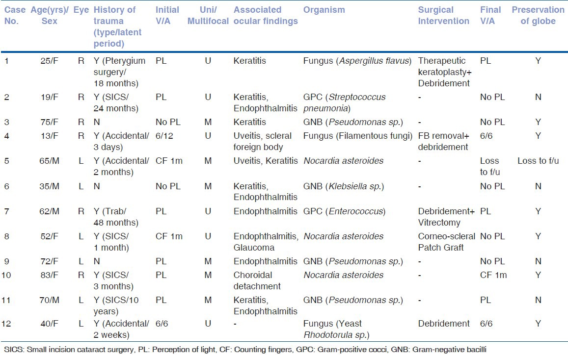
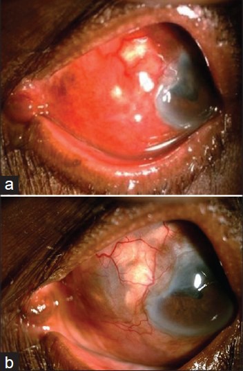
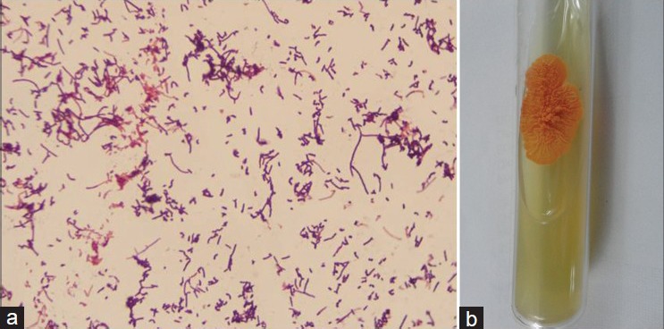
Twelve eyes of 12 patients were identified to have culture-positive infectious scleritis. Demographic data and clinical features are described in Table 1. All except 3 had a history of either surgical or accidental trauma. Preceding surgical procedures performed included manual small-incision cataract surgery (4), pterygium excision (1), and trabeculectomy (1). None of the patients had a history of fever, joint pain, respiratory illness, skin rashes, etc., One patient (Case 2) had progeria, another had chronic liver disease (Case 6), and 2 patients had diabetes (Case 8 and 10). No other systemic factors which could predispose to scleritis were found. Six cases had multifocal scleral abscesses [Fig. 1a and b]. Patients with multifocal scleritis were associated with growth of either gram-negative bacilli (GNB) or Nocardia on culture an example of which is shown in Fig. 2a and b.
Table 1.
Demographics, clinical features, organisms, management, and outcomes of infectious scleritis

Figure 1.

(a) Multifocal scleral abscesses owing to Pseudomonas sp. (Case 11). (b) Healed scleritis with minimal scleral thinning 2 months after treatment with topical fortified antibiotics and dexamethasone (Case 11)
Figure 2.

(a) Nocardia asteroides: Gram stain showing filamentous forms fragmented into bacillary and coccoid forms (Magnification = ×1000). (b) Nocardia asteroides: Orange coloured, centrally heaped, dry colony
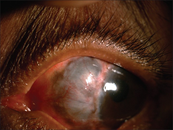
All patients received topical antimicrobials based on the culture and sensitivity report. In addition, most (75%) received systemic antimicrobials. Seven patients were also treated with topical steroids due to severe intra-ocular inflammation. In addition to medical treatment, 5 patients underwent various surgical procedures in an attempt to decrease the load of infective organism or to preserve the globe as shown in Table 1 and Fig. 3. The follow-up ranged from 6 months to 6 years. Of these, only 2 patients had a good final visual acuity (better than 6/18) on the last follow-up visit. We were able to preserve the globe in all but 4 patients.
Figure 3.

Six months after corneo-scleral patch graft following infectious scleritis post-cataract surgery (Case 8)
We determined the prognostic factors of infectious scleritis using the Chi-square test. A poor visual acuity at presentation (less than counting fingers at 1 m) usually resulted in a worse visual outcome (P = 0.005). The causes of poor visual acuity at last follow-up include corneal scarring following keratitis (1), failed corneal graft (1), secondary angle closure glaucoma and optic atrophy (1), choroidal detachment (1), and endophthalmitis (5). The visual outcome was not dependant on any particular organism (P = 0.099) or the presence of multifocal abscesses (P = 0.209). The addition of surgical intervention did not result in a significantly better visual outcome than medical management alone (P = 0.209), but resulted in a higher rate of globe preservation (P = 0.045).
Discussion
A clinical suspicion of infectious scleritis in all cases of scleral inflammation following trauma has been emphasized previously.[1,2,5] Pterygium excision is the most commonly reported surgical procedure preceding infectious scleritis.[1,3,4,6,7] However, in our series, 4 cases were preceded by cataract surgery and only 1 by pterygium surgery. Another case series of Indian eyes also showed maximum number of their cases had cataract surgery prior to an episode of infectious scleritis.[2] This may be due to the large number of scleral-tunnel small incision cataract surgeries performed in India as compared to clear corneal phacoemulsification performed in developed countries.
Pseudomonas aeruginosa is the leading cause of microbial scleritis in reported literature.[1,3,4] In our series, 33.33% had GNB, 25% had Nocardia, and 25% had fungal infection. One of the fungi was identified to be Rhodotorula spp, which we have reported elsewhere.[8] In cases of scleritis following trauma where infection is suspected but routine microbial culture is negative, polymerase chain reaction tests may be used to confirm the diagnosis.[9] If these tests are also negative, the possible diagnosis of surgically induced necrotizing scleritis (SINS) should be considered. SINS is a believed to be a delayed-type hypersensitivity response to any form of surgical trauma.[10] Up to 90% of patients who develop SINS may have an undiagnosed systemic vasculitis and, therefore, a detailed systemic investigation is warranted.[1] This is often seen as late as 9 months after the surgical intervention and, after ruling out an infectious cause, requires treatment with intensive systemic immunosuppression.[1] When infectious scleritis occurs long after surgery, it has been hypothesized to be due to an underlying SINS with secondary microbial infection, which can make diagnosis and treatment difficult.[2] These patients may benefit from a systemic evaluation to rule out an autoimmune vasculitis, which will require appropriate management.
There were 6 patients in our series with associated endophthalmitis. Some presented with scleritis and developed secondary intraocular spread, which has been described in other reports.[9,11,12] Others had scleritis, uveitis, and endophthalmitis at presentation (e.g., Case 7 where the sclera in the area of the bleb was necrotic with vitreous inflammation). The wide use of anti-metabolites at the time of trabeculectomy increases risk of scleral melt and inflammation, which may have resulted in secondary infection and endophthalmitis. However, in these cases, determining where the infection originated is difficult.
Infectious scleritis often presents with multifocal nodules. Pseudomonas is most often associated with this presentation, which has been attributed to an intra-scleral dissemination facilitated by its proteolytic products and motility.[3,4,6] In contrast, Jain et al.[2] found that all cases of multifocal scleritis were due to fungi. Our series had 6 cases of multifocal scleral abscesses, 3 of which were due to P. aeruginosa, 2 due to Nocardia, and 1 due to Klebsiella sp. A review of past literature[2,3,4,5,6] suggests that multifocal scleritis can be caused by several organisms and may be related to factors such as the load of infection, rather than specific organisms.
The visual acuity at presentation is the most important prognostic factor determining the final visual acuity, and this has been seen in other studies.[7,13] This suggests that early diagnosis should help preserve the patient's remaining vision if effective treatment is initiated.
There is a lot of discrepancy among researchers regarding the effect of surgical debridement on the outcome of infectious scleritis. Some showed that surgical debridement shortens the course of treatment.[3,6] Others found early debridement improved visual acuity.[14] A large series showed that despite adjuvant surgery in 46 of 56 eyes, approximately 50% lost functional vision.[13] They did not, however, examine the effect of surgery on globe preservation. We found anatomical outcome was better in the cases which underwent surgical procedures. However, unlike other studies,[3,7] we did not find any difference in the final vision between the medically and surgically treated groups.
In conclusion, manual small incision cataract surgery is a common predisposing factor to infectious scleritis in developing countries. The visual acuity at presentation is the most important prognostic factor determining the final visual outcome. Aggressive surgical intervention improves the chance of globe preservation but does not improve the visual outcome.
Footnotes
Source of Support: Nil,
Conflict of Interest: None declared.
References
- 1.Okhravi N, Odufuwa B, McCluskey P, Lightman S. Scleritis. Surv Ophthalmol. 2005;50:351–63. doi: 10.1016/j.survophthal.2005.04.001. [DOI] [PubMed] [Google Scholar]
- 2.Jain V, Garg P, Sharma S. Microbial scleritis – experience from a developing country. Eye (Lond) 2009;23:255–61. doi: 10.1038/sj.eye.6703099. [DOI] [PubMed] [Google Scholar]
- 3.Huang FC, Huang SP, Tseng SH. Management of infectious scleritis after pterygium excision. Cornea. 2000;19:34–9. doi: 10.1097/00003226-200001000-00008. [DOI] [PubMed] [Google Scholar]
- 4.Hsiao CH, Chen JJ, Huang SC, Ma HK, Chen PY, Tsai RJ. Intrascleral dissemination of infectious scleritis following pterygium surgery. Br J Ophthalmol. 1998;82:29–34. doi: 10.1136/bjo.82.1.29. [DOI] [PMC free article] [PubMed] [Google Scholar]
- 5.Chung PC, Lin HC, Hwang YS, Tsai YJ, Ngan KW, Huang SC, et al. Paecilomyces lilacinus scleritis with secondary keratitis. Cornea. 2007;26:232–4. doi: 10.1097/01.ico.0000248384.16896.7d. [DOI] [PubMed] [Google Scholar]
- 6.Lin CP, Shih MH, Tsai MC. Clinical experiences of infectious scleral ulceration: A complication of pterygium surgery. Br J Ophthalmol. 1997;81:980–3. doi: 10.1136/bjo.81.11.980. [DOI] [PMC free article] [PubMed] [Google Scholar]
- 7.Cunningham MA, Alexander JK, Matoba AY, Jones DB, Wilhemus KR. Management and outcome of microbial anterior scleritis. Cornea. 2011;30:1020–3. doi: 10.1097/ICO.0b013e31820967bd. [DOI] [PMC free article] [PubMed] [Google Scholar]
- 8.Pradhan ZS, Jacob P. Management of Rhodotorula scleritis. Eye (Lond) 2012;26:1587. doi: 10.1038/eye.2012.181. [DOI] [PMC free article] [PubMed] [Google Scholar]
- 9.Biswas J, Aparna AC, Annamalai R, Vaijayanthi K, Bagyalakshmi R. Tuberculous scleritis in a patient with rheumatoid arthritis. Ocul Immunol Inflamm. 2012;20:49–52. doi: 10.3109/09273948.2011.628195. [DOI] [PubMed] [Google Scholar]
- 10.Morley AM, Pavesio C. Surgically induced necrotizing scleritis following three-port pars plana vitrectomy without scleral buckling: A series of three cases. Eye (Lond) 2008;22:162–4. doi: 10.1038/sj.eye.6702708. [DOI] [PubMed] [Google Scholar]
- 11.Choudhry S, Rao SK, Biswas J, Madhavan HN. Necrotizing nocaridal scleritis with intraocular extension: A case report. Cornea. 2000;19:246–8. doi: 10.1097/00003226-200003000-00023. [DOI] [PubMed] [Google Scholar]
- 12.Sawant SD, Biswas J. Fungal scleritis with exudative retinal detachment. Ocul Immunol Inflamm. 2010;18:457–8. doi: 10.3109/09273948.2010.507129. [DOI] [PubMed] [Google Scholar]
- 13.Hodson KL, Galor A, Karp CL, Davis JL, Albini TA, Perez VL, et al. Epidemiology and visual outcomes in patients with infectious scleritis. Cornea. 2013;32:466–72. doi: 10.1097/ICO.0b013e318259c952. [DOI] [PubMed] [Google Scholar]
- 14.Tittler EH, Nguyen P, Rue KS, Vasconcelos-Santos DV, Song JC, Irvine JA, et al. Early surgical debridement in the management of infectious scleritis after pterygium excision. J Ophthalmic Inflamm Infect. 2012;2:81–7. doi: 10.1007/s12348-012-0062-1. [DOI] [PMC free article] [PubMed] [Google Scholar]


