

Abstract
Upper gastrointestinal symptoms like vomiting, abdominal pain, abdominal distention may be caused by many conditions like complicated peptic/duodenal ulcer, gastritis or hiatal hernia. However, these symptoms are uncommonly produced by superior mesenteric artery (SMA) syndrome. SMA syndrome is triggered when there is narrowing of the mesenteric angle and shortening of the aortomesenteric distance. We report a case of 42-year-old female who presented with features of gastric outlet obstruction which on further investigation was revealed as SMA syndrome. The aetiology, presentation, diagnosis and management of this unusual condition are discussed.
Keywords: Conventional radiography, Gastrointestinal outlet obstruction, Superior mesenteric artery syndrome, Upper gastrointestinal symptoms
Introduction
Superior mesenteric artery (SMA) syndrome was first described in 1861 by Von Rokitansky but remained an unknown entity until 1927 when Wilkie published the first comprehensive series of 75 patients.[1] SMA syndrome is a rare entity characterized by compression of the third portion of the duodenum between the aorta and the SMA. This results in partial or acute duodenal obstruction.[2]
Case Report
A 42-year-old female patient presented with abdominal pain and vomiting after every meal since 3 months. The pain was colicky in nature relieved on lying down on left lateral position. Patient had history of weight loss of about ten kilogram in the last 3 months; she weighed 30 kg and looked cachexic. She had past history for appendicular perforation 3 years back for which she was operated.
Laboratory investigations revealed PCV - 46%, Hb - 11.1 g/dl, MCV - 71.63 cu.mm (80-90) - microcytic, total leucocytes count of 11,400 cu.mm with Neutrophils - 62%. Upper gastrointestinal endoscopy was attempted however probe could not be negotiated through third part of duodenum. Barium meal follow through showed dilated proximal duodenum with linear vertical filling defect across proximal third part of duodenum was suggestive of SMA syndrome [Figures 1-3]. A provisional diagnosis of SMA syndrome was made. For further investigation patient was advised computed tomography (CT) scan, however patient could not afford it.
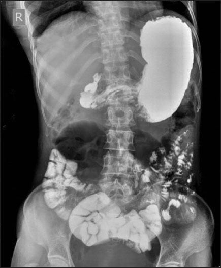
Figure 1.

Barium study (erect) showed distended hypotonic stomach suggestive of prolonged gastric outlet obstruction
Figure 3.
Barium study (performed after change of posture) shows passage of barium into the small bowel loops showing partial obstruction
Figure 2.

Barium study (supine view) shows with a linear vertical filling defect across proximal third part of duodenum (white arrow) with consequent dilatation of second part of duodenum which was suggestive of superior mesenteric artery syndrome. A trifoliate (clover leaf) deformity in duodenal bulb (black arrow) is also noted which is pathognomic of a healed duodenal ulcer
A nasogastric tube was inserted and the patient was started on parenteral nutrition. Conservative measures failed, and 1 week later the patient was subjected to laparotomy, where dilatation of the duodenum proximally to the superior mesenteric vessels was observed. The third part of the duodenum was exposed and mobilized and a loop duodenojejunostomy was performed, approximately 10 cm distally to the ligament of Treitz. Her post-operative course was uneventful.
Discussion
SMA syndrome was first described in 1861 by Von Rokitansky, who proposed that might responsible for causing obstruction of the third part of the duodenum as a result of aortomesenteric compression. Some studies reported the incidence of SMA syndrome to be 0.1-0.3%.[3] Approximately 0.013-0.78% of barium upper GI studies evaluating for SMA syndrome confirm the diagnosis.[4,5] Multidetector CT scan is the best modality for the diagnosis of SMA syndrome.[6]
Predisposing factors of SMA syndrome are lean body build, exaggerated lumbar lordosis, abdominal wall laxity and depletion of the mesenteric fat caused by rapid severe weight loss as result of cancer, surgery, burns, trauma, psychiatric problems etc., The use of Harrington rods in corrective surgery of scoliosis is an important cause of SMA syndrome[1] and abnormally high position of ligament of Treitz can cause SMA syndrome.
The SMA usually forms an angle of approximately 45° (range: 38-56°) with the abdominal aorta and the third part of the duodenum. Any factor that sharply narrows the aortomesenteric angle to approximately 6-25° can cause entrapment of the third part of the duodenum and thus leading to duodenal compression.[7,8]
The patient often presents with chronic upper abdominal symptoms such as epigastric pain, nausea, bilious vomiting, postprandial discomfort and sometimes-subacute small bowel obstruction. The symptoms are typically relieved when the patient is in the knee-to-chest position because it reduces the small bowel mesenteric tension at the aortomesenteric angle.[9] A Hayes maneuver in which pressure is applied below the umbilicus in cephalad and dorsal directions elevates the root of small-bowel mesentery and may also relieve obstruction temporarily.
Plain radiograph demonstrates a dilated, fluid- and gas-filled stomach suggestive of SMA syndrome. Fluoroscopic findings suggestive of SMA syndrome include dilation of the first and second portions of the duodenum with an abrupt narrowing at the third portion, delayed gastroduodenal emptying, and anti-peristaltic waves proximal to the obstruction. Additionally, the obstruction of the duodenum may be relieved by a change in position, especially left lateral decubitus position.[6]
CT scan is useful in the diagnosis of SMA syndrome, showing an aortomesenteric angle of < 22° and an aortomesenteric distance of < 8-10 mm. CT can also identify other problems that may require intervention, like compression of the left renal vein that results in renal vein thrombosis.[6]
Treatment is initially conservative, including the insertion of a nasogastric tube, mobilization of the patient to a prone, left lateral decubitus position, administration of parenteral nutrition and fluid-electrolyte balance correction. In our patient, conservative treatment failed, and surgery was undertaken in order to avoid the risk of duodenal atony and massive dilatation. There is no clear time point to operate, but if the patient remains symptomatic after 2-12 days of conservative treatment, the surgical option should be considered.[10]
Surgical treatment of SMA syndrome involves a wide range of procedures, including Treitz ligament division (Strong's operation), gastrojejunostomy, subtotal gastrectomy and Billroth II gastrojejunostomy, duodenojejunostomy and anterior reposition of the duodenum. Although duodenojejunostomy is still the most frequently conducted procedure for SMAS, it has recently been reported that it does not alleviate vomiting possibly due to the reversed peristalsis which in some cases is greater than the direct peristalsis or because of gastroparesis following correction surgery.[11] Finally, laparoscopic techniques have made SMA Syndrome correction surgery feasible.[12]
Footnotes
Source of Support: Nil.
Conflict of Interest: None declared.
References
- 1.Welsch T, Büchler MW, Kienle P. Recalling superior mesenteric artery syndrome. Dig Surg. 2007;24:149–56. doi: 10.1159/000102097. [DOI] [PubMed] [Google Scholar]
- 2.Gerasimidis T, George F. Superior mesenteric artery syndrome. Wilkie syndrome. Dig Surg. 2009;26:213–4. doi: 10.1159/000219330. [DOI] [PubMed] [Google Scholar]
- 3.Shiu JR, Chao HC, Luo CC, Lai MW, Kong MS, Chen SY, et al. Clinical and nutritional outcomes in children with idiopathic superior mesenteric artery syndrome. J Pediatr Gastroenterol Nutr. 2010;51:177–82. doi: 10.1097/MPG.0b013e3181c7bdda. [DOI] [PubMed] [Google Scholar]
- 4.Baltazar U, Dunn J, Floresguerra C, Schmidt L, Browder W. Superior mesenteric artery syndrome: An uncommon cause of intestinal obstruction. South Med J. 2000;93:606–8. [PubMed] [Google Scholar]
- 5.Unal B, Aktaş A, Kemal G, Bilgili Y, Güliter S, Daphan C, et al. Superior mesenteric artery syndrome: CT and ultrasonography findings. Diagn Interv Radiol. 2005;11:90–5. [PubMed] [Google Scholar]
- 6.Agrawal GA, Johnson PT, Fishman EK. Multidetector row CT of superior mesenteric artery syndrome. J Clin Gastroenterol. 2007;41:62–5. doi: 10.1097/MCG.0b013e31802dee64. [DOI] [PubMed] [Google Scholar]
- 7.Merrett ND, Wilson RB, Cosman P, Biankin AV. Superior mesenteric artery syndrome: Diagnosis and treatment strategies. J Gastrointest Surg. 2009;13:287–92. doi: 10.1007/s11605-008-0695-4. [DOI] [PubMed] [Google Scholar]
- 8.Kyslan K, Barla J, Kyslan K, Stanislayová M. Superior mesenteric artery (SMAS/AMS) syndrome and its management. Rozhl Chir. 2008;87:255–8. [PubMed] [Google Scholar]
- 9.Smith BG, Hakim-Zargar M, Thomson JD. Low body mass index: A risk factor for superior mesenteric artery syndrome in adolescents undergoing spinal fusion for scoliosis. J Spinal Disord Tech. 2009;22:144–8. doi: 10.1097/BSD.0b013e31816b6b9a. [DOI] [PubMed] [Google Scholar]
- 10.Cheshire NJ, Glezer G. Diverticula, volvuluvs, superior mesenteric artery syndrome, and foreign bodies. In: Zinner MJ, Schwartz SI, Ellis H, editors. Maingot's Abdominal Operations. 9th ed. I. East Norwalk Conn: Appleton and Lange; 1990. pp. 587–91. [Google Scholar]
- 11.Yang WL, Zhang XC. Assessment of duodenal circular drainage in treatment of superior mesenteric artery syndrome. World J Gastroenterol. 2008;14:303–6. doi: 10.3748/wjg.14.303. [DOI] [PMC free article] [PubMed] [Google Scholar]
- 12.Palanivelu C, Rangarajan M, Senthilkumar R, Parthasarathi R, Jani K. Laparoscopic duodenojejunostomy for superior mesenteric artery syndrome. JSLS. 2006;10:531–4. [PMC free article] [PubMed] [Google Scholar]