

A 19-year-old male presented with asymptomatic, skin-colored mass (25 × 15 cm) on the lower back since birth [Figure 1]. The patient gave history of increase in the size of the mass over a period of time. Similar lesion was present over his left foot [Figure 2]. No history of similar lesion was present in the family members. Histopathology showed groups and strands of fat cells embedded in the collagen bundles of the mid or lower dermis [Figure 3].
Figure 1.
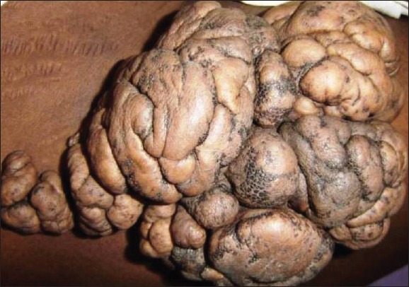
Clustered group of soft, fleshy, skin-colored to yellow nodules with smooth, wrinkled, cerebriform appearance over lower back
Figure 2.

Skin-colored nodules with smooth surface over left foot
Figure 3.
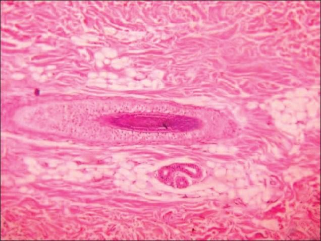
Hematoxylin and eosin stain showed ectopic fat cells in the dermis (H and E, ×100)
Nevus lipomatosus cutaneous superficialis (NCLS) is a rare hamartoma of adipose tissue. It is divided into classical form and solitary form. It usually presents as a clustered group of soft, fleshy, skin-colored or yellowish nodules, which are either sessile or pedunculated growths with a smooth, wrinkled or cerebriform surface.[1] The most common sites are pelvic girdle, buttocks, back or abdomen. They are usually present at birth or emerge during the first two decades of life.[2] It was first reported by Hoffmann and Zurhelle in 1921.[3]
Histopathologically, it is characterized by the presence of ectopic mature fat in the dermis mostly around the perivascular area and varies from 10% to 50% of the lesion. Epidermis may show acanthosis, basket weave hyperkeratosis, rete ridges obliteration with focal elongation, and increased basal pigmentation. There may be increased density of the collagen fibers, number of fibroblast with decreased elastic tissue along with perivascular mononuclear cell, and spindle-shaped cell infiltration in the dermis.[4] Electron microscopy has shown that lipocytes are closely associated with capillaries, and it has been suggested that they may originate from pericytes.[5] Recently a 2p24 deletion was found in nevus lipomatosis superficialis.[6]
There are reports showing its co-localization with lipedematous scalp[7] and Michelin tire baby[8] appearance. The closest differential diagnosis of solitary form is segmental neurofibromatosis.[9] Wide excision with skin grafting remains the treatment of choice.
Footnotes
Source of Support: Nil
Conflict of Interest: Nil.
REFERENCES
- 1.Yap FB. Nevus lipomatosus superficialis. Singapore Med J. 2009;50:e161–2. [PubMed] [Google Scholar]
- 2.Khandpur S, Nagpal SA, Chandra S, Sharma VK, Kaushal S, Safaya R. Giant nevus lipomatosus cutaneous superficialis. Indian J Dermatol Venereol Leprol. 2009;75:407–8. doi: 10.4103/0378-6323.53149. [DOI] [PubMed] [Google Scholar]
- 3.Hoffmann E, Zurhelle E. UBER einen Naevus lipomatodes cutaneaous superficialis der linken Glutaalgegend. Arch Dermatol Syphilol. 1921;130:327. [Google Scholar]
- 4.Umashankar T, Prasad T, Rajeshwari SH. Naevus lipomatosus superficialis: Clinicopathological study of a case. Indian J Pathol Microbiol. 2003;46:444–5. [PubMed] [Google Scholar]
- 5.Dotz W, Prioleau PG. Nevus lipomatosus cutaneus superficialis. A light and electron microscopic study. Arch Dermatol. 1984;120:376–9. [PubMed] [Google Scholar]
- 6.Cardot-Leccia N, Italiano A, Monteil MC, Basc E, Perrin C, Pedeutour F. Naevus lipomatosus superficialis: A case report with a 2p24 deletion. Br J Dermatol. 2007;156:380–1. doi: 10.1111/j.1365-2133.2006.07622.x. [DOI] [PubMed] [Google Scholar]
- 7.Mansur AT, Yasar S, Aydingöz IE, Göktay F, Ozdemir N, Sungurlu F. Colocalization of lipedematous scalp and nevus lipomatosus superficialis: A case report. J Cutan Pathol. 2007;34:342–5. doi: 10.1111/j.1600-0560.2006.00610.x. [DOI] [PubMed] [Google Scholar]
- 8.Palit A, Inamadar AC. Circumferential skin folds in a child: A case of Michelin tire baby syndrome. Indian J Dermatol Venereol Leprol. 2007;73:49–51. doi: 10.4103/0378-6323.30654. [DOI] [PubMed] [Google Scholar]
- 9.Armstrong DK, Walsh MY, Bingham A, McMillan C. Naevus lipomatosus cutaneous superficialis. Australas J Dermatol. 1997;38:88–90. doi: 10.1111/j.1440-0960.1997.tb01115.x. [DOI] [PubMed] [Google Scholar]