

History and clinical signs
A 1-year-old, intact male, German shepherd was referred to the ophthalmology service at the Western College of Veterinary Medicine (WCVM) for evaluation of a progressively enlarging mass at the right side of the nose that had first been noted 5 mo previously. The dog's vaccinations were current, it was not receiving any form of medication, and it was in training as a working police dog.
On physical examination, there were no abnormal findings other than the mass adjacent to the medial canthus of the right eye (Figure 1). Results of the neuro-ophthalmic examination, including menace responses, direct and consensual pupillary light reflexes, palpebral reflexes, and oculocephalic reflexes, were normal. Schirmer tear test (Schirmer Tear Test Strips; Alcon Canada, Mississauga, Ontario) values were 16 mm/min for the right eye and 18 mm/min for the left eye. Applanation tonometry (Tonopen XL; Biorad Ophthalmic Division, Santa Clara, California, USA) revealed normal intraocular pressures of 21 mmHg and 19 mmHg in the right and left eye, respectively. There was no uptake of fluorescein dye (Fluor-I-Strip AT; Ayerst Laboratories, St. Laurent, Quebec) on either cornea. Ophthalmic examination confirmed a 2-cm diameter, round, raised, smooth, nonulcerated, fluctuant subcutaneous mass near the right medial canthus at the level of the lacrimal sac, with no evidence of palpebral conjunctival extension (Figure 1). No fluorescein dye stain was visualized from the right nares or pharynx after 5 min. A nasolacrimal flush, using a 22-gauge IV catheter and 3 mL of eye wash (Eye Stream; Alcon Canada), injected through each dorsal punctum revealed normal fluid passage via the ventral punctum of each eye and the left and right nares. The medial canthal mass of the right eye remained unchanged in size. Slit lamp biomicroscopy (Kowa SL-14; Kowa, Tokyo, Japan) and indirect ophthalmoscopy (Heine Omega 200; Heine Instruments Canada, Kitchener, Ontario) revealed no abnormalities in either eye.
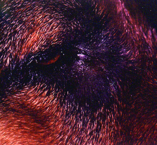
Figure 1. Photograph of the right eye at presentation demonstrating the round mass at the medial canthal region.
Discussion
Differential diagnoses for this fluctuant medial canthal mass were a dacryops (1,2,3) (a cyst of lacrimal gland origin), cysts of the orbital adnexal tissue (canaliculi; lacrimal sac, nasolacrimal duct, or both; conjunctiva, frontal or nasal sinus mucosa) (2,4,5), abscess (6), dacryocystitis (7), granuloma (8), congenital dermoid cyst (9), and neoplasm (2,10).
Diagnostic work-up, including skull radiographs, dacryocystorhinography, and fine needle aspiration of the mass for cytologic examination, with or without bacterial cultures, was advised. A complete blood cell (CBC) count and serum biochemical profile were performed prior to anesthesia; the results were within normal limits. The dog was premedicated and induced routinely, and general anesthesia was maintained with halothane (Halothane, B.P.; MTC Pharmaceuticals, Cambridge, Ontario). A transcutaneous fine needle aspirate of the mass was obtained; it revealed 0.3 mL of moderately cloudy, yellowish fluid. Cytologic examination and aerobic and anaerobic cultures were performed on the fluid. The cytologic examination revealed mainly neutrophils, some of which were karyorrhectic, and fewer macrophages filled with intensely turquoise-blue material. Prussian blue staining indicated that the phagocytosed material in the macrophages was not iron and, thus, not hemosiderin. The cytologic findings were consistent with a glycoprotein-rich, nonseptic, neutrophilic inflammation. However, a Staphylococcus sp. was detected on subculture. Lateral, dorsoventral, lateral oblique, and openmouthed skull radiographs were taken; they failed to reveal any bony abnormalities. Dacryocystorhinography was performed via cannulation of the right ventral punctum by using a 22-gauge IV catheter. Iodinated contrast material (Omnipaque; Nycomed Imaging AS, Oslo, Norway) was injected both with and without digital dorsal punctal occlusion and revealed a normal nasolacrimal system with no apparent communication with the mass. Iodinated contrast material (0.5 mL) was injected via a 25-gauge needle into the mass; it revealed complete filling of a 2-cm round, contained, cystic structure with no connections to other tissues or evidence of a foreign body (Figure 2).

Figure 2. Lateral radiograph of the skull following injection of the medial canthal mass with iodinated contrast agent. Note the 2-cm round, contained, cystic structure with no connections to other tissues or evidence of a foreign body.
Due to the proximity of the cyst to the dorsal and ventral canaliculi, its surgical excision under an operating microscope was planned, and performed the following day. The dog was sedated and induced routinely; general anesthesia was maintained via halothane (Halothane, B.P., MTC Pharmaceuticals). The dog was positioned in sternal recumbency with its head under the operating microscope. The dorsal and ventral canaliculi of the right eye were cannulated with 3-0 monofilament nonabsorbable suture material (Novafil; Sherwood Medical, St. Louis, Missouri, USA) to identify them during the resection of the cyst. The cyst did not communicate with the lacrimal sac. It was not adherent to the canaliculi but was directly overlying them. The cyst, once removed, was placed in 10% neutral buffered formalin and submitted for histologic examination. The subcutaneous tissues and skin incision were closed routinely. The medial canthal incision site was cold-packed for the first 20 min following surgery to lessen postoperative swelling. An Elizabethan collar was placed to prevent self-trauma.
Histologic examination of the surrounding tissue revealed bundles of striated muscle fibers with multifocal signs of necrosis and degeneration, including loss of striation, floccular degeneration, and hyalinization. There were multifocal areas of neutrophils, eosinophils, and a few macrophages surrounding the muscle fibers and vessels. This was consistent with a surrounding pyogranulomatous cellulitis. The cellulitis was likely due to irritation from the iodinated contrast material, cystic contents or both, from violation of cyst integrity 1 d prior to surgery. The cyst was likely missed in processing, as the microscopic examination did not reveal any glandular acini or a cyst wall.
The dog experienced multiple postoperative episodes of hemorrhage from the medial canthal incision site over the next week. A coagulopathy was suspected. Diagnostic tests, including a CBC count, prothrombin and activated partial thromboplastin times (PT and APTT, respectively), buccal mucosal bleeding time, and von Willebrand's factor (vWF), were performed. These evaluations confirmed von Willebrand's disease. The vWF results revealed a value of 36% (normal reference range, 45% to 80%). Testing of the breeding kennel for vWF was advised. Medial canthal incisional dehiscence occurred due to pressure necrosis from the severe swelling that developed as a result of the recurrent hemorrhage. The wound gradually healed by 2nd intention and resulted in granulation tissue at the site and mild medioventral ectropion.
The clinical diagnosis was a congenital medial canthal cyst. Skull radiographs and dacryocystorhinography confirmed that there was no evidence of bony involvement and no connection with the nasolacrimal system, respectively. Cytologic examination of the fluid obtained from the cyst was compatible with a dacryops (2,3). The growth of a Staphylococcal sp. on subculture was likely due to contamination from the transcutaneous fine needle aspiration and was not considered significant. Intramass injection of contrast material outlined a solitary, intact cyst with no evidence of foreign body, or nasal or frontal sinus communication.
Cysts of the periorbital region are rare in domestic animals (2). Periorbital cysts may arise from several glandular or ductular tissues, including the orbital lacrimal gland, accessory gland of the third eyelid, zygomatic salivary gland, lacrimal sac, canaliculi and/or nasolacrimal duct, conjunctival goblet cells, or the nasal or frontal sinus mucosa (2,4,5). The etiologies of these cystic structures include congenital malformations, trauma-induced glandular or ductular disruption, inflammation, and neoplasia (2,4). Given that the location and causes of periocular cysts can vary considerably, the anatomic location and lesions in adjacent tissues are most critical in determining their origin (2). Occasionally, some cysts develop at sites distant from their origins (5). In this case, history, age, clinical findings, and cytologic examination were most consistent with a congenital lacrimal gland cyst (dacryops), most likely arising from embryonically misplaced lacrimal gland or duct tissue. No obvious connection to the gland of the third eyelid was noted and the third eyelid was clinically normal. Cystic lesions of the lacrimal system have been associated with a history of inflammation (1). The pericystic inflammation noted histologically in this case was most likely due irritation from subcutaneous contrast leakage, cystic content leakage, or both, following disruption of cyst wall integrity.
Given the close approximation of this cyst to the canaliculi and lacrimal sac, a thorough evaluation of the nasolacrimal system, in the form of a dacryocystorhinogram, was necessary. Dacryocystorhinography not only evaluated the patency of the nasolacrimal system but also revealed its lack of communication with the cystic structure and its normal anatomic location (11). Ultrasonography of the mass could also have been useful in confirming its cystic structure and in ruling-out a foreign body. A zygomatic sialogram was not completed due to the lack of concurrent orbital signs and the location of the cyst, making a zygomatic salivary cyst or mucocele less likely. No connection to the zygomatic salivary gland was detected intraoperatively or with intralesional contrast injection.
References
- 1.Playter RF, Adams LG. Lacrimal cyst (dacryops) in 2 dogs. J Am Vet Med Assoc 1977;171:736–737. [PubMed]
- 2.Martin CL, Kaswan RL, Doran CC. Cystic lesions of the periorbital region. Compend Contin Educ Pract Vet 1987;9:1022–1029.
- 3.Grahn BH, Mason RA. Epiphora associated with dacryops in a dog. J Am Anim Hosp Assoc 1995;31:15–19. [DOI] [PubMed]
- 4.Davidson HJ, Blanchard GL. Periorbital epidermoid cyst in the medial canthus of three dogs. J Am Vet Med Assoc 1991;198: 271–272. [PubMed]
- 5.Van der Woerdt A, Wilkie DA, Gilger BC, et al. Surgical treatment of dacryocystitis caused by cystic dilatation of the nasolacrimal system in three dogs. J Am Vet Med Assoc 1997;211:445–447. [PubMed]
- 6.Koch SA, Buell BE. Medial orbital abscess in a Collie dog. J Am Vet Med Assoc 1970;156:1905–1906. [PubMed]
- 7.Lavach JD, Severin GA, Roberts SM. Dacryocystitis in dogs: a review of 22 cases. J Am Anim Hosp Assoc 1984;20:463–467.
- 8.Collins BK, MacEwan EG, Dubielzig RR, et al. Idiopathic granulomatous disease with ocular adnexal and cutaneous involvement in a dog. J Am Vet Med Assoc 1992;201:313–316. [PubMed]
- 9.Walde I, Hittmair K, Henninger W, et al. Retrobulbar dermoid cyst in a Dachshund. Vet Comp Ophthalmol 1997;7:239–244.
- 10.Madewell BR, Priester WA, Gillette EL, et al. Neoplasms of the nasal passages and paranasal sinuses in domestic animals as reported by 13 veterinary colleges. Am J Vet Res 1976;37:851–856. [PubMed]
- 11.Gelatt KN, Cure TH, Guffy MM, et al. Dacryocystorhinography in the dog and cat. J Small Anim Pract 1972;13:381–397. [DOI] [PubMed]