Although “localized” or “regional” eruptions of lymphomatoid papulosis (LyP) have been reported in the literature, specific references to distinct grouped lesions in a circumscribed patch have been reported in the context of persistent agminated lymphomatoid papulosis (PALP).1–4 It has been suggested that an “agminated CD30+ lymphoproliferative disorder … be considered a lymphoma,”1(p1011) owing in part to the potential for disease progression to mycosis fungoides–like patches if untreated, and that PALP be treated aggressively with ablation therapy, specifically radiation therapy.1 Of the 9 reported cases of PALP that we found in the literature,1–4 only 1 case, in a 27-year-old man, evolved to disseminated classic LyP.2 We report herein a case of steroid-responsive agminated LyP in a pediatric patient that quickly evolved to disseminated disease, thus providing further evidence to support aggressive treatment of agminated LyP.
Report of Case
A 9-year-old boy presented with a 3-week history of a nontender, pruritic, well-circumscribed crop of waxing and waning papulonodules on his right upper chest. Treatment with oral antibiotics, antifungal creams, and topical antibiotics had failed to resolve the disease. Excision of the 2.5-cm lesion on the right upper chest revealed a CD30+, ALK-1− infiltrate, diagnosed as lymphomatoid papulosis type A. Findings of T-cell receptor gene rearrangement studies were positive, but a karyotype analysis showed no abnormalities.
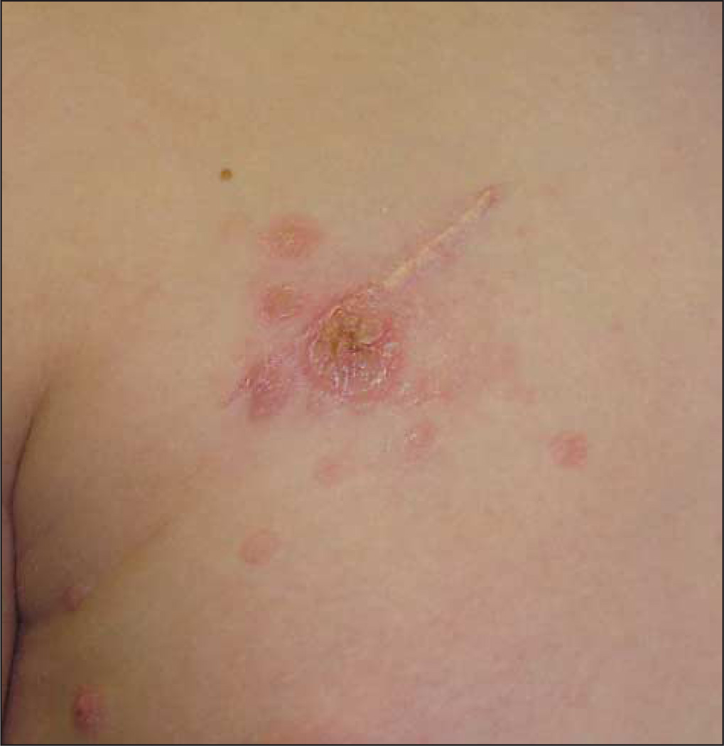
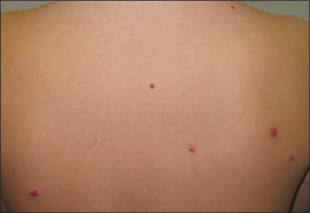
Over the 3 to 4 weeks after excision, a crop of nodules arose around the excision site (Figure 1), and subsequently, generalized lesions on the rest of his trunk (Figure 2) and extremities erupted, crusted, and then regressed before flaring again. The patient was treated with topical fluocinonide, and the lesions cleared, including the lesions around the original agminated plaque. Six months later, occasional crops of new lesions continued to erupt on his trunk and extremities.
Figure 1.
Agminated papulonodules with crusting and ulceration are seen on the patient’s right upper chest with additional secondary lesions outside the agminated patch.
Figure 2.
Evolution of the initial agminated papulonodules to generalized lesions on the back of the patient.
Comment
Our case differs from other published cases in that agmination did not necessarily correlate with persistence of the lesion; the original lesion completely resolved after treatment with topical steroids. To our knowledge, all previously published cases of agminated LyP have presented as PALP. Of the 9 reported cases, 7 were treated with radiation therapy, and remissions of at least 2 years were achieved in 6 of these 7 cases. An eighth case was treated with topical steroids and was in remission 1 year later.4 A ninth case progressed to multifocal lesions and appeared to behave more like classic LyP and was treated successfully with methotrexate.2
Among the 9 reported cases of PALP, only 1 also disseminated, with a significantly longer 9-month period of localized agmination prior to dissemination, and this was in a nonpediatric patient. We note that methotrexate was used successfully to treat the other case of disseminated LyP, but its effect on preventing dissemination is unclear. Our case shows that PALP can disseminate in children, and although aggressive control of PALP is not currently the standard of care, previously described locally ablative therapies such as radiotherapy and topical carmustine therapy that have induced long-term remission of PALP in its locoregional phase prior to possible dissemination should be further studied.1
Acknowledgments
Funding/Support: This work was supported in part by National Institutes of Health (NIH) T32 grant AR007569 to the NIH Training Program in Investigative Dermatology at Case Western Reserve University (Dr Chan).
Footnotes
Financial Disclosure: None reported.
Additional Contributions: Kevin D. Cooper, MD, reviewed and provided helpful comments on this manuscript.
References
- 1.Heald P, Subtil A, Breneman D, Wilson LD. Persistent agmination of lymphomatoid papulosis: an equivalent of limited plaque mycosis fungoides type of cutaneous T-cell lymphoma. J Am Acad Dermatol. 2007;57(6):1005–1011. doi: 10.1016/j.jaad.2007.05.046. [DOI] [PubMed] [Google Scholar]
- 2.Romero-Maté A, Martín-Fragueiro L, Miñano-Medrano R, Martinez-Morán C, Arias-Palomo D, Borbujo J. Persistent agmination of lymphomatoid papulosis evolving to classical lesions of lymphomatoid papulosis. J Am Acad Dermatol. 2009;61(6):1087–1088. doi: 10.1016/j.jaad.2008.12.018. [DOI] [PubMed] [Google Scholar]
- 3.Steinhoff M, Assaf C, Sterry W. Persistent agmination of lymphomatoid papulosis: not a new entity, but localized lymphomatoid papulosis. J Am Acad Dermatol. 2008;59(1):164–165. doi: 10.1016/j.jaad.2007.12.039. [DOI] [PubMed] [Google Scholar]
- 4.Torrelo A, Colmenero I, Hernández A, Goiriz R. Persistent agmination of lymphomatoid papulosis. Pediatr Dermatol. 2009;26(6):762–764. doi: 10.1111/j.1525-1470.2009.01035.x. [DOI] [PubMed] [Google Scholar]


