Abstract
To enable in vivo and in vitro competitive fitness comparisons among West Nile viruses (WNV), three reference viruses were genetically marked by site-directed mutagenesis with five synonymous nucleotide substitutions in the envelope gene region of the genome. Phenotypic neutrality of the mutants was experimentally assessed by competitive replication in cell culture and genetic stability of the substituted nucleotides was confirmed by direct sequencing.
Luminex® technology, quantitative sequencing and quantitative RT-PCR (qRT-PCR) were compared in regard to specificity, sensitivity and accuracy for quantitation of wildtype and genetically marked viruses in mixed samples based on RNA obtained from samples of known viral titers. While Luminex® technology and quantitative sequencing provided semi-quantitative or qualitative measurements a sequence-specific primer extension approach using a specific reverse primer set in singleplex qRT-PCR demonstrated the best quantitation and specificity in the detection of RNA from wildtype and mutants viruses.
Keywords: West Nile virus, genetic marker, Luminex® technology, quantitative sequencing, qRT-PCR, fitness competition
1. Introduction
West Nile virus (WNV) is a single-stranded, positive-sense RNA virus belonging to the family Flaviviridae. The WNV genome is approximately 11,000 nucleotides in length and encodes a single polyprotein that is cleaved into three structural proteins (C, prM, E) and seven non-structural proteins (NS1, NS2A, NS2B, NS3, NS4A, NS4B, NS5) (Rice et al., 1985).
As an arthropod-borne virus, WNV naturally perpetuates in a two-host transmission cycle between birds and Culex spp. mosquitoes with occasional tangential infection of humans and other vertebrates. To maintain sufficient fitness in both insect and vertebrate hosts, the genome of flaviviruses seems to be conserved, likely due to alternation between host and vector, and purifying selection in the avian host (Jerzak et al., 2008; Coffey et al., 2008). When mutations occur, they may be detrimental and lost rapidly, neutral and maintained, or beneficial by altering significantly the phenotype and being selected positively (Domingo et al., 1996).
Direct sequencing is the method of choice to identify nucleotide substitutions, study phylogenetic relationships and determine genotypic evolution. However, relating genotypic variation to significant phenotypic change is difficult and often requires intensive reverse engineering experiments. Alternatively, to investigate phenotypic change, in vitro and in vivo fitness competition experiments may be used to compare the replicative capacity of two viruses concurrently competing in the same host (Weaver et al., 1999). Such studies can be used to compare field isolates to a reference or founding strain while controlling for inter-host variability. However, the high degree of genetic conservation among WNV isolates complicates the development of genotype-specific primers and probes for RT-PCR. Therefore, a phenotypically neutral genetic marker is required to label the reference strain for competition against wildtype isolates of interest. The analysis of samples mixed with two genetically similar viral populations that may have markedly different titers is challenging as it requires highly specific and quantitative detection based on one or a few nucleotide differences.
The aim of the current study was to engineer a WNV reference strain containing a stable and fitness neutral genetic marker that would facilitate developing quantitative and specific detection methods to track concurrently genetically marked and wildtype viruses in competition assays. Three strains of WNV were marked genetically by site-directed mutagenesis of either one or five synonymous nucleotide substitutions in the E gene between nucleotide positions 2449–2454 to serve as reference viruses for in vitro and in vivo fitness competition studies. In an effort to develop a suitable detection method, three approaches were compared: Luminex® technology, quantitative sequencing and quantitative real-time RT-PCR.
The Luminex® xTAG® protocol uses a liquid suspension microarray platform to detect individually sequence-tagged, color-coded microspheres with a flowcytometric laser detection system (Luminex® Corporation, Austin, TX, USA). It allows for high-throughput, multiplex testing for as many as 100 different nucleic acid sequences in one sample and previously has been used for host identification of Culex spp. mosquito blood meals (Thiemann et al., 2012) and for detection of several subtypes of influenza A viruses (Smith et al., 2012). While one laser interrogates microsphere identity, the other quantifies the fluorescence derived from the biotinylated template resulting in a cummulative signal used for template quantitation.
Quantitative sequencing is used to measure quantitatively multiple single nucleotide polymorphisms from sequencing chromatograms (Hall and Little, 2007). The peak area and/or height from individual, overlapping (polymorphic) sequence chromatograms is measured with PHRED software and PolySNP, a PERL script, and utilized to estimate the relative template frequency in pooled DNA samples based on the proportions of the overlapping peaks (Hall and Little, 2007). This approach previously has been used in fitness competitions of WNV (Fitzpatrick et al., 2010; Deardorff et al., 2011) and dengue virus (Groat-Carmona et al., 2012).
Finally, TaqMan ® (Applied Biosystems [ABI], Foster City, California, USA) is a quantitative one-step real-time RT-PCR approach that can be multiplexed and is used to distinguish closely related RNA templates with sequence-specific primer and probe sets. A specific single nucleotide polymorphism can be targeted using individual allele-specific TaqMan® probes containing distinct fluorescent dyes and primer sets that uniquely align with the genome providing high specificity for the allele of interest (ABI, USA).
In the current study, Luminex® technology, quantitative sequencing and TaqMan® qRT-PCR were compared with regard to their specificity and ability to accurately quantify a wide range of titers of wildtype and mutant WNV RNA in mixed samples during co-infection experiments. Each method is described and the most successful approach, a novel singleplex quantitative qRT-PCR assay using a sequence-specific reverse primer set is presented.
2. Material and methods
2.1 Viruses and construction of mutants
Plasmids containing WNV genomic information from three wildtype strains of WNV were used to engineer mutants containing either one or five synonymous nucleotide (nt) substitutions in the carboxy terminus area of the E gene (nt 967–2469). A previously generated infectious clone-derived virus based on a field isolate of the NY99 strain (Kinney et al., 2006), was marked genetically by either a single nucleotide transition from C to T at position nt 2454 (1nt-mutation) or by five nucleotide substitutions from CTCTCC to TTGAGT (5nt-mutation) at positions nt 2449 and 2451–2454 (Table 1). The same 5nt-mutations were introduced into a previously generated infectious clone-derived virus of COAV997 (Andrade et al., 2011) containing the amino acid substitutions identified in the founding California 2003 strain, and in a natural field isolate derived from a moribund common raven in Tabasco State, Mexico, TWN354, which was referred to as TM171-03 pp5 in previous publications (Beasley et al., 2004; Langevin et al., 2011). The generated mutants were termed NY99-1nt when containing the 1nt-mutation and NY99-5nt, COAV997-5nt and TWN354-5nt when genetically marked with the 5nt-mutations (Table 1). The synonymous transitions were designed to have minimal effect on melting temperature profiles of the primers and to not incorporate codon usage bias differences that could alter fitness independent of amino acid alterations. Sets of specific mutagenesis primers and capture-probes were designed for site directed mutagenesis of wildtype viruses with QuikChange® Site-directed Mutagenesis Kit and PfuUltra® High-Fidelity DNA polymerase (Stratagene) as shown in Table 1. Synonymous mutations were generated in the 5’ plasmid (1–2495 nt) and then ligated with the 3’ plasmid prior to in vitro transcription to generate the mutant infectious RNA according to Kinney et al. (2006) followed by transfection of transcribed viral RNA into baby hamster kidney (BHK) cells (ATCC, no. CCL-10). Supernatant from transfected cultures was harvested at 3 days post transfection upon observation of cytopathic effect. Viruses then were titered using a 10-fold serial dilution plaque assay in Vero cells (ATCC, no. CCL-81) as previously described (Brault et al., 2004). After confirming the presence of infectious virus, virus stocks were propagated through a single passage in Vero cells for 3–4 days at 37°C. Viral RNA was extracted from Vero cell culture supernatant utilizing a MagMAX™ magnetic particle processor and MagMAX™ −96 Viral RNA isolation Kit (ABI, USA) according to the manufacturer’s instructions. Full-length consensus sequencing of all mutant viruses was performed to confirm presence of introduced mutations and to ensure that spurious mutations were not incorporated during the generation of infectious WNV cDNA clones.
Table 1. Construction of mutants.
Primers and probes designed for site-directed mutagenesis of NY99, COAV997 and TW354 viruses, 5- and 1-nucleotide engineered mutant sequences and thermocycler profiles used for the mutagenesis reaction are shown.
| Primers, probes and viruses | Sequence (5′ → 3′)a |
|---|---|
| One nucleotide substitution (C → T) | |
| WN.2454.mutF | CTGCTCTTCCTCTCTGTGAACGTGCACG |
| WN.2454.mutR | GCGTGCACGTTCACAGAGAGGAAGAGC |
| Five nucleotide substitutions (CTCTCC → TTGAGT) | |
| WN.2449–2454.mutF | GTTGGAGGAGTTCTGCTCTTCTTGAGTG |
| WN.2449–2454.mutR | AGTGTCAGCGTGCACGTTCACACTCAAG |
| Capture probes | |
| WNV.WT.Capt.Probe (+) | AminoC6-GCTCTTCCTCTCCGTGAA |
| WNV.MUT.Capt.Probe (+) | AminoC6-GCTCTTCTTGAGTGTGAA |
| Wildtype and mutant sequencesb | |
| Wildtype (NY99, COAV997, TW354) | GCAGTTGGAGGAGTTCTGCTCTTCCTCTCTGG |
| 1-nucleotide mutant (NY99-1nt) | GCAGTTGGAGGAGTTCTGCTCTTCCTCTCTGG |
| 5-nucleotide mutants (NY99-5nt, COAV997-5nt, TW354-5nt) | GCAGTTGGAGGAGTTCTGCTCTTCTTGACTGG |
| Reactions and thermocycle profiles | |
| Mutagenesis | |
| 95°C/1 min → 17× [95°C/50 s → 60°C/50 s → 68°C/5.7 min] → 68°C/7 min → hold at 4°C | |
Bold nucleotides indicate polymorphic region.
Sequences represent a 53 nucleotide amplicon located in the envelope and NS1 gene at nucleotide positions 2427–2479.
2.2 Luminex xTAG® microsphere array
Two sets of two carboxylated fluorescent microspheres were labeled uniquely for the detection of wildtype WNV, 1nt- and 5nt-mutants (Table 2). Microsphere LUA75 for NY99- 1nt (C → T) and LUA10 for wildtype WNV were selected having a net mean fluorescence intensity (MFI) of 5617 and 6851, respectively. For the detection of the 5nt-mutants (CTCTCC → TTGAGT), LUA40 (net MFI 3286) and LUA50 for wildtype WNV (net MFI 6749) were assigned. The net MFI values for the selected microspheres were kept within a 2.5× range for the same set as recommended by the manufacturer. Each microsphere was attached covalently to an anti-tag sequence extending into the individual wildtype or mutant sequence at its 3′-end (Table 2). Primers for the allele-specific primer extension (ASPE) reaction were designed to contain a universal tag sequence on their 5′-end, being complementary to the corresponding anti-tag sequence of the microsphere (Table 2). Primers and sequences (Eurofins MWG Operon, Huntsville, AL, USA) were summarized in Table 2. Two sets of primers, WNV.2387.F/WNV.2530.R (amplicon length nt 143) and WNV.2296.F/WNV.2786.R (amplicon length nt 490) were designed for the reverse transcription and PCR amplification of viral cDNA (Table 2) and compared regarding detection of products in downstream applications using both LUA40/50 and LUA10/75 sets (Table 4).
Table 2. Luminex® xTAG® microsphere array.
Primers, tag and anti-tag sequences designed for Luminex® xTAG® microsphere array are presented including thermocycler profiles used for all Luminex® xTAG® reactions.
| Primers sets, tags and anti tags | Sequence (5′ → 3′)a |
|---|---|
| Reverse transcription and PCR primers | |
| WNV.2387.F | TCAATGCTCGTGATAGGTCCAT |
| WNV.2530.R | TGAACACTCCACTTCCACATCT |
| WNV.2296.F | CAAGTGTTCGGAGGAGCATTC |
| WNV.2786.R | GCGGTGAGGCGTTTAGGT |
| Allele-specific primer extension (ASPE) primers | |
| 2454.WT.ASPE | agttctgctcttcctctcC |
| 2454.MUT.ASPE | agttctgctcttcctctcT |
| 2449–2454.WT.ASPE | gagttctgctcttcCTCTCC |
| 2449–2454.MUT.ASPE | aggagttctgctcttcTTGAGT |
| Anti-tags coupled to microspheres | |
| 2454.WT.LUA10 | ATCATACATACATACAAATCTACAagttctgctcttcCTCTCC |
| 2454.MUT.LUA75 | AATCATACCTTTCAATCTTTTACAagttctgctcttcCTCTCT |
| 2449–2454.WT.LUA50 | CAATATACCAATATCATCATTTACgagttctgctcttcCTCTCC |
| 2449–2454.MUT.LUA40 | CTTTCTACATTATTCACAACATTAaggagttctgctcttcTTGAGT |
| Biotinylation primer | |
| WNV.2530.R biotinylated | biotin-TGAACACTCCACTTCCACATCT |
| Reactions and thermocycle profiles | |
| Reverse transcription and PCR amplification | |
| 50°C/30 min → 94°C/2 min → 40× [94°C/15 s → 55°C/30 s → 72°C/1 min] → 72°C/10 min → hold at 4°C | |
| Allele-specific primer extension (ASPE) and biotinylation | |
| 96°C/2 min → 94°C/30 s → 30× [55°C/1 min → 74°C/2 min] → hold at 4°C | |
| ExoSAP-it purification | |
| 37°C/30 min → 80°C/15 min → hold at 4°C | |
| Hybridization | |
| 96°C/90 sec → 37°C/30 min → hold at 37°C | |
Lowercase nucleotides indicate the complementary part of the universal tag and anti-tag sequences located at the 5′-end of the ASPE primers and microsphere anti-tag, respectively. Bold nucleotides represent the polymorphic nucleotides located at the 3′-end of the ASPE primers and microsphere anti-tags. Uppercase letters at the 3′-end indicate nucleotides that originated from the wildtype WNV backbone and at the 5′-end they indicate the non-complementary part of the tag and anti-tag sequences. WT; wildtype. MUT; mutant.
Table 4. Mean fluorescence intensity (MFI) values of NY99, NY99-1nt and NY99-5nt after separate amplification with primer set WNV2387F/2530R and WNV2296F/2786R.
Mean MFI values and standard deviations of NY99 (LUA10 and LUA50), NY99-1nt (LUA75) and NY99-5nt (LUA40) are shown after separate amplification with PCR primer set WNV2387F/2530R and WNV2296F/2786R. All non-template controls were yielding a MFI value below 11 which was considered as background cut-off.
| Primer set | WNV2387F/2530R (amplicon length: 143 nt) mean fluorescence intensity ± SD |
WNV2296F/2786R (amplicon length: 490 nt) mean fluorescence intensity ± SD |
|||||||
|---|---|---|---|---|---|---|---|---|---|
| Virus-microsphere mixa | LUA 10 | LUA 75 | LUA 50 | LUA 40 | LUA 10 | LUA 75 | LUA 50 | LUA 40 | |
| NY99 | LUA 10 | 2342 ± 519 | 122 ± 59 | 26 ± NA | 0 | 5 ± 1 | 6 ± 4 | 0 | 0 |
| NY99-1nt | LUA 75 | 494 ± 363 | 4311 ± 2873 | 24 ± NA | 0 | 8 ± 1 | 60 ± 5 | 0 | 0 |
| NY99 | LUA 50 | 0 | 0 | 5528 ± 114 | 18 ± 3 | 0 | 0 | 13 ± 2 | 9 ± 1 |
| NY99-5nt | LUA 40 | 0 | 0 | 86 ± 10 | 3290 ± 39 | 0 | 0 | 5 ± 2 | 39 ± 4 |
NY99 was tagged with LUA 10 when tested with NY99-1nt (LUA 75) and with LUA50 when tested with NY99-5nt (LUA40).
Bold numbers indicate correct identification. Numbers were rounded up and represent mean values of duplicates with ± SD. NA, not applicable.
The manufacturer’s xTAG® washed assay format was utilized for all experiments (http://www.luminexcorp.com). Briefly, cDNA and PCR amplicons were generated from viral RNA using SuperScript™ One-Step RT-PCR with Platinum© Taq (Invitrogen, Carlsbad, CA, USA) and primer set WNV.2387.F/WNV.2530.R followed by a purification step with ExoSAP-IT® (USB) per manufacturer’s instructions (Table 2). During the subsequent multiplex ASPE-reaction (Table 2), the specific 3′-end of the ASPE-primers 2449–2454.WT.ASPE/2449–2454.MUT.ASPE were attached to the specific portion of the previously generated amplicons for extension into biotinylated products containing the universal tag sequence at their 3′-end using Platinum® Genotype Tsp DNA polymerase (Invitrogen, Carlsbad, CA, USA). From the ASPE-product, 5µL was used for hybridization of the tag portion to the complementary anti-tag sequences (Table 2) using a total of 2,500 microspheres each per set and reaction in 25µL 2× Tm hybridization buffer (0.4 M NaCl, 0.2 M Tris, 0.16% Triton X-100, pH 8.0). Reaction volume was adjusted to 50µL with DEPC-treated water (Ambion) and samples were washed three times with 100µL of 1× Tm hybridization buffer (0.2 M NaCl, 0.1 M Tris, 0.08% Triton X-100, pH 8.0) by vacuum manifold filtration through a prewet 1.2µm PVDF filter microtiter plate (Millipore, Billerica, MA, USA). Microspheres were resuspended in 60µL of 1X Tm hybridization buffer containing 2µg/mL Streptavidin-R-phycoerythrin. From each reaction, 50µL was transferred into wells of a Luminex® 96-well plate, incubated for 15 min at 37°C and analyzed on a Luminex 200® analyzer with a two-laser detection system. Serial 10-fold dilutions of wildtype NY99, NY99-1nt and NY99-5nt RNA were used for experiments including non-template control (water) for determination of background reactivity.
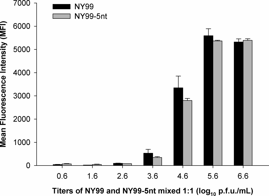
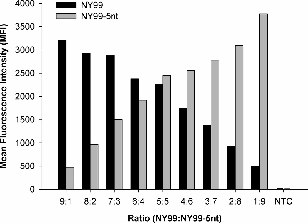
To compare specific distinction of wildtype from mutant template by microsphere sets LUA 40/50 (for 5nt-mutants) and LUA 10/75 (for 1nt-mutants), 1:1 dilutions of NY99 with either NY99-1nt or NY99-5nt were analyzed using primer sets WNV.2387.F/WNV.2530.R and WNV.2296.F/WNV.2786.R (Table 2) according to the above protocol. The resulting MFI values are presented in Table 4. The manufacturer’s recommended temperature for hybridization of ASPE-product to microspheres is 37°C (Table 2). To optimize hybridization, a temperature gradient between 36°C and 38°C was run on a thermocycler (36.0, 36.1, 36.2, 36.3, 36.6, 36.9, 37.2, 37.5, 37.7, 37.9, 38.0) using 1:1 mixtures of NY99 and NY99-5nt. Equal mixtures (1:1) of NY99 and NY99-5nt were diluted 10-fold serially based on viral titers ranging from 6.6 to 0.6 log10 plaque-forming units (p.f.u.)/mL and analyzed according to the protocol described above (Fig. 1). Different ratios of NY99 and NY99-5nt were analyzed (Fig. 2) by combining decreasing ratios of NY99 with increasing proportional ratios of NY99-5nt based on the DNA concentration (9:1, 8:2, 7:3, 6:4, 5:5, 4:6, 3:7, 2:8, 1:9).
Figure 1. Mean fluorescence intensity (MFI) of 1:1 mixtures of equal titers of NY99 and NY99-5nt.
Viral titers of NY99 and NY99-5nt were diluted 10-fold (0.6 to 6.6 log10 p.f.u./mL), mixed 1:1 as shown on the x-axis and analyzed on the Luminex® 200 analyzer in duplicate. The MFI values of NY99 (black bars) and NY99-5nt (gray bars) are shown on the y-axis with corresponding standard deviation. The MFI of NY99-5nt (LUA40) of 3286 was normalized to the MFI of NY99 (LUA50) of 6749 using a multiplication factor of 2.053864881.
Figure 2. Mean fluorescence intensity (MFI) of unequal mixtures of NY99 and NY99-5nt based on DNA template ratios.
The DNA concentration of NY99 and NY99-5nt was measured by Nanodrop spectrophotometer, adjusted and diluted 10-fold. Increasing ratios of NY99 (black bars) were mixed 1:1 with decreasing ratios of NY99-5nt (gray bars) to produce unqual mixtures. The MFI of NY99-5nt (LUA40) of 3286 was adjusted to the MFI value of NY99 (LUA50) of 6749 using a multiplication factor of 2.053864881. The non-template control is indicated as NTC.
2.3 Quantitative sequencing
The programs polySNP (Little and Hall, 2006) and PHRED (academic license, courtesy of Brent Ewing) were employed to measure overlapping chromatogram peak and height identified in extracted polymorphic sequences (Hall and Little, 2007). Two reference alignments in FASTA format were imported into polySNP each constituting a 143nt sequence centered on the polymorphic region (Table 1). Serial 10-fold dilutions of viral RNA were transcribed and amplified with the primer set WNV.2387.F/WNV.2530.R (Table 2) and SuperScript™ One-Step RT-PCR with Platinum® Taq (Invitrogen, Carlsbad, CA, USA). For NY99 and NY99-5nt mixtures, 25 or 30 PCR cycles were performed, whereas COAV997 and COAV997-5nt were amplified for 40 cycles (Table 2). Amplicons were purified using ExoSAP-IT® (USB) (Table 2) and run on a capillary 3730 DNA Analyzer utilizing BigDye Terminator v3.1 sequencing chemistry (ABI, USA). Resulting chromatograms were viewed with Sequencher version 4.9 and overlapping chromatograms were identified as the polymorphic region in samples containing wildtype and 5nt-mutant template by an ambiguity code of YTSWSY. The height and area under the chromatogram peak is proportional to the corresponding template concentration at the start of the sequencing reaction (Hall and Little, 2007). Each base within overlapping chromatograms was called in reference to both imported alignments (Table 2) and measured as the proportion of the chromatogram peak area or height resulting in a number from 0 to 1.
2.4 Allele-specific quantitative RT-PCR (qRT-PCR)
Sets of specific reverse primers were designed to contain the wildtype or 5nt-mutation at their 3′-ends, middle and 5′-ends. Forward primers and probes were designed to bind in the adjacent parental portion of the sequence. All sequences (ABI, USA) were designed using Primer Express v.2.0 software and are shown in Table 3.
Table 3. Allele-specific qRT-PCR.
Design of different sets of probes, forward and sequence-specific reverse primers for use in qRT-PCR. Forward primers and probes bind to the same sequence of wildtype and 5nt-mutant viruses. Specific reverse primers cover the nt 2449–2545 sequence polymorphism.
| Primers sets and probes | Sequence (5′ → 3′)a | Region |
|---|---|---|
| Forward primers | ||
| WNV.TAQ.2375.F | TGTGGATGGGCATCAATGC | 2375–2393 |
| WNV.TAQ.2393.F b | CTCGTGATAGGTCCATAGCTCTCA | 2393–2415 |
| 5′-end wildtype reverse primers | ||
| WNV.TAQ.WT.2456.R | ACGGAGAGGAAGAGCAGAACTC | 2456–2435 |
| 3′-end wildtype reverse primers | ||
| WNV.TAQ.WT.2464.R b | GCACGTTCACGGAGAGGAA | 2464–2446 |
| WNV.TAQ.WT.2469.R.ART.1 | CGTGCACGTTCACGaAGAG | 2469–2449 |
| WNV.TAQ.WT.2466.R.ART.2 | AGCGTGCACGTTCACGtAGAG | 2466–2449 |
| 5′-end 5nt-mutant reverse primers | ||
| WNV.TAQ.5nt.2454.R | ACTCAAGAAGAGCAGAACTCCTCC | 2454–2432 |
| WNV.TAQ.5nt.2456.R | ACACTCAAGAAGAGCAGAACTCCTC | 2456–2432 |
| 3′-end 5nt-mutant reverse primers | ||
| WNV.TAQ.5nt.2467.R | CGTGCACGTTCACACTCAAGA | 2467–2447 |
| WNV.TAQ.5nt.2467.R-alt b | CGTGCACGTTCACACTCAA | 2465–2447 |
| Probes | ||
| WN.2399–2415.FAM | 6FAM-ATAGGTCCATAGCTCT- MGBNFQ | 2399–2415 |
| WN.2417–2431.FAM b | 6FAM-CGTTTCTCGCAGTTG-MGBNFQ | 2417–2431 |
| Reactions and thermocycler profiles | ||
| Reverse transcription and PCR amplification | ||
| 52°C/30 min → 95°C/10 min → 50× [95°C/15 s → 60°C/1 min] | ||
Nucleotides in bold indicate polymorphic region. Lowercase letters represent artificial nucleotides.
Primers and probe selected for use in final qRT-PCR protocol.
The 5’ plasmids of NY99 and NY99-5nt were generated and in vitro transcribed to produce viral RNA standards for standard curve quantitation by qRT-PCR. Briefly, 5′ plasmids (Kinney et al., 2006) were transformed into and replicated in E. coli (One Shot® Top 10 chemically competent cells, Invitrogen). Purified plasmid DNA was digested with NgoMIV (New England BioLabs), treated with proteinase K (Invitrogen) and extracted using phenol-chloroform/isoamyl alcohol. Following ethanol precipitation, DNA served as template for in vitro RNA transcription (AmpliScribe™ T7 Transcription kit, EpiCentre). Viral RNA was purified (RNeasy Mini Kit, Invitrogen) and quantified by NanoDrop spectrophotometer and aliquots frozen at −80°C.
The above described primers, probe, RNA standards and the TaqMan® One-Step RT-PCR Master Mix Reagents Kit (ABI, USA) were used to develop a qRT-PCR protocol (Table 3). Samples and standards were run in duplicate with a final volume of 25 µL per reaction and tested concurrently in singleplex format on two machines for separate wildtype and mutant quantification.
To increase assay specificity, concentrations of 3 µM, 4.5 µM and 7.5 µM of forward and specific reverse primers were tested with RNA template concentrations of 2 µL, 5 µL and 8 µL.
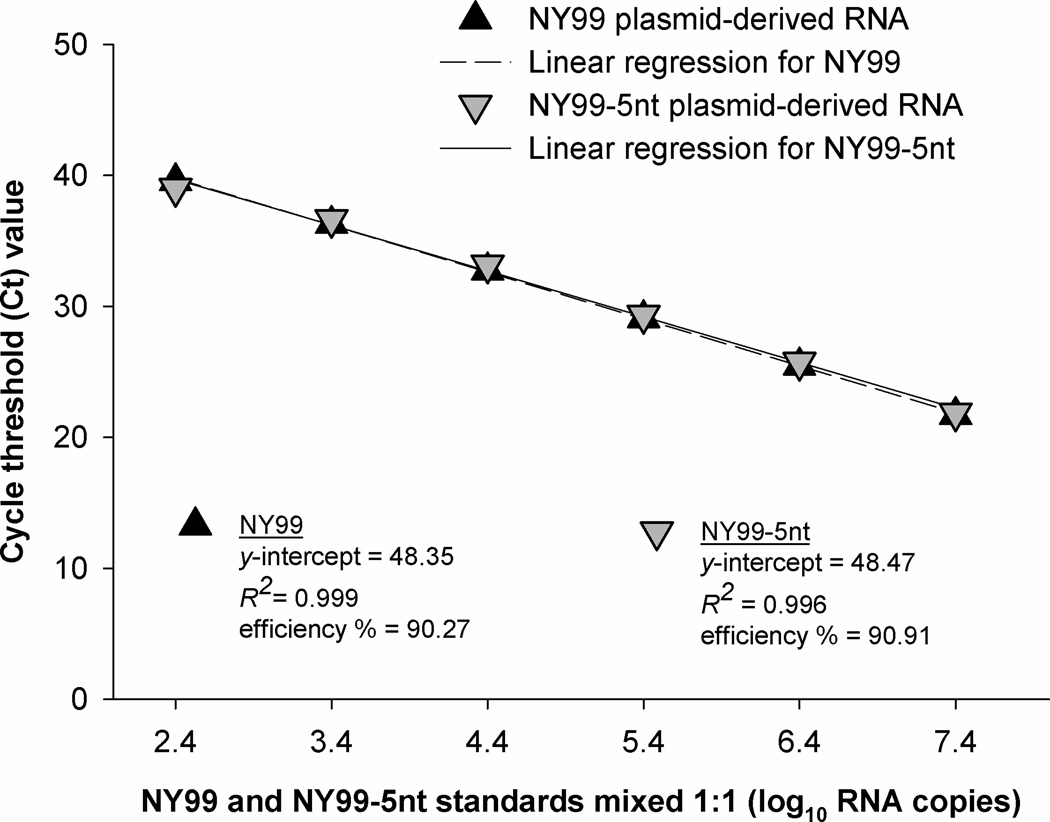
Plasmid-derived RNA standards and Vero cell culture supernatant of NY99 and NY99-5nt were diluted 10-fold based on viral titer and run in parallel as singles and 1:1 mixtures for side-by-side comparison. The suitability of the RNA to be used as standards was determined by comparing PCR efficiency and sensitivity (Fig. 6).
Figure 6. Detection of 1:1 mixtures of plasmid-derived in vitro transcribed NY99 and NY99-5nt RNA by allele-specific qRT-PCR.
Linearity, amplification efficiency (%) and correlation coefficient (R2) are presented for NY99 (black triangles) and NY99-5nt (gray triangles) used in a 1:1 mixture to serve as standards for standard curve quantitation. RNA copies are shown on the x-axis as log10 and cycle threshold (Ct) values on the y-axis.
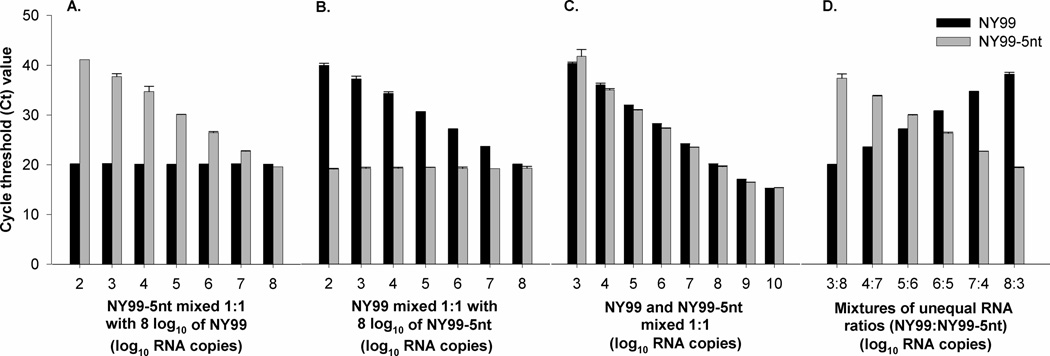
To analyze the specificity and quantitative accuracy of the allele-specific qRT-PCR, different mixtures of log10 RNA copies of NY99 and NY99-5nt were made: equal 1:1 mixtures of 3 – 10 log10 RNA copies of NY99 and NY99-5nt (Fig. 7A), constant concentrations of 8 log10 RNA copies of NY99 and NY99-5nt mixed 1:1 with 2 – 8 log10 RNA copies of NY99-5nt and NY99, respectively (Fig. 7B–C), and unequal mixtures using increasing concentrations of 3 – 8 log10 RNA copies of NY99 combined with decreasing concentrations of 8 – 3 log10 RNA copies of NY99-5nt (Fig. 7D).
Figure 7. Detection of different mixtures of 10-fold diluted RNA of NY99 (black bars) and NY99-5nt (gray bars) by allele-specific qRT-PCR.
Values represent mean cycle threshold (Ct) values calculated from duplicates including standard deviation on top of bars. A. Detection of 1:1 mixtures of 3 – 10 log10 RNA copies of NY99 and NY99-5nt. B. Detection of 2 – 8 log10 RNA copies of NY99 each mixed 1:1 with 8 log10 RNA copies of NY99-5nt. C. Detection of 2 – 8 log10 RNA copies of NY99-5nt each mixed 1:1 with 8 log10 RNA copies of NY99. D. Detection of 3 – 8 log10 RNA copies of NY99-5 mixed 1:1 with 8 – 3 log10 RNA copies of NY99-5nt.
2.5 Replication of NY99, NY99-5nt, COAV997 and COAV997-5nt in Vero and C6/36 cells
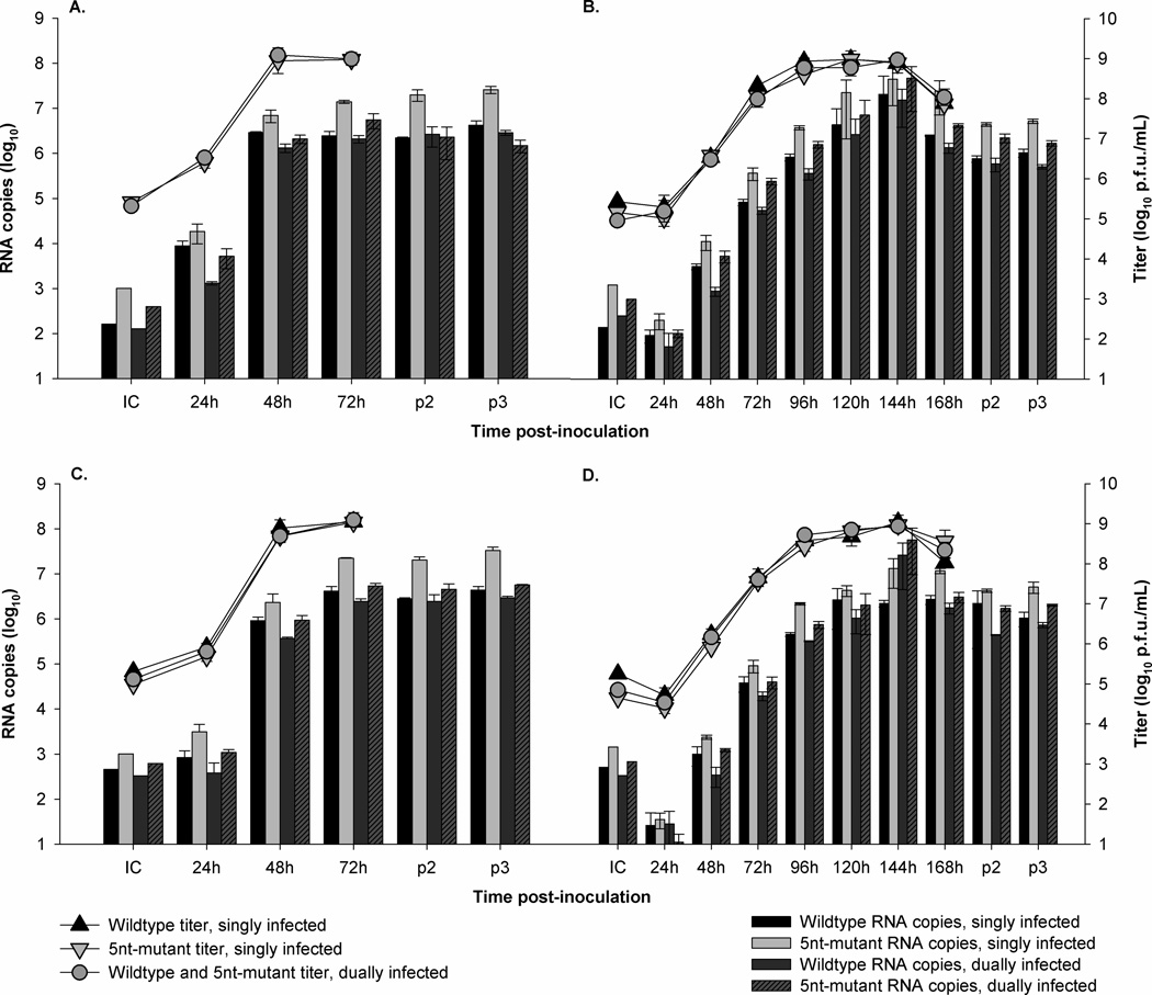
To test for fitness neutrality of the engineered NY99-5nt, equal titers of NY99 and NY99-5nt were determined by plaque assay titration (Brault et al., 2004), and allowed to replicate singly and competitively (mixed 1:1) in Vero and C6/36 (Aedes albopictus-derived mosquito cells) cell cultures in triplicate. Subconfluent, 24 hours-old monolayers in 25cm2 flasks of each cell line were washed twice with PBS (pH 7.4, GIBCO®) and inoculated with a total multiplicity of infection (MOI) of 0.1. After an adsorption period of 2 hours, the inocula were removed and monolayers washed once with PBS before adding 7 mL of growth medium. Vero cell cultures were incubated at 37°C and C6/36 cells at 28°C, both in a 5% CO2 atmosphere. From the first passage (p1), 500 µL of supernatant were harvested at 24, 48 and 72 hours from both cell lines and additionally at 96, 120, 144 and 168 hours post inoculation from C6/36 cell cultures. Supernatant taken at 72 and 120 hours from Vero and C6/36 cells, respectively, was diluted 1:100 and used for inoculation of fresh cell cultures for two additional passages from which supernatant was collected at the end of each passage (p2 and p3). The same procedure was used to assess replication of COAV997 and COAV997-5nt and mock-infected cell cultures were included for all experiments.
The cell culture supernatants were clarified from debris by centrifugation and frozen at −80°C. All inocula and p1 supernatants were titered by plaque assay (Brault et al., 2004) (Figs. 8A–D) and RNA was extracted from all samples and initial inocula using MagMAX™ magnetic particle processor and MagMAX™ −96 Viral RNA isolation Kit (ABI, USA). Viral RNA was analyzed by allele-specific qRT-PCR described in 2.4 (Figs. 8A–D; Fig. 9). The presence of the five nucleotide substitutions was confirmed via direct sequencing of virus collected from the supernatant after three rounds of competition.
Figure 8. Replication of NY99, NY99-5nt, COAV997 and COAV997-5nt in Vero, and C6/36 cells.
Results from singly infected cell cultures are shown in black for NY99 and COAV997 and in light grey for NY99-5nt and COAV997-5nt. Competitive replication (mixed 1:1) is presented in dark grey for viral titer and wildtype RNA and in striped dark grey for 5nt-mutant RNA. Triangles show the infectious virus titer (right y-axis) as determined by plaque assay titration and bars present the number of RNA copies (left y-axis) measured by allele-specific qRT-PCR of the initial inocula (IC) and the cell culture supernatant collected at hourly (h) time intervals post-inoculation (x-axis) during passage 1 and at the end of passages 2 and 3 (p2, p3). Lines on top of bars and triangles show standard deviation calculated from the mean value of triplicate samples. A. Replication of NY99 and NY99-5nt in Vero cells. B. Replication of NY99 and NY99-5nt in C6/36 cells. C. Replication of COAV997 and COAV997-5nt in Vero cells. D. Replication of COAV997 and COAV997-5nt in C6/36 cells.
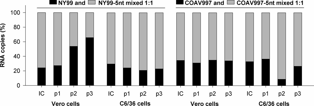
Figure 9. RNA production in 1:1 mixed samples of NY99 with NY99-5nt and COAV997 with COAV997-5nt in Vero and C6/36 cells.
The percentage of RNA copies (y-axis) of wildtype viruses (black) and 5nt-mutants (light grey) in 1:1 mixed samples is shown in staked bars at the time of initial inoculation (IC) and at the end of three passages (p1, p2, p3) for Vero and C6/36 cell cultures (x-axis).
3. Results
3.1 Luminex xTAG® microsphere array
Equal concentrations of NY99, NY99-1nt and NY99-5nt were transcribed and PCR amplified singly with either WNV.2296.F/WNV.2786.R or WNV.2387.F/WNV.2530.R (Table 2). Transcripts amplified with primer set WNV.2296.F/WNV.2786.R resulted in MFI values between 5 and 60 which were below the MFI expected for each microsphere (Table 4). In contrast, the use of WNV.2387.F/WNV.2530.R resulted in higher MFI values of 2342 and 4311 for LUA10/75 and a correct MFI quantification of LUA50/40 with 5528 and 3290, respectively (Table 4). Therefore, only primer set WNV.2387.F/WNV.2530.R was used for further reactions.
The detection of microsphere set LUA10/75 for identification of NY99 and NY99-1nt, respectively, was not quantitative or specific and generated nonspecific signals with LUA50 (Table 4). The detection of LUA50/40 for NY99 and NY99-5nt, was within the desired MFI and produced limited cross-reactivity of 86 and 18 MFI between the two microspheres (Table 4). Therefore only NY99-5nt and microsphere set LUA50/40 were used for further analysis.
Detection of equal mixtures of 10-fold diluted titers of NY99 and NY99-5nt by LUA50/40 yielded linear MFI values for titers between 3.6 – 5.6 log10 p.f.u./mL (Fig. 1). Equal mixtures with titers between 0.6 – 2.6 log10 p.f.u./mL were not detected (Fig. 1). Although 5.6 log10 p.f.u./mL yielded mean MFI values of 5593 and 2612 for NY99 and NY99-5nt, respectively, similar mean MFI values of 5318 and 2625 were detected for 6.6 log10 p.f.u./mL of the same viruses. However, detection of different ratios of the DNA concentrations of NY99 with NY99-5nt was proportional (Fig 2). No differences in MFI values were observed using the different hybridization temperatures described in 2.2 (data not shown).
3.2 Quantitative sequencing
All five bases of the COAV997 (CTCTCC) and COAV997-5nt (TTGAGT) sequences were called individually using chromatogram peak area or height and compared to the corresponding reference sequence. Identification of bases in samples of known titers of singly diluted COAV997 and COAV997-5nt are shown in Figs. 3A–D, 1:1 mixtures of equal titers of the two viruses are presented in Figs. 4A–B, and 1:1 mixtures of unequal titers of the two viruses are summarized in Figs. 5A–B.
Figure 3. Quantitative sequencing of single viral titers of COAV997 and COAV997-5nt through measurement of the total chromatogram peak area (A, C) and height (B, D).
Viral titers (log10 p.f.u./mL) of COAV997 and COAV997-5nt were serially 10-fold diluted. All five bases of COAV997 (CTCTCC; black triangles) and COAV997-5nt (TTGAGT; gray triangles) were individually called and matched to the imported reference sequences. Peak proportions were calculated for each base as the proportion of total chromatogram peak area or height yielding values between 0.0 and 1.0. Viral titers are presented on the x-axis, peak proportion on the y-axis. A. Peak area measurement of 1 – 8 log10 p.f.u./mL of COAV997. B. Peak height measurement of 1 – 8 log10 p.f.u./mL of COAV997. C. Peak area measurement of 1 – 8 log10 p.f.u./mL of COAV997-5nt. D. Peak height measurement of 1 – 8 log10 p.f.u./mL of COAV997-5nt.
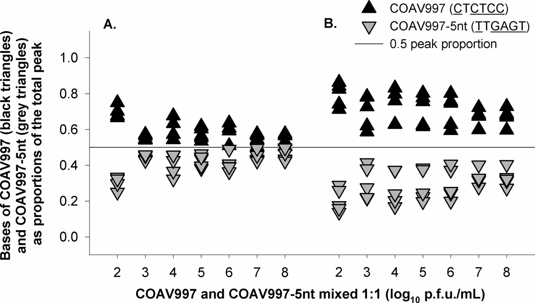
Figure 4. Quantitative sequencing of 1:1 mixtures of equal viral titers of COAV997 and COAV997-5nt through measurement of the total chromatogram peak area (A) and height (B).
A. Peak area measurement of 1:1 mixtures of 2 – 8 log10 p.f.u./mL of COAV997 and COAV997-5nt. B. Peak height measurement of 1:1 mixtures of 2 – 8 log10 p.f.u./mL of COAV997 and COAV997-5nt.
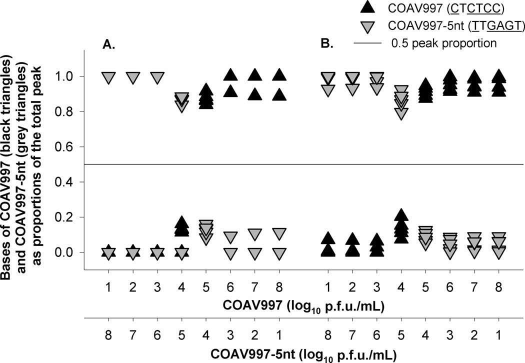
Figure 5. Quantitative sequencing of 1:1 mixtures of unequal viral titers of COAV997 and COAV997-5nt through measurement of the total chromatogram peak area (A) and height (B).
A. Peak area measurement of increasing 10-fold diluted titers of COAV997 mixed with decreasing titers of COAV997-5nt. B. Peak height measurement of increasing 10-fold diluted titers of COAV997 mixed with decreasing titers of COAV997-5nt.
In single viral dilutions, the chromatogram proportion of all five bases was expected to be 1.0 for the virus present and 0.0 for the absent virus. All five bases of COAV997 were identified as 1.0 of the peak area in titers 7, 4 and 3 log10 p.f.u./mL (Fig. 3A). In titers 8, 6 and 1 log10 p.f.u./mL cytosine was miscalled partially as either thymine or guanine, and in dilutions of 5 and 1 log10 p.f.u./mL no cytosine and thymine were detected yielding 0.0 of the total peak area, respectively (Fig. 3A). Using information from chromatogram peak height, all bases of COAV997 were identified correctly after the second call, but none of the base calls resulted in a 1.0 peak proportion, indicating that all bases also were matched falsely with the COAV997-5nt reference sequence to some extent (Fig. 3B). The first thymine was not detected in any of the single viral dilutions of COAV997-5nt using peak area information and in titers 5, 3 and 1 log10 p.f.u./mL the last thymine also was not called (Fig. 3C). Adenine was miscalled partially as thymine in COAV997-5nt titers of 8 and 4 log10 p.f.u./mL and both guanine bases were mismatched additionally as cytosines in the 2 log10 p.f.u./mL dilution using peak area (Fig. 3C). When extracting peak height information three to five bases within each viral dilution of COAV997-5nt showed semi-mismatches with the wildtype sequence, although all bases were identified correctly (Fig. 3D).
In 1:1 mixtures of equal titers of COAV997 and COAV997-5nt a chromatogram proportion of 0.5 is expected for the bases of both viruses. Bases were called correctly in reference to their individual sequence, but all bases of COAV997 yielded proportions above 0.5 and those of COAV997-5nt yielded proportions below 0.5 using both total peak area (Fig. 4A) and height (Fig. 4B).
In 1:1 mixtures of varying titers of COAV997 and COAV997-5nt, the chromatogram proportions are expected to reflect quantitatively the titer of each virus used for that mixture. Results using peak area and height showed that titers of 1, 2 and 3 log10 p.f.u./mL of one virus mixed with titers of 8, 7 and 6 log10 p.f.u./mL supernatant of the other virus, respectively, yielded results (Figs. 5A–B) similar to only one virus being present in the sample (Figs. 3A–D). Respective mixtures of titers 5 log10 p.f.u./mL with 4 log10 p.f.u./mL reflected the presence of both viruses, but bases of the 5 log10 p.f.u./mL virus titer were quantified disproportionally higher than those of the 4 log10 p.f.u./mL diluted virus (Figs. 5A–B).
Calling bases of NY99 and NY99-5nt using the same protocol produced results comparable to those of COAV997 and COAV997-5nt described above, therefore data are not shown.
3.3 Allele-specific quantitative RT-PCR (qRT-PCR)
In vitro transcription of the 5’ plasmids of NY99 and NY99-5nt yielded 2.15×10¹² and 2.33×10¹² RNA copies/µL, respectively. To make qRT-PCR standards both NY99 and NY99-5nt RNA were diluted to 9 log10 RNA copies, mixed 1:1, diluted serially 10-fold in DEPC-treated water (Ambion), aliquoted into RNase-free 200 µL strip tubes and frozen at −80°C for single use.
Side-by-side testing of primers and probes (Table 3) showed that probe WN.2417–2431.FAM worked best with forward primer WNV.TAQ.2393.F and the specific reverse primers designed to contain the wildtype and 5nt-mutant region at their 3′-end. Primer sets having the polymorphic region at their 5′-end and primers designed with artificial nucleotides did not amplify specifically their respective targets (data not shown).
Genomic RNA extracted from NY99 and NY99-5nt singly infected Vero cell culture supernatants and plasmid-derived, in vitro transcribed RNA showed comparable qRT-PCR efficiency and specificity. To improve standardization, the plasmid-derived, in vitro transcribed RNA was used for template quantification in qRT-PCR.
After side-by side comparison of different primer, probe and template concentrations, a final qRT-PCR protocol was developed using the following concentrations and volumes per reaction: 0.5 µL of WN.2417–2431.FAM (100 nM), 0.05 µL WNV.TAQ.2393.F (25 nM), 12.5 µL TaqMan® One-Step RT-PCR Master Mix 2X (ABI, USA), 0.6 µL of TaqMan® One-Step RT-PCR RNase Inhibitor, and 1.3 µL of DEPC-treated water (Ambion). For specific wildtype and 5nt-mutant detection, 0.05 µL of either WNV.TAQ.WT.2464.R (25 nM) or WNV.TAQ.5nt.2467.R-alt were added to two separate master mixes, respectively (Table 3). Samples and standards were run in duplicate with a final volume of 25 µL per reaction and tested concurrently in singleplex format on two ViiA™7 Real-Time PCR System (ABI, USA) machines for separate wildtype and 5nt-mutant WNV quantification according to protocol in Table 3.
To assess analytical sensitivity, amplification efficiency and linearity of the two allele-specific qRT-PCRs, 1:1 mixed, serial 10-fold dilutions of the plasmid-derived, in vitro transcribed NY99 and NY99-5nt RNA ranging from 7.4 to 1.4 log10 RNA copies were tested. As shown in Fig. 6, both assays yielded positive results with cycle threshold (Ct) values <50. Although 1.4 log10 RNA copies were not detected by the assays with a calculated detection limit of < 250 RNA copies, they both showed a linear dynamic detection range of at least 6 log10 (Fig. 6) without cross-reactivity. An amplification efficiency of 90.3% and 90.9% was calculated assuming linearity, with a goodness of fit expressed as the square of the correlation coefficient or R2, of 0.999 and 0.996 for wildtype and 5nt-mutant specific qRT-PCR, respectively (Fig. 6). This demonstrated that both assays were sensitive with similar amplification efficiencies.
Quantitation of RNA copies in 3 – 10 log10 equal 1:1 mixtures of NY99 and NY99-5nt was linear and not significantly different (p=1.00) between the two viruses per dilution (Fig. 7C). Quantitation of RNA copies also was linear and specific for NY99 and NY99-5nt in 2 – 8 log10 dilutions when mixed with constant concentrations of 8 log10 of NY99-5nt (Fig. 7A) and NY99 (Fig. 7B), respectively, as well as in unequal mixtures (Fig. 7D). In addition, specificity was confirmed by testing 10-fold log10 RNA copy dilutions of NY99 and NY99-5nt singly with both assays yielding negative results (data not shown).
3.4. Phenotypic neutrality and genetic stability of NY99-5nt and COAV997-5nt
Growth profiles and RNA copy production of NY99 compared to NY99-5nt and COAV997 compared to COAV997-5nt after three passages in singly and dually infected Vero and C6/36 cells are presented in Figs. 8A–D. A summary of the RNA output of 1:1 mixed competition samples over the course of three passages is presented in Fig. 9.
When tested by a 2-way repeated measures analysis of variance (ANOVA) with virus, cell culture and time post-inoculation as main effects (Hintze et al., 2008), there was no difference (P>0.05) in mean titers among wildtype and 5nt-mutants singly or mixed 1:1 in Vero or C6/36 cell cultures. However, 5nt-mutants consistently produced a significantly greater (P<0.01) mean number of RNA copies in both Vero and C6/36 cell cultures than the remaining virus groups, which did not differ significantly. Based on infectious titer, it was concluded that the five nucleotide substitutions did not influence the replication of NY99-5nt and COAV997-5nt in vitro compared to their parental strains (fitness neutrality). The genetic stability of the five nucleotide substitutions in NY99-5nt was confirmed in passage three supernatants by direct sequencing (data not shown).
4. Discussion
This study introduces a novel approach to detect single nucleotide polymorphisms (SNPs) as genetic markers in the envelope gene region of WNV using different reverse primers for allele-specific RT-PCR quantitation. Other approaches including Luminex xTAG® microsphere array and quantitative sequencing were evaluated for their ability to quantitatively distinguish the engineered mutant viruses from wildtype WNV.
The substitution of five nucleotides (CTCTCC → TTGAGT) at positions 2449 and 2451–2454 was genetically stable and did not significantly influence viral replication as demonstrated by similar growth characteristics of wildtype compared to 5nt-mutant viruses in singly and 1:1 mixed cell cultures (Figs. 8A–D). However, the presence of higher RNA copy numbers in NY99-5nt and COAV997-5nt infected cell cultures compared to their parental viruses (Figs. 8A–D, Fig. 9) could result from deficient viral particle formation during replication. This discrepancy will need to be taken into account when interpreting qRT-PCR results from future fitness competitions by comparing the RNA ratios between initial inocula and final sample.
The use of one nucleotide substitution as a genetic marker in NY99-1nt was not sufficient to guarantee a sensitive, specific and quantitative detection by Luminex xTAG® microsphere array (Tables 1, 4) compared to the five nucleotide changes made in NY99-5nt (Tables 1, 4). Therefore only NY99-5nt, COAV997-5nt and TWN354-5nt were used for assay development and subsequent fitness competitions. The shorter amplicon of 143nt amplified with primer set WNV2387/2530 was more reproducibly quantifiable by Luminex xTAG® microsphere array compared to the 490nt amplicon produced by primer set WNV2387/2530 (Table 4). Because the five nucleotide marked viruses were intended for use in in vivo and in vitro fitness competitions with wildtype WNV isolates, it was considered necessary to develop an accurate RNA quantitation method of templates derived from infectious viral titers. Although the Luminex xTAG® microsphere array protocol provided specific quantitation of different DNA concentration ratios of NY99 and NY99-5nt, this was not observed for viral titers where a <2 log10 p.f.u./mL titer range was accurately measured (Fig. 1). Due to limited quantitation based on viral titer, Luminex xTAG® microsphere technology was not used further, but would be of interest when multiplexed identification of more than two targets per reaction is desired.
Chromatogram peak area measurements using quantitative sequencing provided higher specificity through more accurate quantitation of the peak proportion compared to peak height in single viral titer dilutions, because proportions of 0.0 or 1.0 were observed for most bases (Figs. 3A–D). However, peak area measurements were less sensitive because of false base calls on several occasions and failure to detect thymine in most dilutions of COAV997-5nt. Chromatogram peak height measurements were less specific due to a scattering of most bases between 0.0 – 0.2 or 0.8 – 1.0 peak proportion (Figs. 3B, 3D, 4B, 5B). Therefore, a non-specificity of up to 20% was expected using peak height information irrespective of viral titer. However, peak height measurements were more sensitive because all bases were called correctly, including all thymines.
Both viruses were identified in all 1:1 mixtures of equal titers of COAV997 and COAV997-5nt, but the measurements were not quantitative as they never resulted in an accurate 0.5 proportion of the total chromatogram peak (Figs. 4A–B). COAV997 yielded consistently higher peak proportions in all tested viral titers compared to COAV997-5nt when measuring both area and height (Figs. 4A–B). This bias could be addressed by controlling for the amount of template present at the start of the sequencing reaction by reducing the number of PCR cycles or diluting the products prior to sequencing. The use of an in vitro RNA transcript to generate a standard curve also could be used to regress the amount of viral template added to the PCR reaction for each base onto peak area or height of wildtype and 5nt-mutant sequences to minimize the error of the concentration estimate.
Peak area and height measurements of 1:1 mixtures of unequal titers did not produce quantitative results (Figs. 5A–B). Mixtures with titers of 1 – 3 log10 p.f.u./mL and 6 – 8 log10 p.f.u./mL were detected as 0.0 or 1.0 peak proportion, respectively, indicating the presence of only one virus (Figs. 5A–B). Mixtures between 4 – 5 log10 p.f.u./mL resulted in 0.0 – 0.2 and 0.8 – 1.0 peak proportion which corresponded to the non-specificity observed in height measurements of single virus dilutions (Figs. 5A–B).
According to Little and Hall (2007), who used 35 PCR cycles for template amplification, the DNA concentration at the start of the sequencing reaction is proportional to the measured chromatogram peak. The peak measurements of single virus dilutions of NY99, NY99-5nt, COAV997 and COAV997-5nt showed no difference after 25, 30 and 40 PCR cycles (data not shown). However, the reduction to 25 and 30 cycles for 1:1 mixtures of equal viral titers slightly helped improve the quantification of bases which yielded peaks that were closer to proportions of 0.5 for both sequences compared to 40 cycles (data not shown).
In summary, quantitative sequencing using PolySNP and PHRED did not give quantitative results over the range of viral titers used in the current study. Either both viruses were detected when present in equal amounts or one of the two viruses was identified when present in >1 log10 p.f.u./mL titer difference, indicating that this approach would be more suitable for studies investigating co-existence or exclusion of viruses. However, the use of biased input proportions would be a possible way to offset this limitation if one wished to use this method.
Allele-specific qRT-PCR using an individual set of reverse primers showed specific, quantitative, linear and sensitive amplification of wildtype WNV and 5nt-mutants (Fig. 6, Figs. 7A–D). The design and optimization of a multiplex qRT-PCR can be extensive and would require specific primers and probe sets that were not available in the current study due to the location of the genetic markers. However, specific amplification of two targets was accomplished with as few as five nucleotide differences given that amplification is carried out in a singleplex format and the polymorphic region is designed to be at the 3′-end of the reverse primer. In addition, the presented qRT-PCR has wide applicability and is an economic and high-throughput alternative to currently available quantitative methods.
The Luminex xTAG® microsphere array and quantitative sequencing were semi-quantitative or qualitative in the detection and distinction of wildtype and 5nt-mutants based on log10 titers of WNV. The allele-specific qRT-PCR provided an accurate and quantitative detection and will be used for future analysis of mixed samples from in vivo and in vitro fitness competitions between wildtype WNV and NY99-5nt, COAV997-5nt and TWN354-5nt viruses. Future applications of the genetic marker will include studies tracking the spatio-temporal evolution and potential adaptation of WNV to differing biomes and possible fitness changes associated with WNV outbreaks.
Highlights.
Development of a fitness-neutral genetic marker in the E gene of West Nile virus.
Comparison of methods used to quantitatively detect marked strain and wildtype virus.
Allele-specific qRT-PCR using unique reverse primers quantified both strains.
qRT-PCR was superior to Luminex® and quantitative sequencing due to detection range.
qRT-PCR is now used in fitness competitions in cell culture, birds and mosquitoes.
Acknowledgements
The authors acknowledge Dr. Brent Ewing for providing an academic license of PolySNP and PHRED. Special thanks to Dr. Gregory Ebel and Dr. Gerod Hall for their advice on the implementation of the quantitative sequencing. G. Worwa was supported by the Swiss National Science Foundation (SNSF; PBBEP3_128345) and the Swiss Foundation for Grants in Biology and Medicine (SFGBM; PASMP3_137034 / 1). C. Andrade was supported by the National Institutes of Health training grant (NIH; T32 AI074550). Funding for these studies was provided by the Pacific Southwest Regional Center for Excellence (PSWRCE; U54 AI065359), Biomedical Advanced Research and Development Authority (BARDA), and the National Institutes of Allergy and Infectious Diseases (NIH; RO1-AI55607).
Footnotes
Publisher's Disclaimer: This is a PDF file of an unedited manuscript that has been accepted for publication. As a service to our customers we are providing this early version of the manuscript. The manuscript will undergo copyediting, typesetting, and review of the resulting proof before it is published in its final citable form. Please note that during the production process errors may be discovered which could affect the content, and all legal disclaimers that apply to the journal pertain.
Conflict of interest statement
None of the authors of the publication ”Use of allele-specific primer amplification to quantitatively detect single nucleotide polymorphisms as genetic markers for West Nile virus” (Gabriella Worwa, Christy Andrade, Tara Thiemann, Bborie Park, Payal Maharaj, Michael Anishchenko, Aaron Brault, William Reisen) has any financial or personal relationships with other people or organizations that could inappropriately influence this work.
References
- Andrade CC, Maharaj PD, Reisen WK, Brault AC. North American West Nile virus genotype isolates demonstrate differential replicative capacities in response to temperature. J. Gen. Virol. 2011;92:2523–2533. doi: 10.1099/vir.0.032318-0. [DOI] [PMC free article] [PubMed] [Google Scholar]
- Beasley DW, Davis CT, Estrada-Franco J, Navarro-Lopez R, Campomanes-Cortes A, Tesh RB, Weaver SC, Barrett AD. Genome sequence and attenuating mutations in West Nile virus isolate from Mexico. Emerg. Infect. Dis. 2004;12:2221–2224. doi: 10.3201/eid1012.040647. [DOI] [PMC free article] [PubMed] [Google Scholar]
- Brault AC, Langevin SA, Bowen RA, Panella NA, Biggerstaff BJ, Miller BR, Komar N. Differential virulence of West Nile strains for American crows. Emerg. Infect. Dis. 2004;10:2161–2168. doi: 10.3201/eid1012.040486. [DOI] [PMC free article] [PubMed] [Google Scholar]
- Coffey LL, Vasilakis N, Brault AC, Powers AM, Tripet F, Weaver SC. Arbovirus evolution in vivo is constrained by host alternation. Proc. Natl. Acad. Sci. USA. 2008;105:6970–6975. doi: 10.1073/pnas.0712130105. [DOI] [PMC free article] [PubMed] [Google Scholar]
- Deardorff ER, Fitzpatrick KA, Jerzak GV, Shi PY, Kramer LD, Ebel GD. West Nile virus experimental evolution in vivo and the trade-off hypothesis. PLoS Pathog. 2011;7:e1002335. doi: 10.1371/journal.ppat.1002335. [DOI] [PMC free article] [PubMed] [Google Scholar]
- Domingo E, Escarmis C, Sevilla N, Moya A, Elena SF, Quer J, Novella IS, Holland JJ. Basic concepts in RNA virus evolution. FASEB J. 1996;10:859–864. doi: 10.1096/fasebj.10.8.8666162. [DOI] [PubMed] [Google Scholar]
- Fitzpatrick KA, Deardorff ER, Pesko K, Brackney DE, Zhang B, Bedrick E, Shi PY, Ebel GD. Population variation of West Nile virus confers a host-specific fitness benefit in mosquitoes. Virology. 2010;404:89–95. doi: 10.1016/j.virol.2010.04.029. [DOI] [PMC free article] [PubMed] [Google Scholar]
- Groat-Carmona AM, Orozco S, Friebe P, Payne A, Kramer L, Harris E. novel coding-region RNA element modulates infectious dengue virus particle production both mammalian and mosquito cells and regulates viral replication in Aedes aegypti mosquitoes. Virology. 2012;432:511–526. doi: 10.1016/j.virol.2012.06.028. [DOI] [PMC free article] [PubMed] [Google Scholar]
- Langevin SA, Bowen RA, Ramey WN, Sanders TA, Maharaj PD, Fang Y, Cornelius J, Barker CM, Reisen WK, Beasley DWC, Barrett ADT, Kinney RM, Huang CYH, Brault AC. Envelope and pre-membrane protein structural amino acid mutations mediate diminished avian growth and virulence of a Mexican West Nile virus isolate. J. Gen. Virol. 2011;92:2810–2820. doi: 10.1099/vir.0.035535-0. [DOI] [PMC free article] [PubMed] [Google Scholar]
- Little DP, Hall GS. polySNP: an analysis tool for quantitative sequencing. Program distributed by the authors. 2006 http://www.nybg.org/files/scientists/dlittle/polySNP.html. [Google Scholar]
- Hall GS, Little DP. Relative quantitation of virus population size in mixed genotype infections using sequencing chromatograms. J. Virol. Methods. 2007;146:22–28. doi: 10.1016/j.jviromet.2007.05.029. [DOI] [PMC free article] [PubMed] [Google Scholar]
- Hintze J. NCSS Statistical Software [computer program] Kaysville, UT: NCSS; 2008. [Google Scholar]
- Jerzak GV, Brown I, Shi PY, Kramer LD, Ebel GD. Genetic diversity and purifying selection in West Nile virus populations are maintained during host switching. Virology. 2008;374:256–260. doi: 10.1016/j.virol.2008.02.032. [DOI] [PMC free article] [PubMed] [Google Scholar]
- Kinney RM, Huang CY, Whiteman MC, Bowen RA, Langevin SA, Miller BR, Brault AC. Avian virulence and thermostable replication of the North American strain of West Nile virus. J. Gen. Virol. 2006;87:3611–3622. doi: 10.1099/vir.0.82299-0. [DOI] [PubMed] [Google Scholar]
- Rice CM, Lenches EM, Eddy SR, Shin SJ, Sheets RL, Strauss JH. Nucleotide sequence of yellow fever virus: implications for flavivirus gene expression and evolution. Science. 1985;229:726–733. doi: 10.1126/science.4023707. [DOI] [PubMed] [Google Scholar]
- Smith J, Sammons D, Toennis C, Butler MA, Blachere F, Beezhold D. Semi-quantitative analysis of influenza samples using the Luminex xTAG (®) respiratory viral panel kit. Toxicol. Mech. Methods. 2012;22:211–217. doi: 10.3109/15376516.2011.610387. [DOI] [PMC free article] [PubMed] [Google Scholar]
- Thiemann TC, Brault AC, Ernest HB, Reisen WK. Development of a high-throuput microsphere-based molecular assay to identify 15 common bloodmeal hosts of Culex mosquitoes. Mol. Ecol. Resour. 2012;12:238–246. doi: 10.1111/j.1755-0998.2011.03093.x. [DOI] [PMC free article] [PubMed] [Google Scholar]
- Weaver SC, Brault AC, Kang W, Holland JJ. Genetic and fitness changes accompanying adaptation of an arbovirus to vertebrate and invertebrate cells. J. Virol. 1999;73:4316–4326. doi: 10.1128/jvi.73.5.4316-4326.1999. [DOI] [PMC free article] [PubMed] [Google Scholar]


