Figure 3.

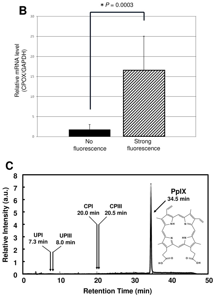
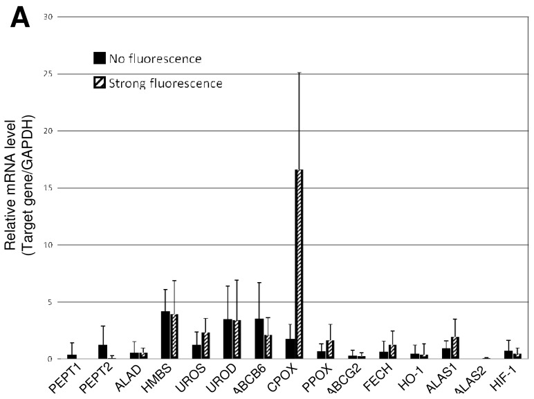
Enhanced expression of CPOX gene in glioblastoma with strong red fluorescence. (A) Comparison of the mRNA levels of PEPT1, PEPT2, ALAD, HMBS, UROS, UROD, ABCB6, CPOX, PPOX, ABCG2, FECH, HO-1, ALAS1, ALAS2, and HIF-1 genes between the “no fluorescence” and “strong fluorescence” groups. Gene names: PEPT1, oligopeptide transporter 1; PEPT2, oligopeptide transporter 2; ALAD, delta-aminolevulinate dehydratase; HMBS, hydroxymethylbilane synthase; UROS, uroporphyrinogen III synthase; UROD, uroporphyrinogen decarboxylase; ABCB6, ABC transporter B6; CPOX, coproporphyrinogen oxidase; PPOX, protoporphyrinogen oxidase; ABCG2, ABC transporter G2 (BCRP); FECH, ferrochelatase; HO-1, heme oxygenase 1; ALAS1, 5-aminolevulinate synthase 1; ALAS2, 5-aminolevulinate synthase 2; HIF-1, hypoxia inducible factor-1 alpha subunit. Total RNA was extracted from brain tumor areas, and the first strand cDNA was prepared from the extracted total RNA in a reverse transcriptase (RT) reaction. Quantitative PCR was performed as described previously [33]. The mRNA levels of these genes are normalized to the mRNA level of glyceraldehyde-3-phosphate dehydrogenase (GAPDH). Data are expressed as means ± SD (N = 10) [33]; (B) Statistical analysis of CPOX mRNA levels between the “no fluorescence” and the “strong fluorescence” groups. Data are expressed as means ± SD (N = 10) [33]. (C) HPLC elution profile of porphyrins in brain tumor samples with strong red fluorescence. HPLC analysis was performed as described previously [34]. As the standards, uroporphyrin 1 (UPI), Uroporphyrin III (UPIII), Coproprophyrin I (CPI), Coproprophyrin III (CPIII), and protoporphyrin IX (PpIX) were eluted at retention times of 7.3, 8.0, 20.0, 205, and 34.5 min, respectively.

