

Abstract
We demonstrate a compact, inexpensive, and reliable fiber–coupled light source with broad bandwidth and sufficient power at 1300 nm for high resolution optical coherence tomography (OCT) imaging in real-time applications. By combining four superluminescent diodes (SLEDs) with different central wavelengths, the light source has a bandwidth of 145 nm centered at 1325 nm with over 10 mW of power. OCT images of an excised stage 30 embryonic chick heart acquired with our combined SLED light source (<5 μm axial resolution in tissue) are compared with images obtained with a single SLED source (~10 μm axial resolution in tissue). The high resolution OCT system with the combined SLED light source provides better image quality (smaller speckle noise) and a greater ability to observe fine structures in the embryonic heart.
Introduction
Optical coherence tomography (OCT) can perform micron scale, noninvasive imaging of biological tissue morphology and has been successfully applied in the clinic. Since OCT is based on low-coherence interferometry, the axial image resolution is determined by the bandwidth and center wavelength of the light source. Superluminescent diode (SLED) light sources are preferred for standard OCT systems, because they are inexpensive, compact, reliable, and have low noise. When imaging opaque tissue with OCT, SLEDs centered at 1300 nm are preferred because they penetrate deeper into the tissue than SLEDs centered at shorter wavelengths. Unfortunately, the bandwidth of single SLED centered around 1300 nm is limited to approximately 40–75nm, resulting in axial OCT image resolution of 12–15 μm in air (9–12 μm in tissue).
High resolution OCT with an axial resolution less than 5 μm in tissue at 1300 nm has been demonstrated using different method [1–7]. OCT images with higher axial resolution clearly show improved image quality due to smaller speckle size and the ability to display finer structures with higher contrast, which can possibly improve diagnosis in the clinic [5]. Normally broadband light sources for OCT imaging at 1300 nm are generated by pumping a fiber with either a femotosecond laser or a high power continuous wave (CW) laser [2, 4, 8]. The broadband spectrum manifests from nonlinear effects that occur in the fiber also known as supercontinuum generation (SCG). However, SCG has several limitations for clinical applications. First, the fesmotosecond laser is bulky, expensive and requires careful maintenance for smooth running. Second, the femotosecond pulses output from the bulky lasers have to be coupled into the fiber in free space, which makes the system inconvenient and stationary. Third, some SCG light sources have strong excess noise [4, 9] which limits the sensitivity of the system. Fiber femotosencond lasers will probably provide a good solution in the future, if an appropriate fiber is used for pumping [6, 7, 10]. However, the performance of current fiber femotosecond lasers is limited for this application.
Schmitt et al proposed improving OCT image quality by combining light emitting diodes (LED) with offset center wavelenghths around 1300 nm, and demonstrated OCT imaging using two LEDs [11]. This demonstration resulted in OCT imaging with approximately 10 μm axial resolution in air but optical power output was low. We noticed that a broadband light source combining four SLEDs has been recently developed commercially (SuperlumDiodes Ltd), however to our knowledge, a demonstration of this light source for OCT imaging has not been reported. In this paper, we demonstrate the use of an OCT light source centered at ~1300 nm for high resolution OCT imaging that consists of four SLEDs with offset center wavelenghths optimally combined using fiber couplers and current tuning. With ~145 nm of bandwidth and over 10 mW of output power, the combined SLED is fiber based, reliable, compact and can be easily integrated into an OCT system. OCT images of a stage 30 embryonic chick heart using the combined light source and a single SLED are compared using identical instrumentation and sample location.
Method and results
The new combined broadband SLED source was generated by combing four SLEDs which were independently driven. To generate a broadband spectrum with the smallest ripples possible, each of the SLEDs was chosen according to their central wavelength and output power. The layout of the combination is shown in Fig. 1. S1325 (Inphenix, USA) has a central wavelength of 1325 nm, a full width half maximum (FWHM) bandwidth of about 70 nm, and an output power of 15 mW. S1325 has similar specifications as the SLEDs popularly used singly in OCT systems. SLED S1355 (Denselight, Singapore) has a central wavelength of 1350 nm, a FWHM bandwidth of 50 nm, and an output power of 15 mW. The third and fourth SLEDs (S1265 and S1380) were custom built by WT&T (Canada) and have center wavelength at 1265 nm and 1380 nm. The center wavelength of S1265 and S1380 can be tuned over a 10 nm range. The spectrum of each SLED is shown in Fig. 2(a).
Fig. 1.
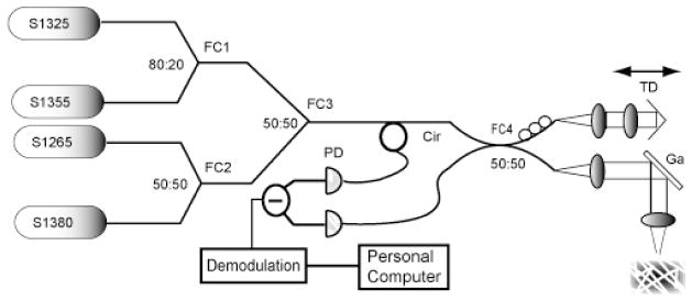
Schematic of the OCT system with a broadband combined SLED light source for high resolution OCT imaging. PD: photodetector; Cir: circulator; TD: optical delay line; FC: fiber coupler; Ga: galvanometer scanner;
Fig. 2.
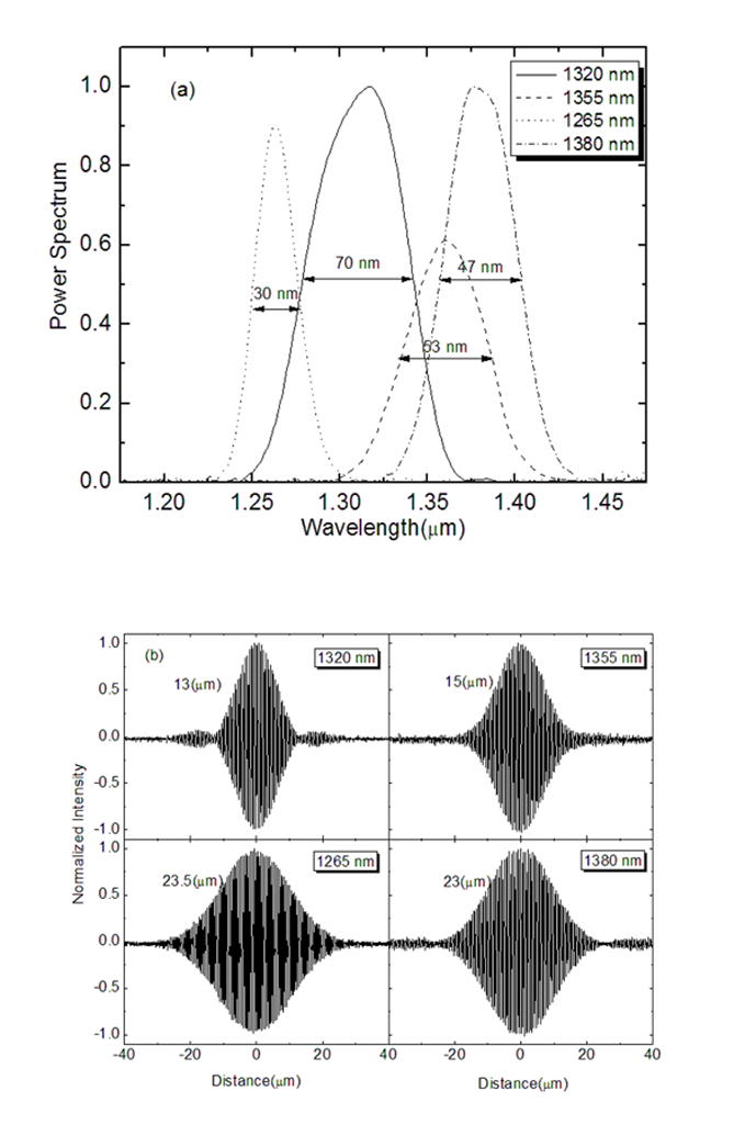
Individual SLEDs. (a) The spectrum of the SLEDs used for combination. The spectrum has been scaled according to output power. (b) Measured point spread functions and coherence length of each SLED.
The four SLEDs were combined using three broadband fiber couplers (FCs). The S1325 and S1355 were combined employing a FC (FC1) with a splitting ratio of 80:20, which is chosen for smooth combined spectrum shape The S1355 was used for filling the gap between S1265 and S1380. S1270 and S1380 were combined using a 50:50 FC (FC2). The output of FC1 and FC2 were then combined by using another 50:50 FC (FC3) to generate the final output spectrum. The combined spectrum is plotted in Fig. 3(a). The final output spectrum had a FWHM bandwidth of 145 nm, an output power of over 10 mW, and ripples less than 1 dB.
Fig. 3.

Combined SLEDs (a) Combined spectrum of SLEDs shown in Fig 1(a) (b) Measured point spread function of combined spectrum. (c) Measured demodulated point spread function in logarithmic scale.
A time domain OCT system (see Fig. 1) was built for coherence function measurement and for demonstrating the performance of the light source for OCT imaging. The combined broadband source was coupled into a 50:50 fiber coupler (FC4), which split the light into a reference arm and a sample arm. In the sample arm, the light was collimated and then focused onto the sample. In the reference arm, a galvanometer was used to generate a time delay. The light returning from the sample and reference arms interfered at the balanced detectors. The OCT signal was amplified, band-pass filtered and logarithmically demodulated before being sampled and displayed. The delay line scanned 3 mm at 15 Hz repetition rate. The center frequency of the filter was set to 300 KHz with 60 KHz of bandwidth.
System performance was characterized by measuring the coherence function by using a mirror as the sample. The coherence function of each SLED was measured and plotted in Fig. 2(b). The shortest coherence length (13 μm) was measured with the S1325, which represents the typical coherence length of SLEDs commonly used for OCT. Fig. 3(a) shows the coherence function of the combined spectrum. A coherence length of 6.5 μm was measured in air, corresponding to an axial point spread function of ~5 μm in tissue. Compared with typical single SLED light sources, the combined SLEDs improves the axial resolution by half and is comparable to the demonstrated resolution of broadband light sources generated by SCG [3–5, 7]. However, the combined SLED light source is more compact, inexpensive, reliable and portable. Although the spectral ripples are small, small side-lobes can be observed on the coherence function. These arise mainly due to the rectangular shape of the combined spectrum. Fig. 3(c) presents the logarithmically demodulated signal. The plot has been scaled to reflect 56 dB attenuation in the sample arm. The power from the reference arm was optimized to maximize the sensitivity, which was −102 dB for 3 mW on the sample. The lateral resolution was approximately 12 μm which is determined by the lens group used in the sample arm scanner.
Figure 4 presents OCT images of an excised embryonic chick heart at stage 30 of development. The arrows in Fig. 4(a) point to trabeculae in the heart. The image in Fig. 4(a) was acquired using the combined broadband light source with an axial resolution of 4.8 μm in the tissue. As a comparison, the OCT image in Fig. 4(b) was obtained using a single SLED (S1325) with an axial resolution of 9.6 μm in tissue. Acquired images were ~ 2 mm × 4 mm (axial × transverse) with 2440 × 1000 pixels. For the single SLED image, the power on the sample was 1.2 mW. In order to keep the same sensitivity for the two acquisitions, the power on the sample with combined SLEDs was attenuated to the same level. Except for the power, the sample and optics instrumentation were kept identical for both acquisitions. Figures 4(9Xa) and 4(9Xb) are 9 times zoomed images of the boxed areas in 4(a) and 4(b) respectively.
Fig. 4.
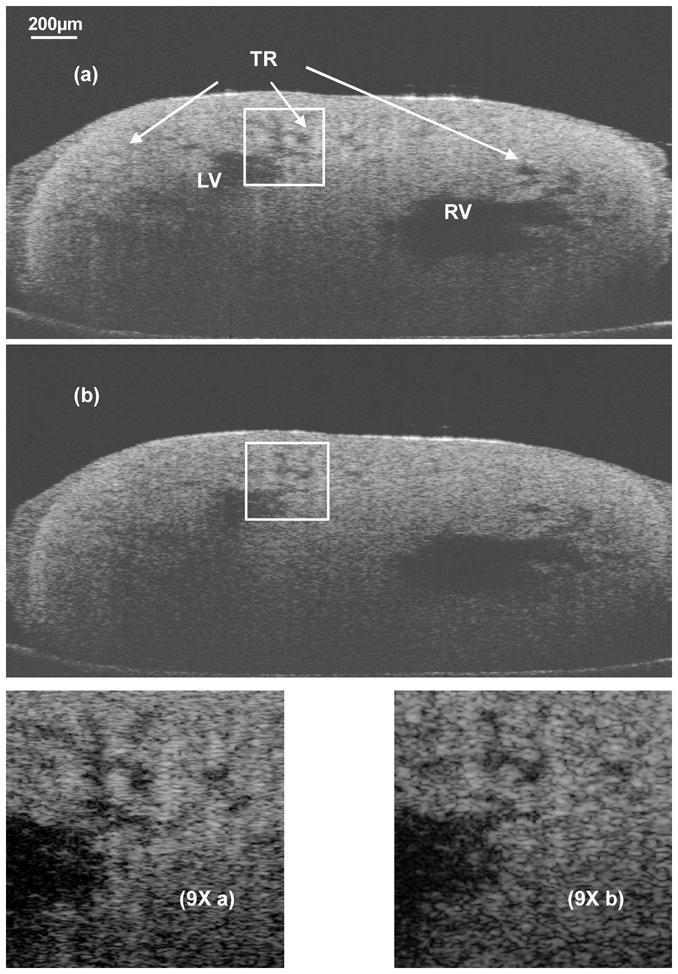
OCT images of an excised embryonic chick heart at stage 30 of development. (a) OCT image acquired with the combined SLED light source (axial resolution of ~5 μm in the tissue). (2440(h)×1000(v) pixels, 2mm(h)×4 mm(v)) (b) OCT image acquired with a single SLED light source (axial resolution of 9.5 μm in the tissue). (2440(h) ×1000(v) pixels, 2mm (h) ×4mm (v) (9Xa) 9 times zoomed image of the boxed area in (a). (9Xb) 9 times zoomed image of the identical boxed area in (b). LV: Left Ventricle; RV: Right Ventricle; Tr: Trabeculae.
The high resolution image in Fig. 4(a) and Fig. (9Xa) demonstrates improved image quality when compared to the lower resolution image in 4(b) and Fig. (9Xb). Specifically, the speckle size in (a) is just about half of that in (b) due to the shorter coherence length, and the fine structure of the trabeculations are better defined with higher contrast and sharp edge. These differences are important for quantitative image analysis, reducing speckle noise and improving segmentation of structures of interest.
Discussion and conclusion
The combined broadband light source demonstrated here was realized with four SLEDs from different vendors. They are all compact, reliable and portable. The main drawback of this light source is the observable side-lobes shown in the coherence function. The side-lobes can be mostly attributed to the rectangular spectral shape achieved when the multiple SLEDs were combined. We optimized the ripple to less than 1dB by carefully selecting the component SLEDs, and adjusting the power and center wavelengths. As demonstrated in Fig. 4, the side-lobes are small enough that they are not apparent when imaging opaque tissue and image contrast was not compromised. If necessary, the side-lobes could be further reduced by digital spectral shaping [12].
Another limitation of this light source is inefficiency. Of necessity, each fiber-optic coupler used to combine the SLEDs imposes on average a 50% loss. The approximately 10 mW output of this source represents approximately 25% of the total optical power output of the four individual SLEDs combined. While 10 mW is lower than the optical output available from some single SLEDs (e.g. S1325), it is sufficient for real time OCT imaging, and it is significantly higher than previously demonstrated broadband LED or SLED combinations.
The bandwidth of the light source could be further extended by integrating more or broader SLEDs at the two sides of the spectrum. However, extending the spectrum longer than 1400 nm is not preferred due to a strong water absorption peak at 1440 nm that will strongly attenuate that spectral range in tissue. The commercial available combined SLEDs (SuperlumDiodes Ltd.) has a shorter coherence length around 4.5 μm in air corresponding to a bandwidth of 230 nm. However, because the center wavelength is approximately 1375nm, a significant component of the spectrum is beyond 1400nm and can be expected to be seriously attenuated in biological tissue. In addition, the optical power is limited to 5mW. Extending the spectrum shorter than 1250 nm would be advantageous, but will depend on the availability of SLEDs at this wavelength range.
In conclusion, we have demonstrated a broadband light source for high resolution OCT by combining four SLEDs. The smooth spectrum has a bandwidth of ~145 nm and over 10 mW of output power. The measured coherence length is approximate 6.5 μm in air (4.8 μm in tissue) which enables imaging at approximately half the axial resolution of OCT imaging with a single SLED. The resolution performance of this light source is similar to previously demonstrated broadband light sources which pump fibers with femtosecond lasers, such as the Cr:fosterite laser, but is less expensive, more compact and more portable.,. For direct comparison, two images with different axial resolution were acquired under identical conditions. High resolution imaging with the new light source reveal improved visibility of fine structures. When implemented in real-time OCT, this light source could be well suited for biomedical applications of OCT imaging, especially for clinic applications.
Acknowledgments
This work is supported by NIH (R24CA110943-01), PTAP, and conducted in a facility constructed with support from Research Facilities Improvement Program Grant Number C06 RR12463-01 from the National Center for Research Resources, National Institutes of Health. The authors acknowledge the technical support of Dr. Ftimofeev from WT&T.
References
- 1.Bouma BE, Tearney GJ, Bilinsky IP, Golubovic B, Fujimoto JG. Self-phase-modulated Kerr-lens mode-locked Cr:forsterite laser source for optical coherence tomography. Opt Lett. 1996;21 doi: 10.1364/ol.21.001839. [DOI] [PubMed] [Google Scholar]
- 2.Hartl I, Li XD, Chudoba C, Ghanta RK, Ko TH, Fujimoto JG, Ranka JK, Windeler RS. Ultrahigh-resolution optical coherence tomography using continuum generation in an air silica microstructure optical fiber. Opt Lett. 2001;26:608–610. doi: 10.1364/ol.26.000608. [DOI] [PubMed] [Google Scholar]
- 3.Herz P, Chen Y, Aguirre A, Fujimoto J, Mashimo H, Schmitt J, Koski A, Goodnow J, Petersen C. Ultrahigh resolution optical biopsy with endoscopic optical coherence tomography. Opt Express. 2004;12:3532–3542. doi: 10.1364/opex.12.003532. [DOI] [PubMed] [Google Scholar]
- 4.Hsiung P-L, Chen Y, Ko T, Fujimoto J, Matos Cd, Popov S, Taylor J, Gapontsev V. Optical coherence tomography using a continuous-wave, high-power, Raman continuum light source. Opt Express. 2004;12:5287–5295. doi: 10.1364/opex.12.005287. [DOI] [PubMed] [Google Scholar]
- 5.Hsiung PL, Pantanowitz L, Aguirre AD, Chen Y, Phatak D, Ko TH, Bourquin S, Schnitt SJ, Raza S, Connolly JL, Mashimo H, Fujimoto JG. Ultrahigh-resolution and 3-dimensional optical coherence tomography ex vivo imaging of the large and small intestines. Gastrointestinal Endoscopy. 2005;62(4):561–574. doi: 10.1016/j.gie.2005.05.006. [DOI] [PubMed] [Google Scholar]
- 6.Wang Y, Tomov I, Nelson JS, Chen Z, Lim H, Wise F. Low-noise broadband light generation from optical fibers for use in high-resolution optical coherence tomography. J Opt Soc Am A. 2005;22:1492–1499. doi: 10.1364/josaa.22.001492. [DOI] [PubMed] [Google Scholar]
- 7.Wang H, Rollins AM. Optimization of dual band continuum light source for ultrahigh resolution optical coherence tomography. Appl Opt. 2007;46:1787–1794. doi: 10.1364/ao.46.001787. [DOI] [PubMed] [Google Scholar]
- 8.Drexler W. Ultrahigh-resolution optical coherence tomography. J Biomed Opt. 2004;9:47. doi: 10.1117/1.1629679. [DOI] [PubMed] [Google Scholar]
- 9.Corwin KL, Newbury NR, Dudley JM, Coen S, Diddams SA, Weber K, Windeler RS. Fundamental Noise Limitations to Supercontinuum Generation in Microstructure Fiber. Phys Rev lett. 2003;90:113904–113901. doi: 10.1103/PhysRevLett.90.113904. [DOI] [PubMed] [Google Scholar]
- 10.Bizheva K, Povazay B, Hermann B, Sattmann H, Drexler W, Mei M, Holzwarth R, Hoelzenbein T, Wacheck V, Pehamberger H. Compact, broad-bandwidth fiber laser for sub-2- mm axial resolution optical coherence tomography in the 1300-nm wavelength region. Opt Lett. 2003;28:707–709. doi: 10.1364/ol.28.000707. [DOI] [PubMed] [Google Scholar]
- 11.Schmitt JM, Lee SL, Yung KM. An optical coherence microscope with enhanced resolving power in thick tissue. Optics Communications. 1997;142(4–6):203–207. [Google Scholar]
- 12.Tripathi R, Nassif N, Nelson JS, Park BH, Boer JFd. Spectral shaping for non-Gaussian source spectra in optical coherence tomography. Opt Lett. 2002;27:406–408. doi: 10.1364/ol.27.000406. [DOI] [PubMed] [Google Scholar]