Abstract
Background:
A prolonged life of fissure sealant has always been the target for preventing caries in vulnerable newly erupted teeth. The use of preparatory techniques including bur introduction to the fissures is considered among such improving steps.
Materials and Methods:
Ninety freshly extracted healthy maxillary premolar teeth were randomly selected for this investigation. Teeth were then divided into three fissure sealant preparatory groups of A: Fissurotomy bur + acid etch; B: Pumice prophylaxis + acid etch and C: Acid etch alone. Sealant was applied to the occlusal fissures of all specimens using a plastic instrument. This was to avoid any air trap under the sealant. Sample teeth were first thermocycled (1000 cycles, 20 s dwell time) and then coated with two layers of nail varnish leaving 2 mm around the sealant. This was then followed by immersion in basic fuchsin 3%. Processed teeth were sectioned longitudinally and examined under a stereomicroscope for microleakage assessment using a score of 0-3. Collected data was then subjected to Kruskall-Wallis Analysis of Variance and Mann-Whitney U-test. P > 0.05 was considered as significant.
Results:
Teeth in fissurotomy bur and pumice prophylaxis groups had significantly reduced level of microleakage than those in acid etch alone (P = 0.005 and P = 0.003, respectively).
Conclusion:
Use of fissurotomy bur and pumice prophylaxis accompanied with acid etching appears to have a more successful reduction of microleakage than acid etch alone.
Keywords: Fissure sealant, fissurotomy, microleakage, permanent teeth, preparation, prevention
INTRODUCTION
Despite global improvements in caries status, dental caries remains to be the most common chronic childhood disease world-wide.[1] Teeth with deep pits and fissures are shown to be more vulnerable to develop caries. In the other hand, fissure sealant application is proved to be able to block pits and fissures in order to prevent occlusal caries development.[2,3,4,5] Any sign of microleakage in sealants is considered as the weak point eventually leading to failure as the inability to isolate pit and fissures would enhance the retention of bacteria, nutrients, and their acidic metabolic products.[6,7,8,9]
The necessity of tooth preparation prior to the sealant application and its effect on microleakage has different perceptions among the researchers. Varying preparation methods have been tested showing different results prior to the sealant application. Hatibovic-Kofman et al. believes that lower microleakage rate is associated with teeth following bur preparation[10] while some studies illustrate opposing results.[7,11,12,13,14] Pumice prophylaxis has long been used prior to the sealant application[15,16] with its effect on microleakage being mostly reported as beneficial.[12,17,18]
In the other hand, the use of acid etching alone or associated with one of the other preparatory methods has shown little to no difference in microleakage level of the teeth.[7,10,19,20,21,22]
The aim of this investigation was to compare the degree of microleakage at the enamel-sealant interface when prepared by a fissurotomy bur or pumice prophylaxis before the acid etchant being applied.
MATERIALS AND METHODS
Ninety freshly extracted healthy human maxillary premolar teeth were allocated for this investigation. Teeth were randomly divided into three separate groups (30 per group).
Occlusal fissures of the specimens in group A were prepared with a Micro STF fissurotomy bur (SS White Burs Inc., Lakewood, NJ, USA) on a high-speed hand piece. For each specimen, the bur was moved along all the occlusal fissures with a gentle force applied. The occlusal surface of the specimens in the group B received a thorough prophylaxis for 20 s with water-based slurry of pumice, using a prophylaxis brush fitted on a slow-speed hand piece. All teeth were then etched for 20 s using 35% phosphoric acid gel (3M ESPE, USA), washed and dried for 20 s each. Specimens in group C received no preparation before the etching procedure. Each sample received an adhesive (Adper Single Bond Plus, 3M ESPE, USA) prior to Fissure Sealant placement. A light-cured sealant (Clinpro Sealant, 3M ESPE, USA) was then applied to all the prepared fissure through a syringe type tip.
Polymerization was performed using an LED Demetron II (Kerr, USA) light-curing unit with an output of 800 mW/cm2 for 20 s.
Specimens were then subjected to a thermo cycle machine with 1000 cycles between 5°C and 55°C with 20 s dwell time.[23] Apices of each sample tooth was then covered with a layer of sticky wax followed by the application of two layers of nail varnish to the surface of each tooth leaving 2 mm around the sealant borders. Specimens were then immersed in a 3% basic fuchsin dye solution for 72 h.[17] Sample teeth were subsequently washed under the tap water for 1 min to remove excess dye from the surfaces. Each was then embedded in acrylic blocks and sectioned in the bucco-lingual plane through the sealant for two identical sections using a water-cooled diamond disk on an Isomet saw (Buehler Ltd., USA).
Three prepared sections with four surfaces from each sample were examined under ×10 magnifications using the XTD series stereomicroscope (Blue Light Inc., USA). All the sections were blindly subjected to an assessment process using a four point scoring system suggested by Overbo and Raadal.[24] Dye penetration scale employed was as: (0) no dye penetration; (1) dye penetration restricted to the outer half of the enamel-sealant interface; (2) dye penetration in the inner half of the enamel-sealant interface; (3) dye penetration into the underlying fissure. Data were analyzed using Kruskall-Wallis Analysis of Variance and Mann-Whitney U-tests for comparing microleakage levels among and between groups. P < 0.05 was considered as significant.
RESULTS
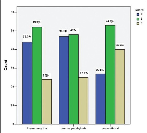
Mean ± standard deviation of microleakage score of the cases in each group was separately calculated [Table 1]. The highest frequency of the microleakage in all groups was for score 0 and 1, with fissurotomy 80%, pumice prophylaxis 79.2% and control group 66.7%. Surprisingly, score 3 was not observed in any of the groups [Figure 1].
Table 1.
Mean±SD of microleakage score of different preparatory groups

Figure 1.

Dye penetration scores in three different tooth preparation methods
Kruskall-Wallis analysis of variance showed significant differences among microleakage levels of different preparation methods (P = 0.003). Mann-Whitney U-test indicated that teeth in fissurotomy bur and pumice prophylaxis groups had significantly reduced level of microleakage than those in acid etch alone (P = 0.005 and P = 0.003, respectively); However, it showed no significant differences of micloleakage level between fissurotomy bur and pumice prophylaxis groups (P = 0.83).
DISCUSSION
Sealing pits and fissures have been proved to have an effective role on preventing teeth from fissure caries in childhood.[2,3,4,5] There are several preparatory methods introduced prior to the placement of the fissure sealant with varying degrees of efficacy on the adhesiveness of material to the prepared surfaces. However, the necessity of the tooth surface preparation prior to the sealant application is still under scrutiny.[12,13,15,17]
As a possible choice, the currently available and specifically designed fissurotomy bur could conservatively widen the fissures providing a larger surface for the adhesion of sealant material. A similar concept has also been reported using fine diamond bur in order to reduce microleakage of the sealants. Fissurotomy bur has been suggested to have a significantly high influence on sealant retention when used along with acid etch.[12] As indicated earlier it seems that bur preparation can provide greater surface area, inducing better sealant adhesion as well as an increased chance for the flow of sealant into the depth of fissures improving its wear resistance.[7,10,25,26,27]
Hatibovic-Kofman et al. reported that quarter round bur preparation with acid etch has a better effect than acid etching alone in reducing microleakage;[10] however, several other studies are also raising doubts on the effectiveness of bur preparation.[7,11,12,13,14] The diversity of the materials and methods used to assess the microleakage may explain the contradictions between the results along with the use of dye agents such as methylene blue or basic fuchsin. Results of the current investigation indicated that pumice prophylaxis along with acid etch as superior to acid etching alone in reducing microleakage around the sealants, supporting earlier reports by Ansari et al.[17]
Despite the introduction of pumice prophylaxis as one of the essential steps of fissure sealing by Cueto and Bunocore,[16,17,28] this application has later been proved ineffective in promoting the sealant effectiveness by lowering the microleakage.[12,18,29] The retention of sealant is believed to be enhanced when adhesive bonding agents are applied prior to the sealant. However, there are also reports suggesting no improvement for microleakage reduction when these adhesive agents are applied.[27,29] The invasive use of bur to open the fissures has not been widely accepted due to its damaging effect on the teeth requiring fissure sealant.[30] Result of this investigation shows some improvement while minimally removing tissues. Interestingly, there was no case with score three dye penetration (to the full depth of the fissure) indicating reasonable flow of the sealant into the depth of fissures.
CONCLUSION
Findings of this study indicate that the use of fissurotomy bur along with etchant gel enhances adhesiveness of sealant to the tooth.
The use of prophylaxis paste would be advantageous in microleakage reduction to the use of etching alone.
ACKNOWLEDGMENTS
Authors would like to thank the research center of Rafsanjan University of Medical Sciences and Mashhad Dental Research Center to fund this research.
Footnotes
Source of Support: Authors would like to thank the research center of Rafsanjan University of Medical Sciences and Mashhad Dental Research Center to fund this research.
Conflict of Interest: None declared.
REFERENCES
- 1.Hicks J, Flaitz CM. Pit and fissure sealants and conservative adhesive restoration: Scientific and clinical rationale. In: Pinkham JR, Casamassimo PS, Mctigue DJ, Fields HW, Nowak AJ, editors. Pediatric Dentistry: Infancy through Adolescence. 4th ed. St. Louis: Elsevier Saunders; 2005. pp. 522–76. [Google Scholar]
- 2.Koh SH, Chan JT, You C. Effects of topical fluoride treatment on tensile bond strength of pit and fissure sealants. Gen Dent. 1998;46:278–80. [PubMed] [Google Scholar]
- 3.Ripa LW. Sealants revisted: An update of the effectiveness of pit-and-fissure sealants. Caries Res. 1993;27(Suppl 1):77–82. doi: 10.1159/000261608. [DOI] [PubMed] [Google Scholar]
- 4.Simonsen RJ. Pit and fissure sealant: Review of the literature. Pediatr Dent. 2002;24:393–414. [PubMed] [Google Scholar]
- 5.Mejàre I, Lingström P, Petersson LG, Holm AK, Twetman S, Källestål C, et al. Caries-preventive effect of fissure sealants: A systematic review. Acta Odontol Scand. 2003;61:321–30. doi: 10.1080/00016350310007581. [DOI] [PubMed] [Google Scholar]
- 6.Going RE, Loesche WJ, Grainger DA, Syed SA. The viability of microorganisms in carious lesions five years after covering with a fissure sealant. J Am Dent Assoc. 1978;97:455–62. doi: 10.14219/jada.archive.1978.0327. [DOI] [PubMed] [Google Scholar]
- 7.Hatibovic-Kofman S, Butler SA, Sadek H. Microleakage of three sealants following conventional, bur, and air-abrasion preparation of pits and fissures. Int J Paediatr Dent. 2001;11:409–16. doi: 10.1046/j.0960-7439.2001.00303.x. [DOI] [PubMed] [Google Scholar]
- 8.Kidd EA. Microleakage: A review. J Dent. 1976;4:199–206. doi: 10.1016/0300-5712(76)90048-8. [DOI] [PubMed] [Google Scholar]
- 9.Askarizadeh N, Norouzi N, Nemati S. The effect of bonding agents on the microleakage of sealant following contamination with saliva. J Indian Soc Pedod Prev Dent. 2008;26:64–6. doi: 10.4103/0970-4388.41618. [DOI] [PubMed] [Google Scholar]
- 10.Hatibovic-Kofman S, Wright GZ, Braverman I. Microleakage of sealants after conventional, bur, and air-abrasion preparation of pits and fissures. Pediatr Dent. 1998;20:173–6. [PubMed] [Google Scholar]
- 11.Mazzoleni S, De Francesco M, Perazzolo D, Favero L, Bressan E, Ferro R, et al. Comparative evaluation of different techniques of surface preparation for occlusal sealing. Eur J Paediatr Dent. 2007;8:119–23. [PubMed] [Google Scholar]
- 12.Blackwood JA, Dilley DC, Roberts MW, Swift EJ., Jr Evaluation of pumice, fissure enameloplasty and air abrasion on sealant microleakage. Pediatr Dent. 2002;24:199–203. [PubMed] [Google Scholar]
- 13.Francescut P, Lussi A. Performance of a conventional sealant and a flowable composite on minimally invasive prepared fissures. Oper Dent. 2006;31:543–50. doi: 10.2341/05-91. [DOI] [PubMed] [Google Scholar]
- 14.Xalabarde A, Garcia-Godoy F, Boj JR, Canalda C. Microleakage of fissure sealants after occlusal enameloplasty and thermocycling. J Clin Pediatr Dent. 1998;22:231–5. [PubMed] [Google Scholar]
- 15.Brockmann SL, Scott RL, Eick JD. A scanning electron microscopic study of the effect of air polishing on the enamel-sealant surface. Quintessence Int. 1990;21:201–6. [PubMed] [Google Scholar]
- 16.Cueto EI, Buonocore MG. Sealing of pits and fissures with an adhesive resin: Its use in caries prevention. J Am Dent Assoc. 1967;75:121–8. doi: 10.14219/jada.archive.1967.0205. [DOI] [PubMed] [Google Scholar]
- 17.Ansari G, Oloomi K, Eslami B. Microleakage assessment of pit and fissure sealant with and without the use of pumice prophylaxis. Int J Paediatr Dent. 2004;14:272–8. doi: 10.1111/j.1365-263X.2004.00565.x. [DOI] [PubMed] [Google Scholar]
- 18.Selecman JB, Owens BM, Johnson WW. Effect of preparation technique, fissure morphology, and material characteristics on the in vitro margin permeability and penetrability of pit and fissure sealants. Pediatr Dent. 2007;29:308–14. [PubMed] [Google Scholar]
- 19.Courson F, Renda AM, Attal JP, Bouter D, Ruse D, Degrange M. In vitro evaluation of different techniques of enamel preparation for pit and fissure sealing. J Adhes Dent. 2003;5:313–21. [PubMed] [Google Scholar]
- 20.Grande RH, Ballester R, Singer Jda M, Santos JF. Microleakage of a universal adhesive used as a fissure sealant. Am J Dent. 1998;11:109–13. [PubMed] [Google Scholar]
- 21.Fuks AB, Grajower R, Shapira J. In vitro assessment of marginal leakage of sealants placed in permanent molars with different etching times. ASDC J Dent Child. 1984;51:425–7. [PubMed] [Google Scholar]
- 22.Rudolph JJ, Phillips RW, Swartz ML. In vitro assessment of microleakage of pit and fissure sealants. J Prosthet Dent. 1974;32:62–5. doi: 10.1016/0022-3913(74)90099-7. [DOI] [PubMed] [Google Scholar]
- 23.Tulunoðlu O, Bodur H, Uçtaþli M, Alaçam A. The effect of bonding agents on the microleakage and bond strength of sealant in primary teeth. J Oral Rehabil. 1999;26:436–41. doi: 10.1046/j.1365-2842.1999.00385.x. [DOI] [PubMed] [Google Scholar]
- 24.Ovrebö RC, Raadal M. Microleakage in fissures sealed with resin or glass ionomer cement. Scand J Dent Res. 1990;98:66–9. doi: 10.1111/j.1600-0722.1990.tb00941.x. [DOI] [PubMed] [Google Scholar]
- 25.Xalabarde A, Garcia-Godoy F, Boj JR, Canaida C. Fissure micromorphology and sealant adaptation after occlusal enameloplasty. J Clin Pediatr Dent. 1996;20:299–304. [PubMed] [Google Scholar]
- 26.Garcia-Godoy F, de Araujo FB. Enhancement of fissure sealant penetration and adaptation: The enameloplasty technique. J Clin Pediatr Dent. 1994;19:13–8. [PubMed] [Google Scholar]
- 27.Shapira J, Eidelman E. Six-year clinical evaluation of fissure sealants placed after mechanical preparation: A matched pair study. Pediatr Dent. 1986;8:204–5. [PubMed] [Google Scholar]
- 28.Pope BD, Jr, Garcia-Godoy F, Summitt JB, Chan DD. Effectiveness of occlusal fissure cleansing methods and sealant micromorphology. ASDC J Dent Child. 1996;63:175–80. [PubMed] [Google Scholar]
- 29.Chan DC, Summitt JB, García-Godoy F, Hilton TJ, Chung KH. Evaluation of different methods for cleaning and preparing occlusal fissures. Oper Dent. 1999;24:331–6. [PubMed] [Google Scholar]
- 30.Kramer N, García-Godoy F, Lohbauer U, Schneider K, Assmann I, Frankenberger R. Preparation for invasive pit and fissure sealing: Air-abrasion or bur? Am J Dent. 2008;21:383–7. [PubMed] [Google Scholar]


