Abstract
Reactive oxygen species (ROS) are cytotoxic. To remove ROS, cells have developed ROS-specific defense mechanisms, including the enzyme Cu/Zn superoxide dismutase (SOD1), which catalyzes the disproportionation of superoxide anions into molecular oxygen and hydrogen peroxide. Although hydrogen peroxide is less reactive than superoxide, it is still capable of oxidizing, unfolding, and inactivating SOD1, at least in vitro. To explore the relevance of post-translational modification (PTM) of SOD1, including peroxide-related modifications, SOD1 was purified from post-mortem human nervous tissue. As much as half of all purified SOD1 protein contained non-native post-translational modifications (PTMs), the most prevalent modifications being cysteinylation and peroxide-related oxidations. Many PTMs targeted a single reactive SOD1 cysteine, Cys111. An intriguing observation was that unlike native SOD1, cysteinylated SOD1 was not oxidized. To further characterize how cysteinylation may protect SOD1 from oxidation, cysteine modified SOD1 was prepared in vitro and exposed to peroxide. Cysteinylation conferred nearly complete protection from peroxide-induced oxidation of SOD1. Moreover, SOD1 that has been cysteinylated and peroxide oxidized in vitro comprised a set of PTMs that bear a striking resemblance to the myriad of PTMs observed in SOD1 purified from human tissue.
Introduction
Reactive oxygen species (ROS) are by-products of aerobic metabolism and are also the primary products of certain oxidoreductases. For example, the incomplete reduction of oxygen to water during mitochondrial respiration can create both hydrogen peroxide (H2O2) and superoxide anion (O2−·). These by-products are harmful to cells because they can alter protein conformation, disrupt enzyme function, and mutate DNA, amongst other things 1–3. A testament to the toxicity of ROS is the monocyte-resident oxidoreductase, NADPH oxidase (NOX), which generates superoxide enzymatically to kill targeted cells, including microorganisms. Cells combat harmful ROS species with a multi-faceted anti-oxidant defense mechanism that includes the metalloenzyme Cu/Zn superoxide dismutase (SOD1). SOD1 catalyzes the disproportionation of the superoxide anion as follows 4:
Loss of SOD1 function leads to an increase in superoxide anions causing negative effects, including cell death under conditions of oxygen stress. One potential mechanism for inactivation of SOD1 is via oxidation by its own reaction product, hydrogen peroxide. Indeed, modification by two (sulfinic acid) or three (sulfonic acid) oxygen atoms on SOD1 Cys111 are well-established peroxide-mediated modifications in vitro 5 and in vivo 6. Residue 111 is situated at the SOD1 dimer interface 5 and is highly conserved, commonly serine. In humans, great apes, and a few other species residue 111 is a cysteine. Cys111 is highly reactive and has been shown to be modified by oxygen, copper 7, glutathione 8–10, and potentially cysteine 7, 11, 12.
Modification by an oxygen atom can be detrimental to SOD1 structure and function 1–3 and has been implicated in diseases such as Amyotrophic Lateral Sclerosis 13. In fact, sulfonic acid modified SOD1 (3 oxygen atoms) is the same form of SOD1 that Bosco et al showed inhibits fast axonal transport in a similar fashion to SOD1 familial Amyotrophic Lateral Sclerosis (FALS) variants 13. Here, we characterize post-translational modifications (PTMs) of SOD1 in situ, including peroxide- and cysteine-related modifications, and provide in vitro evidence that cysteinylation protects SOD1 from oxidative damage.
Methods
SOD1 Purification from Human Tissue
Two purification protocols using distinct elution buffers and antibodies were used to investigate PTMs and their relative amounts in human tissue. The first purification protocol was previously described and used polyclonal rabbit antibodies raised in house against a mixture of native and modified (by both oxygen and sulfur adducts on Cys111) SOD1 purified from human erythrocytes and elution with 5 % acetic acid 14. This protocol provides protein that can be directly infused into a mass spectrometer, avoiding lengthy liquid chromatography. In the second purification SOD1 was isolated from human nervous tissue as previously described 13, using a sheep polyclonal antibody raised against SOD1 from human erythrocytes and Gentle Elution Buffer® (reportedly 3M MgCl2 at roughly neutral pH). Frozen human nervous tissue was homogenized in lysis buffer (25 mM Tris, pH 7.8, supplemented with protease inhibitor cocktail (Roche)) at 4 °C followed by centrifugation at 14,000 RPM and this supernatant was applied to an individual immunoaffinity column. Columns were washed four times with 600 μl (~20 column volumes total) wash buffer (25 mM Tris, 100 mM NaCl, pH 7.8). SOD1 proteins were eluted with 2 × 500 μl of either 5 % acetic acid (purification 1) or gentle antibody elution buffer (GEB), pH 6.6 (Pierce, 21027) (purification 2). To ensure that the purified samples contained a representative sampling of native and modified SOD1, we verified that SOD1 was immunodepleted from the homogenates. Following the first immunopurification the column was re-equilibrated in lysis buffer, the depleted homogenates (flow-through) were re-applied, and the purifications were repeated in this way a total of 3 times. If protein was detected in a repeat purification (using MALDI-TOF MS, only the second purification occasionally contained minor amounts of SOD1) that purification was pooled with the first. Proteins eluted with GEB were buffer exchanged into 25 mM HEPES, pH 7.4 and concentrated to ~100 μl, and the concentrations were determined by western blot and densitometry (ImageJ) analyses with recombinant wild-type SOD1 standards. Proteins eluted with 5 % acetic acid were used as purified. In addition, SOD1 was purified anaerobically, in the presence or absence of iodoacetamide (10mM), iodoacetic acid (4mM) and S-methyl methanethiosulfanate (MMTS) (0.5 mM) to block any unreacted cysteine residues and to scavenge any free cysteine, using an MBraun Unilab glove box with oxygen levels below 10 ppm, monitored by zinc ethane.
Recombinant SOD1 expression and purification
In vitro studies we used SOD1 overexpressed and purified from S. cerevisiae. The construct for expression of human SOD1 in S. cerevisiae was obtained through the generous gift of Dr. P. John Hart, Ph.D. (University of Texas Health Science Center, San Antonio). Expression and purification was carried out as previously described 15, 16. Briefly, each construct in the yeast expression vector YEp-351 was transformed into EGy118ΔSOD1 yeast and grown at 30 °C for 36–48 hours. Cultures were pelleted, lysed using 0.5 mm glass beads and a blender, and subjected to a 60 % ammonium sulfate cut. After ammonium sulfate precipitation the sample was pelleted and the supernatant was diluted with 0.19 volumes of a low salt buffer (50 mM sodium phosphate, 150 mM sodium chloride, 0.1 M EDTA, 0.25 mM DTT, pH 7.0) to a final concentration of 2.0 M ammonium sulfate. This sample was then purified using a phenyl-sepharose 6 fast flow (high sub) hydrophobic interaction chromatography column (GE Life Sciences) using a 300 mL linearly decreasing salt gradient from a high salt buffer (2.0 M ammonium sulfate, 50 mM sodium phosphate, 150 mM sodium chloride, 0.1 M EDTA, 0.25 mM DTT, pH 7.0) to the low salt buffer. Samples containing SOD1 were eluted between 1.6-1.1 M ammonium sulfate, pooled and buffer exchanged to a 10 mM Tris, pH 8.0 buffer using Amicon Ultra-15 centrifugal filter units (Millipore). The protein was then loaded onto a Mono Q 10/100 anion exchange chromatography column (GE Life Sciences) and eluted using a 200 mL linearly increasing salt gradient from a low salt buffer (10 mM Tris, pH 8.0) to a high salt buffer (10 mM Tris, pH 8.0, 1 M sodium chloride). The gradient was run from 0–30 % 10 mM Tris, pH 8.0, 1 M sodium chloride and SOD1 eluted between 5–12 % 10 mM Tris, pH 8.0, 1 M sodium chloride. SOD1 protein was quantified using the Bradford assay with yields of 6 mg/8L (0.75 mg/L) and confirmed by MALDI-TOF and FTMS analysis.
Direct infusion ESI-FTMS and ESI-ion trap MS
Samples in 5 % acetic acid were analyzed by direct infusion (Figure 1 and 2A), and were compared to LC-MS results (Figure 2B). For ESI-FTMS infusion experiments SOD1 was diluted to approximately 1 μM concentration in 50 % acetonitrile (ACN)/49.9 % HPLC grade water/0.1 % formic acid and infused (sprayed directly) into the FTMS using similar instrument acquisition parameters as described below. For ESI-ion trap infusion experiments SOD1 was also diluted to approximately 1 μM concentration in 40 % ACN and infused (sprayed directly) into the Bruker Daltonics HCT Ultra ion trap with capillary voltage = −4000V, Skimmer 1 = 40V, ICC on with a maximum accumulation time of 200,000 μs.
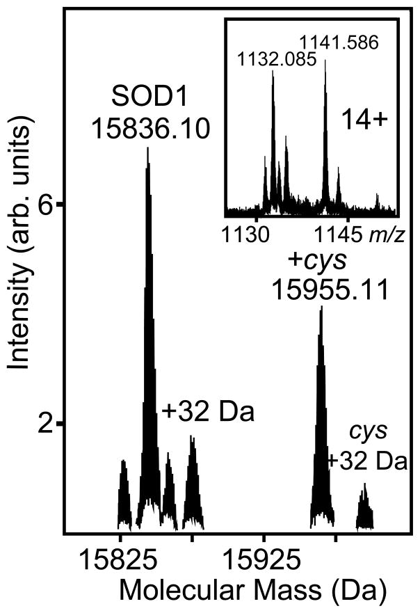
Figure 1. Cysteinylation is a prevalent post-translational modification of SOD1 in human nervous tissue.
SOD1 was isolated from a non-diseased human spinal cord using an SOD1 antibody column, 50 % acetonitrile was added to improve MS signal, and then infused directly into the FTMS14. Deisotoped and deconvoluted data for the entire mass range analyzed showing the monoisotopic mass for unmodified (15836.10 Da) and modified (15955.11 Da) SOD1, the delta mass being 119.01 Da, which is consistent with cysteinylation. Note that the solvents and declustering potentials used were such that native metals were not observed. (Inset) 14+ charge state showing unmodified (m/z 1132.085) and modified (m/z 1141.586) apo SOD1. This data is representative of the 8 human nervous tissue samples analyzed.
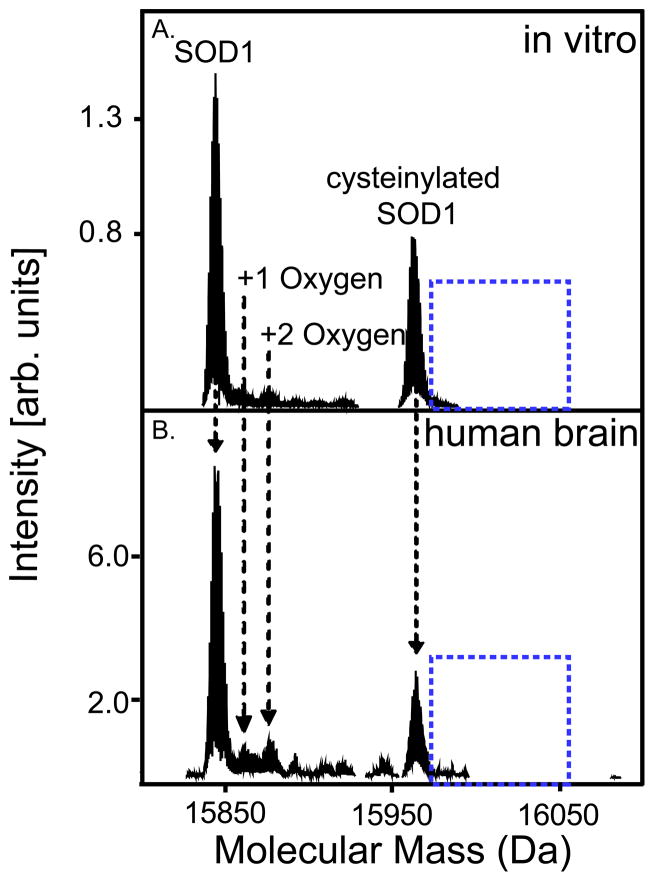
Figure 2. Cysteinylation and peroxide-mediated oxidations accounted for the majority of observed SOD1, purified from human tissue, post-translational modifications.
(A) Spectra of SOD1 modified by cysteine (40 μM) and hydrogen peroxide (100 μM); 3.5 hours after oxidation. SOD1 is modified by multiple oxygen atoms at this time point, whereas cysteine modified SOD1 is not (dotted blue box). (B) Spectra of SOD1 purified from a human brain. SOD1 is oxidized with multiple oxygen atoms, whereas cysteine modified SOD1 is not (dotted box). This is representative of the 4 additional samples analyzed using LC-MS.
RP-HPLC and FTMS
Purified SOD1 protein in GEB was analyzed using reverse-phase liquid chromatography and Fourier transform mass spectrometry as previously described 13. Briefly, reversed-phase liquid chromatography was performed using a two-dimensional nano-flow rate liquid chromatography (Eksigent), a 5-mm, 300-μm ID guard column (LC Packings, Part Number 160454) and a self-packed 14-cm, 100-μm ID column with 5-μm C18 beads (unpacked from a larger Targa column). Buffer A consisted of 0.1 % formic acid (vol/vol) in HPLC grade water and buffer B consisted of 0.1 % formic acid (vol/vol) in 100 % HPLC grade acetonitrile (vol/vol). Samples were diluted to a final formic acid concentration of 0.1 % (v/v) and injected. Following injection, samples were washed on the guard column with 160 column volumes of buffer A (8 μl min−1), and eluted at 650 nl min−1 using a 0–40 % gradient over 30 min. Samples were introduced via a nanospray ion source with a dual ion funnel (Apollo II) connected to a 9.4 T hybrid quadrupole Fourier transform ion cyclotron resonance (FT-ICR, FT-MS) mass spectrometer (apex Qe-94, Bruker Daltonics). External calibration of m/z scale was performed using electrospray tuning mix (Agilent, G2431A) using peaks at m/z 622, 922, 1522, and 2122.
After desolvation, the ions were transferred from a source hexapole to the quadrupole mass filter where isolation could occur in a second hexapole (collision cell). Ions accumulated in the second hexapole were then transferred through the ion optics region of the instrument to the ICR cell. Frequency sweep excitation was followed by image charge detection. Important instrument operation parameters include source declustering potential = 40 V, hexapole 1 accumulation time = 0.1 ms, collision cell accumulation time = 1 s, time of flight = 1.8 ms (D2), sidekick extraction voltages = −1.0 V (EV1, EV2, DEV2), RF excitation voltage = 130 V and ICR trapping potential = 1.2 V. Intact protein masses were reconstructed using the deconvolution function from DataAnalysis (Bruker Daltonics, version 3.4), and monoisotopic masses were determined using the Snap II algorithm (Bruker Daltonics).
In vitro cysteinylation and oxidation
Human SOD1 overexpressed and purified from S. cerevisiae, at either 0.1 μM or 1 μM concentration, was incubated with 40 μM L-cysteine overnight at room temperature and analyzed using direct infusion into the Fourier transform mass spectrometer. Oxidation was performed at room temperature for four hours using 100 μM or 10 mM hydrogen peroxide. Time points were collected every hour and analyzed using direct infusion into the FTMS using similar parameters as described above. All samples were desalted using C18-containing micropipette tips according to the manufacturer’s protocol (Millipore) and diluted 1:3 in HPLC water prior to injection into the mass spectrometer.
Identification of site of cysteinylation using funnel skimmer dissociation (FSD)
Cysteinylated SOD1 (1 μM) in 5 % acetonitrile, 0.1 % formic acid was directly infused into a 9.4 T Bruker Daltonics Fourier transform mass spectrometry in nanospray mode. Skimmer 1 voltage was then increased from 40 V-120 V in ten volt increments in order to fragment the protein using diverse fragmentation channels (at low voltage primarily mobile proton-directed, for example at proline residues, and at higher voltages primarly charge remote, for example at acidic residues 17). Data were analyzed using Bruker Daltonic’s DataAnalysis software.
Results
SOD1 purified from human brain and spinal cord is modified by cysteine and oxygen
SOD1 was first immunopurified using rabbit polyclonal anti-SOD1 (each respective sample individually), from ten non-diseased human nervous tissue samples and mice (8 from human and 2 from mice) overexpressing human SOD1 (brain and spinal cord), eluted using 5 % acetic acid and analyzed by direct infusion-Fourier transform mass spectrometry (Figure 1, human) or by direct infusion-ion trap mass spectrometry (Figure S1, mouse). Cysteinylation of SOD1 was consistently the most prevalent PTM observed as peaks (e.g. the 14+ charge state at m/z 1141.586) corresponding to a deconvoluted and deisotoped monoisotopic mass of 15955.11 Da, which is 119.01 Da larger than unmodified SOD1 (15836.10 Da). A 119.01 Da modification is consistent with the molecular weight of cysteine with the two sulfhydryl hydrogen atoms liberated during disulfide bond formation (theoretical mass 119.01) between Cys111 of SOD1 and free cysteine (Figure 1). In addition to cysteinylation, we observed modifications we putatively assigned as modification with one (15851.40 Da) and two oxygen atoms (15866.38 Da). The abundance of these putative oxidative modifications was too low to permit further characterization - however, oxidative modifications of Cys111 and Trp32 have been described previously14.
To further investigate PTMs by cysteine and oxygen, as well as their relative amounts, an additional four post-mortem human brain samples were analyzed using a different purification method (different homogenization buffer, SOD1 antibodies, wash buffer, and elution buffer) and different preparative conditions (reverse-phase liquid chromatography mass spectrometry [LC-MS], (Figure 2)). Cysteinylation and oxidation were observed in all four samples (Figure 2B), although the percentage of total of Cys modified SOD1 appeared slightly lower in RP-LC-MS analysis (38 % ± 7 % via RPLC-MS versus 48 % ± 6 % via direct infusion). Reverse phase chromatography generally increases the dynamic range of MS analysis, and as a result additional modifications were detected in these samples. Therefore, the overall average cysteinylation observed in human nervous tissue (both direct infusion and RP-LC-MS) was 41 % (maximum cysteinylation observed was 62 %; minimum cysteinylation observed was 22 %). Notably, cysteinylation was not a prevalent modification in SOD1 purified from human blood by the first method 14 but was reportedly observed in other purifications11, 12.
To determine if the extent of cysteine modification or oxidation depended upon purification methods, for example if oxidative addition of free cysteine or thiol disulfide exchanged with free cystine, two additional controls were used. First, SOD1 was purified anaerobically. Second, SOD1 was homogenized anaerobically in the presence of iodoacetamide, iodoacetic acid, and S-methyl methane thiosulfanate (MMTS) to alkylate and scavenge any free cysteine as well as unmodified SOD1 Cys111. Cysteinylation was observed in SOD1 purified anaerobically, however there was approximately a 2-fold reduction in the amount observed compared to samples purified aerobically. The cysteine alkylators and scavengers removed the majority of the cysteinylation, however a small amount was still present.
In summary, to investigate PTMs by cysteine and oxygen, we performed purifications using (1) different antibodies and different elution conditions; (2) with and without liquid chromatography; (3) aerobically and anaerobically; (4) with and without alkylation agent to block endogenous “free” cysteine from binding SOD1 during homogenization; and (5) from different tissues and organisms (human and mouse spinal cord and brain). Under the conditions tested here SOD1 purified from human tissue, but not from yeast, contained cysteinlyated SOD1 (Table 1).
Table 1.
Amount of SOD1 cysteinylated using different purification procedures and from different sources.
| Purification Method | % Cysteinylation | |
|---|---|---|
| Polyclonal rabbit antibody | 48 | Figure 1 |
| Polyclonal sheep antibody | 38 | Figure 2B |
| Elution: 5% acetic acid | 48 | Figure 1 |
| Elution: gentle elution buffer | 38 | Figure 2B |
| With liquid chromatography | 38 | Figure 2B |
| Without liquid chromatography | 48 | Figure 1 |
| Aerobically | 48 | Figure 1, 2B |
| Anaerobically | ||
| With alkylating agent | 1 | |
| Without alkylating agent | 24 | Figure 1, 2B |
| Protein Source | Cysteinlyation | |
| Human nervous tissue | 41 | Figure 1, 2B |
| Mouse nervous tissue | 21 | Supplemental figure 1 |
| Yeast | 0 | Figure 2A, 3A |
The majority of SOD1 modifications observed in human tissue can be created in vitro using cysteine and peroxide
To determine if both the types and relative amounts of modifications of SOD1 purified from human tissue could be recapitulated in vitro, SOD1 was incubated with 40-fold molar excess cysteine, which approximated the cysteinylation levels of post-mortem SOD1. This sample was incubated with a 100-fold molar excess of peroxide (100 μM) in a time course experiment. Following incubation with low levels of peroxide (the amount generated in 100 turnovers of peroxide) for 3.5 hours, these in vitro samples resembled SOD1 purified from human tissue, indicating that many of the modifications observed in SOD1 purified from human samples are the result of modification by cysteine and peroxide (Figure 2).
Cysteine modified SOD1 is protected from oxidation
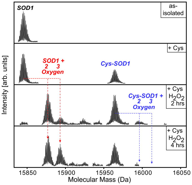
Human SOD1 expressed and purified from yeast cells was analyzed using a Fourier transform mass spectrometer (FTMS), which showed a peak consistent with native SOD1 (Figure 3A, note: acidic buffers and desolvation conditions were such that Cu and Zn could not be detected). Cysteinylated SOD1 was created in vitro as described above. Both native and cysteinylated SOD1 were observed, and the binding stoichiometry was approximately one cysteine per SOD1 dimer (Figure 3B), indicating that cysteinylation of one Cys111 can potentially block cysteinylation of the adjacent Cys111, presumably sterically.
Figure 3. Cysteinylation protected SOD1 from oxidation.
(A) Spectra of unmodified (as-isolated) SOD1 (15835.97 Da) (B) Spectra of SOD1 modified by cysteine (40 μM) (15954.97 Da), which is consistent with the molecular weight of SOD1-cysteine (less two hydrogen atoms, forming cystine) (C and D) Taken from the time course of 10 mM peroxide-mediated oxidization of cysteinylated SOD1. (C) Spectra of cysteine modified, SOD1, oxidized using hydrogen peroxide; 2 hours after oxidation. Most of the native SOD1 protein has been modified by two (sulfinic acid) (15867.97 Da) or three (sulfonic acid) oxygen atoms (15883.96 Da) (dotted red lines), whereas a small amount of cysteine modified SOD1 is oxidized (dotted blue lines). (D) Spectra of SOD1 modified by cysteine and oxidized using hydrogen peroxide; 4 hours after oxidation. All native SOD1 has been oxidized by two (15867.97 Da) or three oxygen atoms (15883.95 Da) (dotted red lines), whereas cysteine modified SOD1 does not appear to be oxidized (dotted blue lines). In addition, the sulfonic acid modified SOD1 is the same form of SOD1 that Bosco et al show inhibits fast axonal transport in a similar fashion to SOD1 FALS variants 13. These experiments were repeated in triplicate.
To determine the extent to which in vitro cysteine modification protected SOD1 from peroxide-mediated modification, a sample containing both cysteine modified SOD1 and the native protein was oxidized using 10 mM hydrogen peroxide in a time course study. After two hours the majority of native SOD1 protein was modified by two or three oxygen atoms. Conversely, only a small amount of cysteine modified SOD1 was oxidized (Figure 3C; red dotted lines versus blue dotted lines), indicating near complete protection. In addition, four hours after peroxide treatment all the native SOD1 had been oxidized by two or three oxygen atoms (native protein is no longer observed), whereas cysteine modified SOD1 was not oxidized (Figure 3D; red dotted lines versus blue dotted lines).
Cysteinylation occured specifically upon SOD1 Cys111
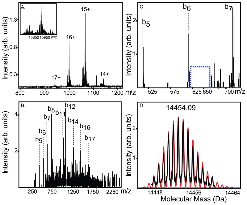
PTMs are typically localized using endoproteinase digestion followed by LC-MS/MS analysis. In both MALDI-TOF fingerprinting and LC-MS/MS experiments, we observed cysteinylated peptides containing Cys111 and localized this modification to Cys111 using MS/MS data (Supplemental Figures 2–3). This approach, however, was not ideal for cysteinylation. Control experiments revealed that, following endoproteinase digestion, rapid scrambling of SOD1 disulfides occurred, including scrambling of the native disulfide (Cys57 and Cys146) with Cys6 and Cys111. This necessitated a “top-down” MS approach, whereby fragmentation occurs within the mass spectrometer such that disulfide scrambling cannot normally occur 18. To localize the site of modification by cysteine, SOD1 was cysteinylated in vitro and analyzed via intact protein dissociation within the FTMS (Figure 4A). Cysteinylated SOD1 was fragmented in the FTMS using collisionally-activated dissociation at the funnel-skimmer interface, yielding an abundant b-ion series (Figure 4B). The b6-ion and larger b-ions (fragments containing the N-terminus and Cys6) fit the theoretical mass of unmodified Cys6 (Figure 4C) and no modified peaks were observed (based upon S/N as little as 2% of modified Cys6 could have been detected). In addition, we observed a y139-ion at a mass of 14454.09 Da. This mass is consistent with a cysteinylated SOD1 y-ion containing C-terminal residues 16-153, and containing the intramolecular disulfide bond between residues 57 and 146 (Figure 4D). In summary, cysteine 57 and 146 are in a disulfide bond and are unable to be cysteinylated; we ruled out cysteinylation of cysteine 6 using MS/MS data; and observed a large cysteinylated C-terminal SOD1 fragment, consistent with cysteinlyation of Cys111. In a sister publication we present the 3-dimensional structure of cysteinylated SOD1, which is consistent with the results presented here, and in addition characterize the binding stoichiometry as 1 cysteinylation per SOD1 dimer.
Figure 4. SOD1 Cys111 is the site of cysteinylation.
(A) Mass spectrum of intact cysteinylated SOD1. (B) Fragmentation spectrum of cysteinlyated SOD1 using funnel skimmer dissociation with the N-terminal b ion series labeled. (C) Zoom in of the b6-ion (contains cysteine 6); no peak corresponding to a cysteinlyated form of cysteine 6 is observed (approximately m/z 632; dotted blue box). (D) C-terminal fragment (y139) of SOD1 (residues 16-153), which is consistent with the mass of a cysteinlyated SOD1 fragment. The red lines are the theoretical isotopic distribution modeled using the simulate isotopic pattern parameter in DataAnalysis (Bruker) of cysteinylated y139 (mass of y139 plus 121.158 Da for cysteine, subtract two hydrogen atoms upon its non-native disulfide formation, and an additional two hydrogen atoms for the intramolecular disulfide).
Discussion
Protein cysteinylation is not well-characterized, partially due to the practice of purifying/treating proteins in the presence of reducing agents such as DTT. It has, however, been observed in transthyretin (TTR), human serum albumin, and the k1 light chain from an amyloid patient19, 20. In addition, cysteinylation has been observed in Bacillus subtilis during oxidative stress treatment. Hochgrafe et al suggest that cysteinylation may play a role in protecting cysteine residues from oxidation and irreversible damage in Bacillus subtilis and possibly other organisms 21.
More than half of all the SOD1 protein isolated here from post-mortem human nervous tissues contained PTMs, predominantly cysteinylation and oxidation. Cysteine modified SOD1 was protected from peroxide mediated oxidation in vitro, and cysteinylated SOD1 purified from human tissue appears also to have been protected from oxidation. SOD1 that had been cysteinylated and peroxide oxidized in vitro was composed of a set of PTMs that bear a striking resemblance to the myriad of PTMs observed in SOD1 purified from human tissue (Figure 1 and 2), indicating peroxide and cysteine are amongst the major modifiers of SOD1.
Putative cysteinylation of SOD1 was observed in vitro 7 and in preparations from blood 11, 12 based upon differences in intact protein mass, and in some cases the modification was labile to reductants. Li et al7 observe a modified SOD1 118 Da heavier than unmodified SOD1, which disappears with DTT treatment and concluded that the modification was cysteine. However, this delta mass of 118 Da is not consistent with the 119 Da increase in mass expected for a cysteine modification. In no previous studies were peptide digest or MSn data provided to confirm that the modification was cysteine or to determine the site of modification. Here, using endoproteinase peptide mapping by MALDI-TOF MS, MSn data of an LC-ESI-ion trap, and top-down MS, cysteinylation of SOD1 on residue 111 was confirmed. In addition, we suggest a possible role for SOD1 cysteinylation, namely protection from peroxide-mediated oxidation.
In anaerobic control experiments in the presence or absence of alkylating/thiol scavenging agents we observed a reduction in the amount of cysteinylation, consistent with some of the modifications we observed from human tissue occurring during the homogenization process. Note that cysteinylation from cystine occurs via thiol-disulfide exchange and as redox neutral, whereas cysteinylation from cysteine requires the loss of two protons and two electrons (oxidation). Although treatment with alkylating agents/cysteine scavengers is a harsh treatment with the potential to remove cysteinylation 22, it did not do so in in vitro control experiments. Cysteinylation remained the most abundant PTM in the anaerobic (with no other chemicals) treatment.
An argument against non-enzymatic cysteinylation of SOD1 occurring during homogenization can be made based upon the cellular ratios of SOD1 to free cysteine/cystine (CSH/CSSC) and glutathione (GSH/GSSG). Whereas the amount of SOD1 is ~10 μM, the amount of cysteine inside the cell is approximately 2.5 μM 23, 24 and the amount of cystine is approximately 0.25–1.3 μM 23, 25–27. Given similarities in redox potential (E0) for CSH/CSSC and GSH/GSSG (− 0.22 V and − 0.24 V 28 respectively) and the 1000-fold higher concentration of glutathione in the brain (~ 1–2 mM GSH and ~8 μM GSSG 29–31), glutathionylation (not observed) is the most likely artifact of our purification. Furthermore, despite concentrations of cystine and cysteine being 50–100 times higher in plasma 32, we do not observe cysteinylated SOD1 in blood preparations 14 (>50 purifications). On the other hand, cysteinylation was putatively assigned, although without MS/MS or protein digest confirmation, in other purifications 11, 12. Thus, based on the protein concentration in the cell, the cysteine concentration, and their redox potential, it is likely cysteinylation can occur in vivo.
Despite treatment with large excesses of cysteine, only one cysteine was detected to be bound per SOD1 dimer, consistent with cysteinylation of SOD1 Cys111 on one monomer blocking cysteinylation on the second monomer. This is probably due to the close proximity (~8–10 Å) of both cysteine residues in the dimer interface of SOD1 7- we were unable to model two cysteine residues per dimer without them overlapping or being strained. A sister publication characterizes the three dimensional structure of cysteinated SOD1. Although the binding of free cysteine to one of SOD1’s cysteine residues blocks the binding of a second cysteine, our data suggested it does not completely protect the adjacent Cys111 (non-cysteinylated Cys111) from oxidation.
Dimer destabilization of SOD1 has been implicated in disease progression for amyotrophic lateral sclerosis (ALS) 15, 33–36. Oxygen-modified cysteine residues are negatively charged 37 and if both Cys111 are oxidized there would be a columbic impetus for destabilizing the SOD1 dimer. We, and others, have shown that oxidation of SOD1 by hydrogen peroxide is capable of destabilizing SOD1 1–3, 13. In addition, the sulfonic acid modified SOD1 (3 oxygen atoms per cysteine) is the same form of SOD1 that Bosco et al show inhibits fast axonal transport in a similar fashion to SOD1 FALS variants 13. Thus, by preventing oxidation of Cys111, cysteinylation could ameliorate SOD1 dimer destabilization and minimize the formation of toxic SOD1 species. Under conditions of both oxidative stress and aging there is both more cystine, which can cysteinylate SOD1 directly, and more oxygen to promote cysteine-SOD1 oxidative coupling. For example, cystine concentrations double with age (av. 52 μM at age 26, av. 105 μM at age 60 22). Cysteinylated SOD1 is therefore more likely to occur under conditions of both oxidative stress and aging and may be an adaptive modification.
Supplementary Material
Acknowledgments
We thank Dr. P. John Hart for the generous gift of the YEP351-SOD1 plasmid and EGy118-( SOD1) yeast cells used to express SOD1 in this study. We also thank the patients who donated their tissue used in these experiments. We also thank members of the Agar and Petsko/Ringe Labs for thoughtful discussions, insights, and critically reviewing this manuscript.
This work was supported in part by a National Institutes of Health 1R01NS065263-01 to J.N.A., 1R01NS067206-02 to D.A.B., ALS Therapy Alliance/CVS Pharmacy to D.A.B., and Fidelity Biosciences Research Initiative (to G.A.P. and D.R.).
Footnotes
Additional figures further characterizing cysteinylation are available as supporting information. This material is available free of charge via the Internet at http://pubs.acs.org.
References
- 1.Keithley EM, Canto C, Zheng QY, Wang X, Fischel-Ghodsian N, Johnson KR. Cu/Zn superoxide dismutase and age-related hearing loss. Hear Res. 2005;209:76–85. doi: 10.1016/j.heares.2005.06.009. [DOI] [PMC free article] [PubMed] [Google Scholar]
- 2.Phillips JP, Tainer JA, Getzoff ED, Boulianne GL, Kirby K, Hilliker AJ. Subunit-destabilizing mutations in Drosophila copper/zinc superoxide dismutase: neuropathology and a model of dimer dysequilibrium. P Natl Acad Sci USA. 1995;92:8574–8578. doi: 10.1073/pnas.92.19.8574. [DOI] [PMC free article] [PubMed] [Google Scholar]
- 3.Woodruff RC, Phillips JP, Hilliker AJ. Increased spontaneous DNA damage in Cu/Zn superoxide dismutase (SOD1) deficient Drosophila. Genome. 2004;47:1029–1035. doi: 10.1139/g04-083. [DOI] [PubMed] [Google Scholar]
- 4.McCord JM, Fridovich I. The reduction of cytochrome c by milk xanthine oxidase. The Journal of biological chemistry. 1968;243:5753–5760. [PubMed] [Google Scholar]
- 5.Fujiwara N, Nakano M, Kato S, Yoshihara D, Ookawara T, Eguchi H, Taniguchi N, Suzuki K. Oxidative modification to cysteine sulfonic acid of Cys111 in human copper-zinc superoxide dismutase. J Biol Chem. 2007;282:35933–35944. doi: 10.1074/jbc.M702941200. [DOI] [PubMed] [Google Scholar]
- 6.Choi J, Rees HD, Weintraub ST, Levey AI, Chin LS, Li L. Oxidative modifications and aggregation of Cu,Zn-superoxide dismutase associated with Alzheimer and Parkinson diseases. J Biol Chem. 2005;280:11648–11655. doi: 10.1074/jbc.M414327200. [DOI] [PubMed] [Google Scholar]
- 7.Liu H, Zhu H, Eggers DK, Nersissian AM, Faull KF, Goto JJ, Ai J, Sanders-Loehr J, Gralla EB, Valentine JS. Copper(2+) binding to the surface residue cysteine 111 of His46Arg human copper-zinc superoxide dismutase, a familial amyotrophic lateral sclerosis mutant. Biochemistry. 2000;39:8125–8132. doi: 10.1021/bi000846f. [DOI] [PubMed] [Google Scholar]
- 8.Redler RL, Wilcox KC, Proctor EA, Fee L, Caplow M, Dokholyan NV. Glutathionylation at Cys-111 induces dissociation of wild type and FALS mutant SOD1 dimers. Biochemistry. 2011;50:7057–7066. doi: 10.1021/bi200614y. [DOI] [PMC free article] [PubMed] [Google Scholar]
- 9.Schinina ME, Carlini P, Polticelli F, Zappacosta F, Bossa F, Calabrese L. Amino acid sequence of chicken Cu, Zn-containing superoxide dismutase and identification of glutathionyl adducts at exposed cysteine residues. Eur J Biochem. 1996;237:433–439. doi: 10.1111/j.1432-1033.1996.0433k.x. [DOI] [PubMed] [Google Scholar]
- 10.Wilcox KC, Zhou L, Jordon JK, Huang Y, Yu Y, Redler RL, Chen X, Caplow M, Dokholyan NV. Modifications of superoxide dismutase (SOD1) in human erythrocytes: a possible role in amyotrophic lateral sclerosis. J Biol Chem. 2009;284:13940–13947. doi: 10.1074/jbc.M809687200. [DOI] [PMC free article] [PubMed] [Google Scholar]
- 11.Nakanishi T, Kishikawa M, Miyazaki A, Shimizu A, Ogawa Y, Sakoda S, Ohi T, Shoji H. Simple and defined method to detect the SOD-1 mutants from patients with familial amyotrophic lateral sclerosis by mass spectrometry. J Neurosci Methods. 1998;81:41–44. doi: 10.1016/s0165-0270(98)00012-0. [DOI] [PubMed] [Google Scholar]
- 12.Shimizu A, Nakanishi T, Miyazaki A. Detection and characterization of variant and modified structures of proteins in blood and tissues by mass spectrometry. Mass spectrometry reviews. 2006;25:686–712. doi: 10.1002/mas.20086. [DOI] [PubMed] [Google Scholar]
- 13.Bosco DA, Morfini G, Karabacak NM, Song Y, Gros-Louis F, Pasinelli P, Goolsby H, Fontaine BA, Lemay N, McKenna-Yasek D, Frosch MP, Agar JN, Julien JP, Brady ST, Brown RH., Jr Wild-type and mutant SOD1 share an aberrant conformation and a common pathogenic pathway in ALS. Nat Neurosci. 2010;13:1396–1403. doi: 10.1038/nn.2660. [DOI] [PMC free article] [PubMed] [Google Scholar]
- 14.Taylor DM, Gibbs BF, Kabashi E, Minotti S, Durham HD, Agar JN. Tryptophan 32 potentiates aggregation and cytotoxicity of a copper/zinc superoxide dismutase mutant associated with familial amyotrophic lateral sclerosis. J Biol Chem. 2007;282:16329–16335. doi: 10.1074/jbc.M610119200. [DOI] [PubMed] [Google Scholar]
- 15.Doucette PA, Whitson LJ, Cao X, Schirf V, Demeler B, Valentine JS, Hansen JC, Hart PJ. Dissociation of human copper-zinc superoxide dismutase dimers using chaotrope and reductant. Insights into the molecular basis for dimer stability. J Biol Chem. 2004;279:54558–54566. doi: 10.1074/jbc.M409744200. [DOI] [PubMed] [Google Scholar]
- 16.Hayward LJ, Rodriguez JA, Kim JW, Tiwari A, Goto JJ, Cabelli DE, Valentine JS, Brown RH., Jr Decreased metallation and activity in subsets of mutant superoxide dismutases associated with familial amyotrophic lateral sclerosis. J Biol Chem. 2002;277:15923–15931. doi: 10.1074/jbc.M112087200. [DOI] [PubMed] [Google Scholar]
- 17.Cobb JS, Easterling ML, Agar JN. Structural characterization of intact proteins is enhanced by prevalent fragmentation pathways rarely observed for peptides. Journal of the American Society for Mass Spectrometry. 2010;21:949–959. doi: 10.1016/j.jasms.2010.02.009. [DOI] [PMC free article] [PubMed] [Google Scholar]
- 18.Kellie JF, Tran JC, Lee JE, Ahlf DR, Thomas HM, Ntai I, Catherman AD, Durbin KR, Zamdborg L, Vellaichamy A, Thomas PM, Kelleher NL. The emerging process of Top Down mass spectrometry for protein analysis: biomarkers, protein-therapeutics, and achieving high throughput. Mol Biosyst. 2010;6:1532–1539. doi: 10.1039/c000896f. [DOI] [PMC free article] [PubMed] [Google Scholar]
- 19.Kleinova M, Belgacem O, Pock K, Rizzi A, Buchacher A, Allmaier G. Characterization of cysteinylation of pharmaceutical-grade human serum albumin by electrospray ionization mass spectrometry and low-energy collision-induced dissociation tandem mass spectrometry. Rapid communications in mass spectrometry: RCM. 2005;19:2965–2973. doi: 10.1002/rcm.2154. [DOI] [PubMed] [Google Scholar]
- 20.Lim A, Wally J, Walsh MT, Skinner M, Costello CE. Identification and location of a cysteinyl posttranslational modification in an amyloidogenic kappa1 light chain protein by electrospray ionization and matrix-assisted laser desorption/ionization mass spectrometry. Anal Biochem. 2001;295:45–56. doi: 10.1006/abio.2001.5187. [DOI] [PubMed] [Google Scholar]
- 21.Hochgrafe F, Mostertz J, Pother DC, Becher D, Helmann JD, Hecker M. S-cysteinylation is a general mechanism for thiol protection of Bacillus subtilis proteins after oxidative stress. The Journal of biological chemistry. 2007;282:25981–25985. doi: 10.1074/jbc.C700105200. [DOI] [PubMed] [Google Scholar]
- 22.Johnson JM, Strobel FH, Reed M, Pohl J, Jones DP. A rapid LC-FTMS method for the analysis of cysteine, cystine and cysteine/cystine steady-state redox potential in human plasma. Clin Chim Acta. 2008;396:43–48. doi: 10.1016/j.cca.2008.06.020. [DOI] [PMC free article] [PubMed] [Google Scholar]
- 23.Sato H, Tamba M, Okuno S, Sato K, Keino-Masu K, Masu M, Bannai S. Distribution of cystine/glutamate exchange transporter, system x(c)-, in the mouse brain. J Neurosci. 2002;22:8028–8033. doi: 10.1523/JNEUROSCI.22-18-08028.2002. [DOI] [PMC free article] [PubMed] [Google Scholar]
- 24.Castagna A, Le Grazie C, Accordini A, Giulidori P, Cavalli G, Bottiglieri T, Lazzarin A. Cerebrospinal fluid S-adenosylmethionine (SAMe) and glutathione concentrations in HIV infection: effect of parenteral treatment with SAMe. Neurology. 1995;45:1678–1683. doi: 10.1212/wnl.45.9.1678. [DOI] [PubMed] [Google Scholar]
- 25.Araki K, Harada M, Ueda Y, Takino T, Kuriyama K. Alteration of amino acid content of cerebrospinal fluid from patients with epilepsy. Acta Neurol Scand. 1988;78:473–479. doi: 10.1111/j.1600-0404.1988.tb03690.x. [DOI] [PubMed] [Google Scholar]
- 26.Arnesano F, Banci L, Bertini I, Martinelli M, Furukawa Y, O’Halloran TV. The unusually stable quaternary structure of human Cu,Zn-superoxide dismutase 1 is controlled by both metal occupancy and disulfide status. J Biol Chem. 2004;279:47998–48003. doi: 10.1074/jbc.M406021200. [DOI] [PubMed] [Google Scholar]
- 27.Lakke JP, Teelken AW. Amino acid abnormalities in cerebrospinal fluid of patients with parkinsonism and extrapyramidal disorders. Neurology. 1976;26:489–493. doi: 10.1212/wnl.26.5.489. [DOI] [PubMed] [Google Scholar]
- 28.Jocelyn PC. The standard redox potential of cysteine-cystine from the thiol-disulphide exchange reaction with glutathione and lipoic acid. European journal of biochemistry/FEBS. 1967;2:327–331. doi: 10.1111/j.1432-1033.1967.tb00142.x. [DOI] [PubMed] [Google Scholar]
- 29.Ratan RR, Murphy TH, Baraban JM. Macromolecular synthesis inhibitors prevent oxidative stress-induced apoptosis in embryonic cortical neurons by shunting cysteine from protein synthesis to glutathione. J Neurosci. 1994;14:4385–4392. doi: 10.1523/JNEUROSCI.14-07-04385.1994. [DOI] [PMC free article] [PubMed] [Google Scholar]
- 30.Slivka A, Mytilineou C, Cohen G. Histochemical evaluation of glutathione in brain. Brain Res. 1987;409:275–284. doi: 10.1016/0006-8993(87)90712-8. [DOI] [PubMed] [Google Scholar]
- 31.Slivka A, Spina MB, Cohen G. Reduced and oxidized glutathione in human and monkey brain. Neuroscience letters. 1987;74:112–118. doi: 10.1016/0304-3940(87)90061-9. [DOI] [PubMed] [Google Scholar]
- 32.Brigham MP, Stein WH, Moore S. The Concentrations of Cysteine and Cystine in Human Blood Plasma. J Clin Invest. 1960;39:1633–1638. doi: 10.1172/JCI104186. [DOI] [PMC free article] [PubMed] [Google Scholar]
- 33.Auclair JR, Boggio KJ, Petsko GA, Ringe D, Agar JN. Strategies for stabilizing superoxide dismutase (SOD1), the protein destabilized in the most common form of familial amyotrophic lateral sclerosis. P Natl Acad Sci USA. 2010;107:21394–21399. doi: 10.1073/pnas.1015463107. [DOI] [PMC free article] [PubMed] [Google Scholar]
- 34.Hornberg A, Logan DT, Marklund SL, Oliveberg M. The coupling between disulphide status, metallation and dimer interface strength in Cu/Zn superoxide dismutase. J Mol Biol. 2007;365:333–342. doi: 10.1016/j.jmb.2006.09.048. [DOI] [PubMed] [Google Scholar]
- 35.Rakhit R, Crow JP, Lepock JR, Kondejewski LH, Cashman NR, Chakrabartty A. Monomeric Cu,Zn-superoxide dismutase is a common misfolding intermediate in the oxidation models of sporadic and familial amyotrophic lateral sclerosis. J Biol Chem. 2004;279:15499–15504. doi: 10.1074/jbc.M313295200. [DOI] [PubMed] [Google Scholar]
- 36.Rakhit R, Robertson J, Vande Velde C, Horne P, Ruth DM, Griffin J, Cleveland DW, Cashman NR, Chakrabartty A. An immunological epitope selective for pathological monomer-misfolded SOD1 in ALS. Nat Med. 2007;13:754–759. doi: 10.1038/nm1559. [DOI] [PubMed] [Google Scholar]
- 37.Yarnell A. Cysteine Oxidation New Chemical tools are poised to help scientists explore the roles of oxidized cysteine residues might play in biology. Chemical and Engineering News. 2009;87:38–40. [Google Scholar]
Associated Data
This section collects any data citations, data availability statements, or supplementary materials included in this article.


