Abstract
Treatment of lung cancer in patients with idiopathic pulmonary fibrosis (IPF) is difficult because the mortality rate after surgery or chemotherapy is high for these patients. Spontaneous regression of cancer is rare, especially in lung cancer. A 62-year-old man, previously diagnosed with IPF, presented with stage IIIC (T2N3M0) non-small cell lung cancer. About 4 months later, spontaneous regression of the primary tumor was observed without treatment. To the best of our knowledge, this is the first report of spontaneous regression of lung cancer in a patient with IPF.
Keywords: Lung Neoplasms; Fibrosis; Neoplasm Regression, Spontaneous
Introduction
Idiopathic pulmonary fibrosis (IPF) is associated with increased risk of lung cancer. Atypical or dysplastic epithelial changes in pulmonary fibrosis can be involved in lung cancer carcinogenesis1. Large, population-based cohort studies report an increased incidence of lung cancer in IPF patients compared to normal subjects2,3. Spontaneous regression (SR) of cancer, defined as a complete or partial disappearance of malignant disease without treatment, is rare4. SR of lung cancer is extremely rare in general, and especially in patients with non-small cell lung cancer (NSCLC) supervening on IPF. We present a rare case of NSCLC in a patient with IPF whose tumor spontaneously regressed without treatment.
Case Report
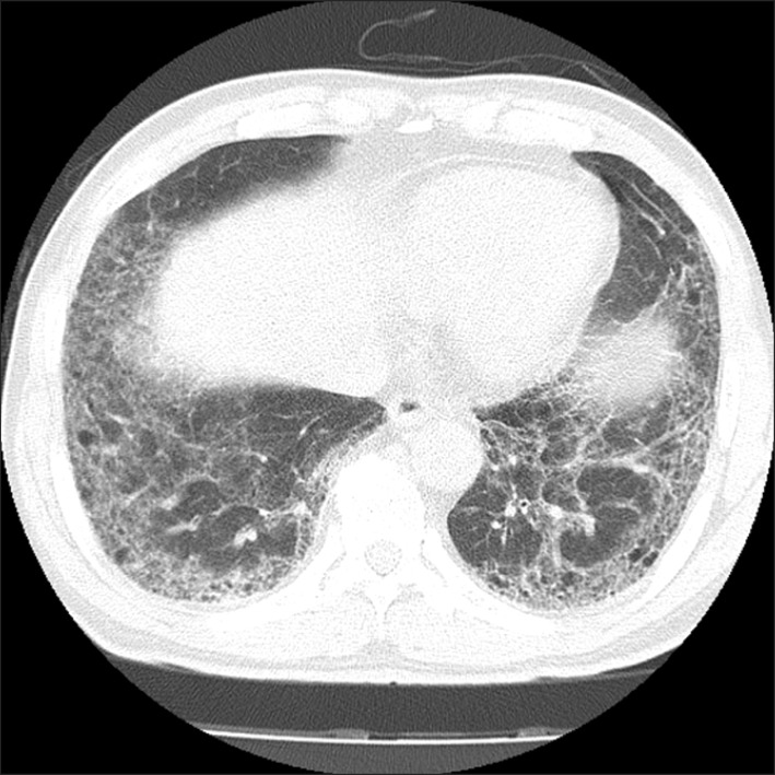
A 62-year-old man complaining of dyspnea was referred to our hospital in November 2011. He had a 70 pack-year history of smoking, and a history of diabetes mellitus. Chest computed tomography (CT) revealed a diffuse subpleural reticular pattern and honeycombing in both lungs without mass-like lesions, suggesting IPF (Figure 1).
Figure 1.
Chest computed tomography in October 2011 showing a diffuse subpleural reticular pattern and honeycomb appearance.
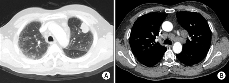
A follow-up chest CT was performed in May 2012. The image revealed a newly developed, 3.2×2.3 cm, irregular mass widely abutting pleura in the left upper lobe (Figure 2A). The CT also revealed multiple enlarged lymph nodes in the paratracheal, subcarinal, prevascular, subaortic, hilar and supraclavicular areas (Figure 2B). Similarly, 18F-fluorodeoxyglucose (FDG) positron emission tomography (PET) showed multiple lymph node enlargements with increased FDG uptake in the hilar, subcarinal, paratracheal, subaortic, prevascular and supraclavicular areas (Figure 3). Carcinoembryonic antigen was 2.41 ng/mL (normal range, <5.0) and cytokeratin 19 fragment was 7.58 ng/mL (normal, <3.3). Endobronchial ultrasound-guided transbronchial needle aspiration and CT-guided fine needle biopsy in June 2012 revealed no malignancy in the left upper lobe tumor, but metastatic NSCLC in a subcarinal lymph node, a right lower paratracheal lymph node, and a left lower paratracheal lymph node (Figure 4). Other metastatic work-up including brain magnetic resonance imaging and whole body bone scan was negative. Consequently, the patient was diagnosed with NSCLC (T2N3M0, stage IIIC). The patient declined palliative chemotherapy, and was discharged from the hospital without treatment.
Figure 2.
(A) Chest computed tomography (CT) in May 2012 showing a 3.2×2.3 cm mass in the left upper lobe. (B) The same CT showing enlargement of multiple mediastinal lymph nodes.
Figure 3.
Positron emission tomography showing enlargement of multiple lymph nodes with increased 18F-fluorodeoxyglucose uptake in the hilar, subcarinal, paratracheal, subaortic, prevascular, and supraclavicular areas, suggesting lymph node metastasis.
Figure 4.
(A) Biopsy specimen from the right lower paratracheal lymph node showing metastatic and poorly differentiated non-small cell carcinoma (H&E stain, ×200). (B) Immunohistochemical stain of the right lower paratracheal lymph node showing positive staining for thyroid transcription factor-1 (×200).
A follow-up outpatient chest CT was performed in October 2012. This image revealed the disappearance of the primary tumor in the subpleural area of the left upper lobe, and a marked decrease in the size of the multiple metastatic lymph nodes in the hilar, subcarinal, paratracheal, subaortic, prevascular and supraclavicular areas (Figure 5). Follow-up chest CT in January 2013 showed no significant change in primary tumor or metastatic lymph nodes.
Figure 5.
(A) Follow-up chest computed tomography (CT) in October 2012 shows the disappearance of the primary tumor in the subpleural area of the left upper lobe. (B) The same CT shows a marked decrease in the size of the multiple metastatic lymph nodes in the hilar, subcarinal, paratracheal, subaortic, prevascular and supraclavicular areas.
Discussion
IPF is a well-known risk factor for lung cancer2,3. Lung cancer treatment in IPF patients is difficult because they have a high mortality rate after surgery or chemotherapy5. Cole and Everson4 defined SR of cancer as complete or partial disappearance of disease without anticancer treatment. Although cancer SR has been documented for several types of malignancies, it is extremely rare for lung cancer. In Korea, only three cases of SR of lung cancer have been reported6-8.
The biological mechanisms of SR remain unclear. Possible mechanisms include apoptosis, immunological response, differentiations, hormone, angiogenesis inhibition, telomerase inhibition, and psychoneuroimmunological response9. In lung cancer, the immunologic response is the most reasonable mechanism for SR. Moriyama et al.10 reported that HLA class I antigen and CD8-positive lymphocytes are increased in lung cancer tissue, and suggested that these lymphocytes might be cytotoxic to tumor cells. Nakamura et al.11 similarly suggested that immunological response to specific antigens such as NY-ESO-1 is a possible mechanism of SR in lung cancer. Pujol et al.12 reported that anti-Hu antibody syndrome is associated with SR of NSCLC.
A limitation of this case is that we failed to find evidence of malignancy in the left upper lobe tumor, but only in the mediastinal lymph nodes. However, the cell type was metastatic NSCLC and there was no abnormal lesions other then the left upper lobe tumor and enlarged mediastinal lymph nodes on PET-CT. We also found that the immunohistochemical staining for thyroid transcription factor-1 was positive in the mediastinal lymph nodes.
The patient refused treatment for lung cancer or IPF. We have performed regular, outpatient follow-up of this patient and no evidence of recurrence has been found by chest CT through January 2013. The cause of tumor remission remains unknown. We propose that immunological response might have led to tumor reduction. Further studies are necessary to explain the association between SR of lung cancer and IPF.
To the best of our knowledge, this is the first report of a complete remission of NSCLC in a patient with IPF. SR of lung cancer is extremely rare and its mechanism remains unclear. More research is needed to explain this unusual phenomenon.
References
- 1.Park J, Kim DS, Shim TS, Lim CM, Koh Y, Lee SD, et al. Lung cancer in patients with idiopathic pulmonary fibrosis. Eur Respir J. 2001;17:1216–1219. doi: 10.1183/09031936.01.99055301. [DOI] [PubMed] [Google Scholar]
- 2.Hubbard R, Venn A, Lewis S, Britton J. Lung cancer and cryptogenic fibrosing alveolitis: a population-based cohort study. Am J Respir Crit Care Med. 2000;161:5–8. doi: 10.1164/ajrccm.161.1.9906062. [DOI] [PubMed] [Google Scholar]
- 3.Le Jeune I, Gribbin J, West J, Smith C, Cullinan P, Hubbard R. The incidence of cancer in patients with idiopathic pulmonary fibrosis and sarcoidosis in the UK. Respir Med. 2007;101:2534–2540. doi: 10.1016/j.rmed.2007.07.012. [DOI] [PubMed] [Google Scholar]
- 4.Cole WH, Everson TC. Spontaneous regression of cancer: preliminary report. Ann Surg. 1956;144:366–383. doi: 10.1097/00000658-195609000-00007. [DOI] [PMC free article] [PubMed] [Google Scholar]
- 5.Isobe K, Hata Y, Sakamoto S, Takai Y, Shibuya K, Homma S. Clinical characteristics of acute respiratory deterioration in pulmonary fibrosis associated with lung cancer following anti-cancer therapy. Respirology. 2010;15:88–92. doi: 10.1111/j.1440-1843.2009.01666.x. [DOI] [PubMed] [Google Scholar]
- 6.Lee YS, Kang HM, Jang PS, Jung SS, Kim JM, Kim JO, et al. Spontaneous regression of small cell lung cancer. Respirology. 2008;13:615–618. doi: 10.1111/j.1440-1843.2008.01294.x. [DOI] [PubMed] [Google Scholar]
- 7.Lee JK, Kim DJ, Won TS, Park SH, Son HS, Cho SJ. A case of spontaneous regression of non-small-cell lung cancer. Tuberc Respir Dis. 2009;66:42–46. [Google Scholar]
- 8.Hong SH, Park SM, Shin TR. A case of partial spontaneous regression of non-small cell lung cancer. Tuberc Respir Dis. 2009;66:132–135. [Google Scholar]
- 9.Kappauf H, Gallmeier WM, Wunsch PH, Mittelmeier HO, Birkmann J, Buschel G, et al. Complete spontaneous remission in a patient with metastatic non-small-cell lung cancer. Case report, review of the literature, and discussion of possible biological pathways involved. Ann Oncol. 1997;8:1031–1039. doi: 10.1023/a:1008209618128. [DOI] [PubMed] [Google Scholar]
- 10.Moriyama C, Yamazaki K, Yokouchi H, Kikuchi E, Oizumi S, Nishimura M. A case of spontaneous remission of large cell carcinoma of the lung with brain metastasis. Jpn J Lung Cancer. 2008;48:112–117. [Google Scholar]
- 11.Nakamura Y, Noguchi Y, Satoh E, Uenaka A, Sato S, Kitazaki T, et al. Spontaneous remission of a non-small cell lung cancer possibly caused by anti-NY-ESO-1 immunity. Lung Cancer. 2009;65:119–122. doi: 10.1016/j.lungcan.2008.12.020. [DOI] [PubMed] [Google Scholar]
- 12.Pujol JL, Godard AL, Jacot W, Labauge P. Spontaneous complete remission of a non-small cell lung cancer associated with anti-Hu antibody syndrome. J Thorac Oncol. 2007;2:168–170. doi: 10.1097/jto.0b013e31802f1c9d. [DOI] [PubMed] [Google Scholar]


