Abstract
The purpose of this study was to use data from a multi-modality image set of males and females representing the 5th, 50th, and 95th percentile (n=6) to examine abdominal organ location, morphology, and rib coverage variations between supine and seated postures. Medical images were acquired from volunteers in three image modalities including Computed Tomography (CT), Magnetic Resonance Imaging (MRI), and upright MRI (uMRI). A manual and semi-automated segmentation method was used to acquire data and a registration technique was employed to conduct a comparative analysis between abdominal organs (liver, spleen, and kidneys) in both postures. Location of abdominal organs, defined by center of gravity movement, varied between postures and was found to be significant (p=0.002 to p=0.04) in multiple directions for each organ. In addition, morphology changes, including compression and expansion, were seen in each organ as a result of postural changes. Rib coverage, defined as the projected area of the ribs onto the abdominal organs, was measured in frontal, lateral, and posterior projections, and also varied between postures. A significant change in rib coverage between postures was measured for the spleen and right kidney (p=0.03 and p=0.02). The results indicate that posture affects the location, morphology and rib coverage area of abdominal organs and these implications should be noted in computational modeling efforts focused on a seated posture.
INTRODUCTION
Motor vehicle crash remains a leading public health problem in the United States, with approximately 31,000 fatalities and 1.5 million injuries each year [NHTSA, 2010]. Additionally, vehicle crash is one of the top three causes of death for individuals between the ages of 5 and 44 years [World Health Organization, 2009]. Abdominal injuries are found to rank third behind head and thoracic injury, but are related to high morbidity and mortality rates [Klinich, Flannagan, Nicholson et al., 2008].
In recent years, finite element computer modeling has become more prevalent in examining injury causation in motor vehicle crash [Shigeta, Kitagawa and Yasuki, 2009; Vavalle, Moreno, Rhyne et al., 2012]. While there have been numerous studies on material models for the constitutive properties of the body’s internal organs [Kemper, Santago, Sparks et al., 2011], bones [Kemper, McNally, Pullins et al., 2007], and ligaments [Funk, Srinivasan, Crandall et al., 2002; Rudd, Crandall, Millington et al., 2004], there has been considerably less attention paid to geometrical considerations of model development [Danelson, Geer, Stitzel et al., 2008; Gayzik, Yu, Danelson et al., 2008; Weaver, Moody, Armstrong et al., 2011; Urban, Maldjian, Whitlow et al., 2012].
It is typical to use medical image data from one image modality, often in the supine position, to develop full body finite element models, despite the fact that the models themselves are often designed to represent seated or standing occupants [Robin, 2001; Ruan, El-Jawahri, Rouhana et al., 2006; Iwamoto, Nakahira, Tamura et al., 2007; Shigeta et al., 2009]. This is generally due to the quality and availability of supine Computed Tomography (CT) and Magnetic Resonance Imaging (MRI) scans and the relative scarcity of image data in any other postures.
Studies have been conducted to examine the effects of posture on organ morphology and the relative position of surrounding vertebral bodies. Using Upright MRI, previous studies [Beillas, Lafon and Smith, 2009; Lafon, Smith and Beillas, 2010] examined location and morphology changes of thoracic and abdominal organs in four various postures. The study showed that postural changes do affect the location and morphology of organs. It has been shown that liver, spleen, and kidney injuries are often associated with rib fracture [Shweiki, Klena, Wood et al., 2001; Al-Hassani, Abdulrahman, Afifi et al., 2010], yet previous studies have not examined the position of the ribs with respect to abdominal organs in the seated, as opposed to supine posture.
MRI images are not often used to segment bone due to its lower water content, but show excellent sensitivity to soft tissue. CT data on the other hand has excellent sensitivity for bone, but less for soft tissue. Upright MRI data, if available, generally has a lower signal to noise ratio than standard 1.5 Tesla or 3.0 Tesla scanners. Therefore because of their complementary strengths, data sets with multiple modalities (CT and MRI) are best positioned for use in the development of human body computational models.
The purpose of this study was to examine data from a multi-modality image set of males and females representing the 5th, 50th, and 95th percentile in terms of height, weight and other anthropometric measurements. CT, MRI, and Upright MRI were used to examine abdominal organ location and morphology, and to quantify the projected area of the ribs in the supine and seated posture.
METHODS
The medical image data used in this study is representative of three males and three females [Table 1]. Details on subject recruitment and a description of imaging protocol can be found in literature [Gayzik, Moreno, Geer et al., 2011; Gayzik, Moreno, Danelson et al., 2012], but will be briefly reviewed here.
Table 1.
Weight and height values for recruited individuals
| Gender | Percentile | Height, cm | Weight, kg |
|---|---|---|---|
| Male | 5th | 160.0 | 56.2± 0.63 |
| Male | 50th | 174.9 | 78.6± 0.77 |
| Male | 95th | 189.5 | 102.1± 1.31 |
| Female | 5th | 149.9 | 48.0± 0.63 |
| Female | 50th | 161.8 | 60.8± 0.79 |
| Female | 95th | 167.0 | 91.7± 0.94 |
The Wake Forest School of Medicine Institutional Review Board (IRB, #5705) approved subject recruitment and imaging protocol. Subjects were recruited to match as closely as possible 5th, 50th, and 95th percentile male and female height and weight values, in addition to fifteen anthropometric measurements provided in the literature [Gordon, Churchill, Clauser et al., 1989]. Computed Tomography (CT), Magnetic Resonance Imaging (MRI), and upright MRI (uMRI) were acquired from each individual. Typical resolution and slice thickness for segmented images was 0.78mm and 2.0mm for supine MRI, 2.1mm and 2mm for uMRI, and 0.98mm and 0.63 mm for CT. Methods for examining organ location, morphology and rib coverage in a single individual can be found in the literature [Hayes, Gayzik, Moreno et al., 2013]. This study expands on these methods by examining additional subjects, to identify trends that can be found in the data set.
Image Acquisition and Composition
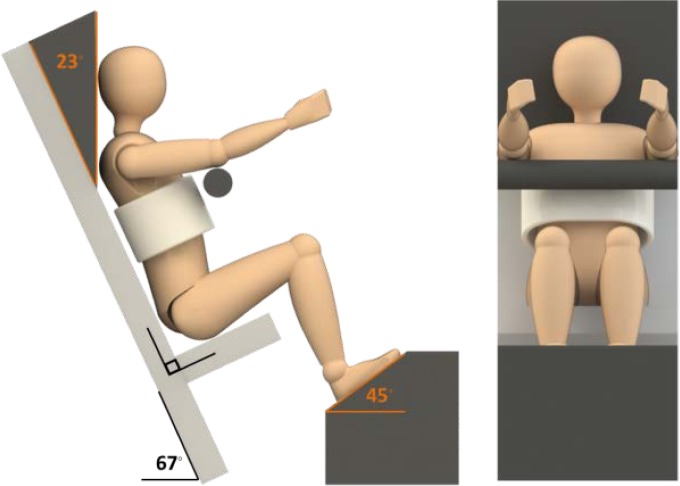
For the supine posture, the subject was lying horizontally, and for the seated posture, the seat back angle was 23 degrees from vertical [Figure 1]. The seated posture was chosen from seating accommodation models [Flannagan, Manary, Schneider et al., 1998] and is representative of a vehicle occupant.
Figure 1.
Seated scan posture for upright MRI.
Supine and upright MRI scans were acquired, and the image data was combined using the software program Amira (Visual Imaging Inc, San Diego, CA) to create a continuous data set from vertebral body T4 to the pelvis. CT images were used for bone segmentation, whereas MRI and uMRI image sets were used in organ segmentation and placement of bone from previously segmented CT images.
Image Segmentation
CT data and the software program Mimics (v. 14, Materialise, Leuven Belgium) were used to segment the sacrum, pelvis, sternum, vertebral bodies T5 through L5, and ribs 5 through 12. Only these respective bones were used due to their proximity to the liver, spleen, and kidneys. Segmented bones were then placed into the MRI supine scan and uMRI scan using repositioning and registration techniques.
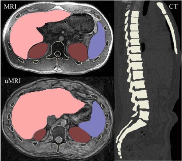
The liver, spleen, and kidneys were segmented in the supine MRI and uMRI scans [Figure 2]. The mask for each organ was used to create a 3D model, and the 3D model was imported into Studio (v.11, Geomagic, Raleigh, NC) for fine adjustments. A final inspection of each organ was conducted in the respective modalities (supine MRI and uMRI) to confirm agreement with scan data.
Figure 2.
Manual segmentation of abdominal organs and placement of bony anatomy in supine MRI (left, top) and uMRI (left, bottom), and manual segmentation of bone in supine CT (right).
Quantifying Rib Coverage
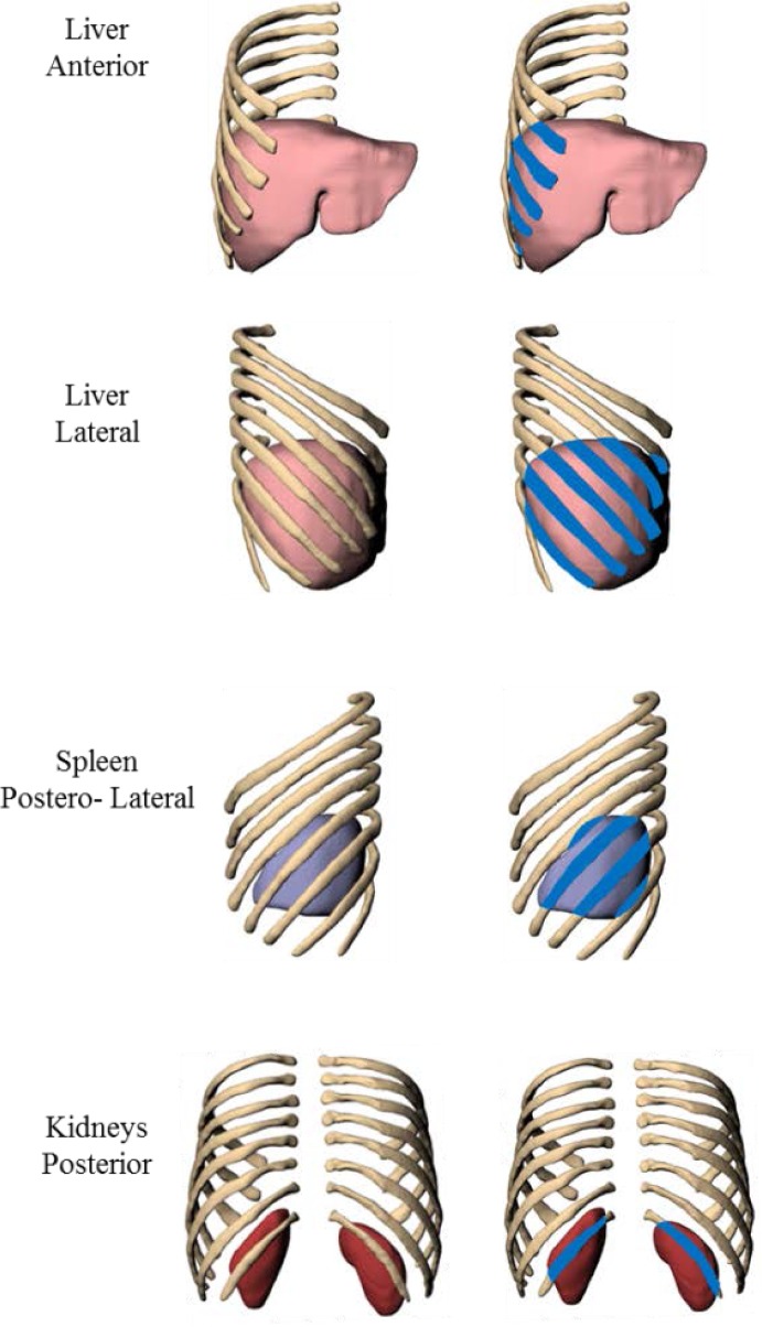
Organ proximity to the rib cage was investigated through rib coverage measurements. Rib coverage is defined as the projected area of the ribs alone onto the respective organ. The projection of ribs 5 through 12 onto the liver, spleen, and kidneys was investigated in the anterior, left, posterior, and right posterior-lateral views for the majority of all scans. Projection views are shown in Figure 3. Views were defined through the scanner acquisition with the exception of the posterior-lateral view, which was defined as approximately 10° rotated posteriorly from the right lateral view along the head-foot axis.
Figure 3.
Projection views and methods for estimating rib coverage. Rib coverage is show in blue. The supine organs of the 95th percentile male subject are shown; the process was repeated for organs in the upright scan for each subject.
Rhinoceros (v4.0, McNeel and Associates, Seattle, WA), a computer-aided software program, was used to quantify area rib coverage. Within each view, polylines were used to define the rib margins. The polylines were projected onto a plane, and the area of coverage was measured. The percent area of rib coverage was calculated by subtracting organ exposure (area not covered by ribs) from the total surface area in the projected view and then dividing that value from the total surface area. Average values between cohorts (supine vs. seated posture) were compared using a Sign Test to identify any significant differences in rib coverage between postures. A p-value less than 0.05 was considered significant.
Organ Location
Vertebral bodies T11 through L2 were used to align the spine and organ structures in the seated posture to the respective anatomy in the supine posture. These bony structures were used due to comparable curvature of the spine in the supine and seated postures. It should be noted that this transformation was only rigid movement, and the relative distance between all anatomical structures was preserved within each individual’s segmented data.
Once the seated anatomy was aligned to the supine anatomy, an analysis was conducted to compare movement of the abdominal organs from the supine to seated position. To examine each organ’s relative location, a local coordinate system was defined [Wu, van der Helm, Veeger et al., 2004; Hayes et al., 2013]. The origin was defined as the midpoint between the center of gravity (CG) of T12 and L1. The x-axis was defined using the most inferior point on the xiphoid process of the sternum, and the z-axis was defined using the CG of vertebral body L2. Differences between the postures were measured, using the CG of each abdominal organ in the local coordinate system.
To examine if CG movement was significant across a single population, bounding box measurements of each organ for all subjects were taken in the anterior-posterior, head-foot, and left-right directions. The bounding box provides the expanse of the organ in the three orthogonal directions of the image space. Using the bounding box measurements of 50th percentile male subject as a reference, scale factors (lambda) were calculated in each direction (anterior-posterior, head-foot, and left-right) for all subjects. The lambda value was defined as the respective bounding box length of the 50th percentile male divided by the bounding box length of each subject. Using the scaled values, a Wilcoxon two-sided rank test was performed to identify any significant effects between postures. The test was performed by comparing the difference between postures to zero. A p-value less than 0.05 was considered significant.
Morphology Comparison
To examine morphology differences, each abdominal organ from the supine and seated postures was imported into a common space, and the organ in the seated position was aligned to the respective organ in the supine posture using a best fit alignment method. Studio was used in for the alignment process and a sample size of 10,000 randomly selected points with a tolerance of 0.1mm was used. Deviation maps were calculated by selecting a reference (supine posture) object and test (seated posture) object. Deviations were reported as the shortest linear distance from the test object to a point on the reference object.
RESULTS
Rib Coverage
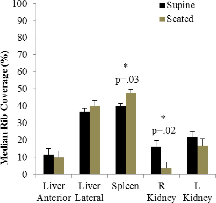
Rib coverage is described in terms of the change from supine to seated posture. On average, rib coverage was found to increase for the liver in the lateral projection and the spleen, but decrease for the kidneys (Table A 2). Minimal change in coverage between postures was seen for the liver in the anterior projection. The percentage of rib coverage for the liver was found to decrease 1% in the anterior view and increase 2.9% in the lateral view. The area of coverage for the spleen increased 7.5%, whereas the right and left kidney had a decrease in rib coverage of 9.1% and 4% (Figure 4). The difference in rib coverage between postures for the spleen and right kidney was found to be significant (p=.03 and p=.02), respectively.
Figure 4.
Median rib coverage between the supine and seated posture. Error bars represent standard error.
Organ Location
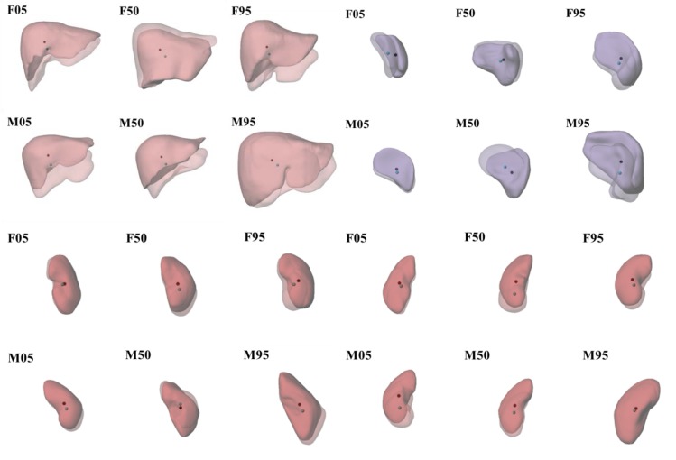
Organ location can be qualitatively examined in Figure 5. The greatest translation for the liver in all subjects was in the head-foot direction, and shows a consistent inferior and medial trajectory. The spleen was seen to translate mostly in the head-foot and medial-lateral directions, with minimal movement in the anterior-posterior direction. Minimal movement was seen in anterior-posterior and head-foot directions for the right and left kidneys with relatively no movement in the medial-lateral direction.
Figure 5.
Postural changes of abdominal organs. The seated posture organs are transparent. The CG is represented by a black circle in the supine posture and blue circle in the seated posture. Liver: top left, spleen: top right, left kidney: bottom left, right kidney: bottom right
Using the local coordinate system, the distance between the CG of each abdominal organ in the supine and seated position was measured in the head-foot, right-left, and anterior-posterior direction. CG movement from the supine to seated position is found in Table A 3, and scaled CG movement is found in Table A 4. While significance was only assessed on the scaled data, a similar response can be found in both data sets. Significant movement in the head-foot direction was seen for all abdominal organs. Right-left movement was significant for the liver and anterior-posterior movement was significant for both the right and left kidney.
Morphology Comparison
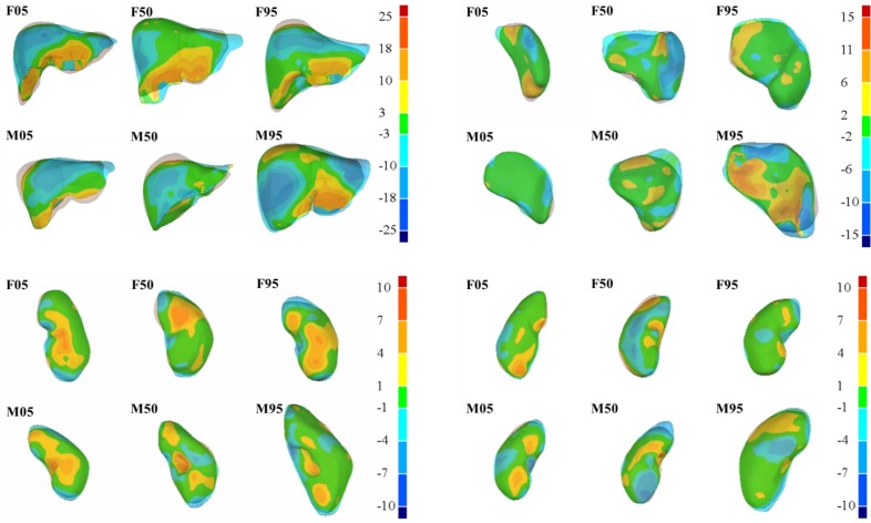
Morphology comparisons between each abdominal organ in the supine and seated postures were also analyzed qualitatively and quantitatively through surface deviation contours. Surface deviation contours of each abdominal organ in the supine position relative to the seated position are shown in Figure 6. The largest deviations from the supine position to the seated were seen in the liver, with surface deviations varying from −14.8mm (compression) to 20.7mm (expansion). Surface deviations of −7.9mm to 7.6mm were seen in the spleen, and similar deviations were seen in the right and left kidneys were variations from −5.2mm to 5.1mm and −5.7mm to 5.3mm.
Figure 6.
Surface deviation analysis between abdominal organs in the supine (contours) and seated (transparent) postures. Positive values indicate expansion (red), and negative values indicate compression (blue). Green indicates minimal or no change in the region. Liver: top left, spleen: top right, left kidney: bottom left, right kidney: bottom right.
DISCUSSION
The purpose of this study was to examine differences in abdominal organ morphology, position and rib coverage due to changes in posture using a multi-modality dataset that contained CT, MRI and uMRI data. The results show that changes from the supine to seated posture significantly affect abdominal organ position for liver, spleen, and kidneys, and rib coverage for the spleen and right kidney. Significant findings relative to abdominal organ movement and spleen and right kidney rib coverage, support data from field studies that show liver, spleen, and kidney injury is prominent in motor vehicle crash [Yoganandan, Pintar, Gennarelli et al., 2000]. Morphology changes are also seen between postures.
The synthesis of multiple modality image sets from each subject allowed for direct quantification of rib coverage. We speculate that rib coverage varies due to relative motion between bones and organs, and morphology changes of the organs themselves between postures. In regards to rib coverage, the kidneys on average had a decrease in coverage, while the liver in the lateral view and spleen had an increase in coverage between postures. The liver in the anterior view had minimal change in coverage between postures. For the spleen, where coverage was seen to increase, there was a significant difference between the supine and seated postures (p=0.03). In addition, coverage between the supine and seated postures for the right kidney were seen to be significance (p=0.02).
Literature studies have shown that the ribs are commonly loaded in a blunt force impact [Siegel, Mason-Gonzalez, Dischinger et al., 1993] and that rib fracture has high association with liver, spleen, and kidney injury [Al-Hassani et al., 2010; Shao, Zou, Li et al., 2013]. Therefore, abdominal organ injury is likely influenced by the proximity of these organs to the chest wall. In the present study we have introduced the quantity of rib coverage as a means of quantifying the surface area of these organs deep to the ribs in a given posture.
While a significant difference was found in rib coverage between postures for the spleen and right kidneys, posture affects rib coverage for the liver and left kidney also. Although a level of significance was not seen in the liver, the findings of the study taken as a whole do suggest changes occur given the significant change in CG location for all organs. It is highly likely for any given simulated impact, the relative placement of the organ with respect to the rib cage will influence the regional deformation, local strains within the organ, and ultimately (if sufficient biomechanical data were available) the prediction of organ injury. While posture-specific scan data is preferred for model development, in the absence of such data, the findings of this study may be used (albeit judiciously) to make minor adjustments to organ position, relative location to the rib cage, and local morphology to account for these changes.
When examining the translation of the CG from the supine to seated posture for the liver, spleen, and kidneys, several significant trends are seen across all subjects [Table A 3, Figure 5]. This analysis was conducted on scaled data per the Methods section, but raw data is also included in the appendix [Table A 4]. For the liver, the largest translation occurs inferiorly, for all subjects. The liver also translates towards the midline, which may be a consequence of spinal flexion. Head-foot and right-left movement of the liver was significant between postures (p=.002 in head-foot direction and p=.04 in right-left direction) [Table A 4]. Although movement of the liver in the anterior-posterior direction was not significant between postures, the dominant trend was movement in the anterior direction.
The right and left kidneys generally moved in the anterior and inferior direction from the supine to seated posture for all subjects (consistent with a flexed spine), and the movement was significant. Movement of the kidneys in the right-left direction was minimal between postures. In regards to the spleen, movement again is inferior with exception to the 50th percentile male subject, and towards the midline with exception to the 5th percentile male who saw minimal movement in the right-left directions. Movement of the spleen in the inferior direction between postures was significant. Similar to the liver, the spleen translation in the anterior-posterior direction was seen to vary, but trend was anterior.
Summarizing, organ CG movement between postures was significant in the left-right and head-foot directions for the liver and head-foot direction for the spleen, whereas significant movement was seen in the anterior-posterior and head-foot directions for the kidneys. The kidneys, which are retroperitoneal, likely had their L-R movement impeded by the spine and lower back muscles. This may in turn cause the greater movement in the anterior-posterior direction than in other organs. The relative distance of the liver and spleen from the spine likely accounts for the significant right-left movement in the liver and spleen that is not seen in the kidneys. All organs showed significant inferior motion which is likely a consequence of the upright posture.
Morphology changes of abdominal organs as a result of posture are in Figure 6. The largest deviations in organ morphology were seen in the liver, with variations up to 25mm between postures. The majority of expansion in the seated liver was seen in the anterior portion of the liver (red), whereas compression occurred mainly in the anterior left and anterior cranial portions of the liver (blue) (Figure 6). The spleen and kidneys in the seated posture also had variations in morphology compared to the supine posture, but were less in magnitude compared to the liver.
Abdominal organ location and morphology changes in the seated posture may affect predicted injury response in computational models used for vehicle crash research. Literature studies report that many abdominal organ injuries in a motor vehicle crash result from interior vehicle components, such as a load from the steering wheel or seat belt [Rouhana and Foster, 1985].. While it is possible to position a full body finite element model within simulation to achieve the proper initial position, these interactions can only be accurately simulated if the internal organ representation is also reasonably representative.
A limitation of this study is that the results are specific to a single individual in each cohort (5th, 50th, or 95th) in terms of height, weight, and anthropometric measurements. However, the data used in this study are not-typical (3 image modalities of full body with roughly 15,000 images collected per individual). In addition, given the volume of data required of each participant in the study, it was not practical to continue this collection for much larger population. These data were collected for use in the development of computational models of various population cohorts, yet the dataset can be used to examine purposes described herein. Despite this limitation, the results indicate the evidence of general trends.
The results show that model developments based on supine images alone are likely to omit significant changes in abdominal organ location, and rib coverage seen in the seated position. As human body computational modeling advances, anatomy changes as a result of posture should be considered to improve the geometrical accuracy of model.
CONCLUSION
Multiple image modalities were acquired from six subjects (3 male and 3 female) representing 3 cohorts (5th, 50th, and 95th percentile) to complete a comparative analysis between the abdominal organs in the supine and seated postures. Segmentation and registration methods were used to complete the comparative analysis. The results indicate that changes in abdominal organ morphology, location and coverage from the ribs should be considered in the development of finite element models in various postures. In regards to organ location, significance of CG movement between postures was seen in the liver, spleen, and kidneys. Head-foot movement was significant for all organs, whereas right-left movement was significant for the liver and anterior-posterior movement for the kidneys. When examining abdominal organ surface contours, morphology changes are seen through local definition of the organs with similar trends observed across subjects. Rib coverage between postures was also found to be significant for the spleen and right kidney. The results of this study show trends across the set, and provide quantitative data for future computational modeling.
Acknowledgments
This study was supported by the Global Human Body Models Consortium (WFU-001). Special thanks to Isma Shah and Amanda Dunn who supported the project through undergraduate work study program at Wake Forest University.
APPENDIX
Table A 1.
Fifteen anthropometric measurements. Subject (S) measurements are in cm and deviations from literature value (Δ) are in %. Positive deviations indicate increase in measurements from literature to subject, and negative deviations indicate decrease in measurements from literature to subject. Average Δ is the absolute average of all measurements for each subject.
| Variable | F05 | F50 | F95 | M05 | M50 | M95 |
|---|---|---|---|---|---|---|
| Sitting Height* | S: 80.0 Δ: +0.6 |
S: 86.4 Δ: +1.4 |
S: 86.4 Δ: −5.1 |
S: 86.8 Δ: +1.6 |
S: 92.1 Δ: +0.7 |
S: 94.1 Δ: −3.1 |
| Hip Breadth* | S: 35.6 Δ: +3.9 |
S: 41.5 Δ: +8.5 |
S: 45.7 Δ: +5.8 |
S: 33.3 Δ: +1.2 |
S: 38.1 Δ: +4.5 |
S: 39.9 Δ: −3.1 |
| Buttock Knee Length* | S: 52.8 Δ: −2.5 |
S: 58.7 Δ: −0.2 |
S: 62.5 Δ: −2.3 |
S: 55.9 Δ: −1.8 |
S: 58.9 Δ: −4.3 |
S: 68.1 Δ: +2.0 |
| Knee Height* | S: 46.0 Δ: −3.0 |
S: 50.8 Δ: −1.2 |
S: 51.4 Δ: −8.2 |
S: 48.8 Δ: −5.2 |
S: 56.1 Δ: +0.6 |
S: 60.5 Δ: −0.2 |
| Bideltoid Breadth* | S: 40.6 Δ: +2.4 |
S: 40.9 Δ: −5.2 |
S: 44.5 Δ: −5.8 |
S: 43.2 Δ: −4.0 |
S: 47.8 Δ: −2.8 |
S: 51.8 Δ: −3.1 |
| Shoulder-Elbow Length† | S: 33.0 Δ: +7.3 |
S: 33.0 Δ: −1.5 |
S: 37.1 Δ: +1.6 |
S: 34.0 Δ: +0.1 |
S: 36.3 Δ: −1.5 |
S: 40.8 Δ: +2.2 |
| Forearm-hand length † | S: 40.4 Δ: −0.6 |
S: 42.7 Δ: −3.5 |
S: 46.0 Δ: −4.7 |
S: 42.2 Δ: −5.8 |
S: 49.8 Δ: +3.1 |
S: 50.8 Δ: −3.1 |
| Waist Circumference † | S: 78.2 Δ: +15.8 |
S: 79.4 Δ: +1.6 |
S: 101.6 Δ: +7.4 |
S: 77.0 Δ: +5.1 |
S: 92.1 Δ: +7.6 |
S: 96.5 Δ: −15.0 |
| Hip Breadth † | S: 31.2 Δ: +1.5 |
S: 34.3 Δ: +0.4 |
S: 37.6 Δ: −1.5 |
S: 30.5 Δ: −1.6 |
S: 33.8 Δ: −1.0 |
S: 35.8 Δ: −4.9 |
| Foot Length † | S: 22.6 Δ: +0.8 |
S: 23.4 Δ: −4.4 |
S: 24.0 Δ: −9.3 |
S: 23.5 Δ: −5.5 |
S: 27.2 Δ: +0.9 |
S: 27.9 Δ: −4.3 |
| Head Breadth | S: 14.5 Δ: +5.9 |
S: 15.0 Δ: +3.9 |
S: 15.0 Δ: −1.8 |
S: 13.7 Δ: −4.1 |
S: 16.4 Δ: +8.0 |
S: 16.0 Δ: −0.5 |
| Head Length | S: 18.8 Δ: +6.6 |
S: 20.3 Δ: +8.4 |
S: 19.8 Δ: +0.4 |
S: 19.1 Δ: +2.7 |
S:19.8 Δ: +0.5 |
S: 20.8 Δ: −0.1 |
| Head Circumference | S: 53.6 Δ: +2.6 |
S: 57.4 Δ: +5.1 |
S: 56.5 Δ: −0.9 |
S: 52.7 Δ: −2.9 |
S: 57.8 Δ: +1.8 |
S: 59.7 Δ: +0.6 |
| Chest Circumference | S: 83.8 Δ: +3.0 |
S: 87.6 Δ: −2.7 |
S: 112.7 Δ: +10.2 |
S: 88.6 Δ: +3.7 |
S: 99.7 Δ: +1.0 |
S: 106.7 Δ: −4.1 |
| Neck Circumference | S: 30.5 Δ: +4.3 |
S: 31.0 Δ: −1.5 |
S: 35.6 Δ: +3.9 |
S: 34.9 Δ: +1.7 |
S: 36.2 Δ: −4.4 |
S: 40.0 Δ: −3.3 |
| Average Δ, % | 4.1 | 3.3 | 4.6 | 3.1 | 2.9 | 2.6 |
: Measured in seated postured
: Measured in the standing posture
Table A 2.
Percent rib coverage for each subject in the supine and seated postures, dimensions in %.
| Subject | Liver Anterior | Liver Lateral | Spleen Postero-Lateral | Right Kidney Posterior | Left Kidney Posterior | |||||
|---|---|---|---|---|---|---|---|---|---|---|
| Supine | Seated | Supine | Seated | Supine | Seated | Supine | Seated | Supine | Seated | |
| F05 | 13.2 | 10.1 | 37.1 | 34.1 | 38.1 | 55.3 | 17.8 | 14.8 | 22.6 | 3.7 |
| F50 | 6.4 | 6.6 | 32.0 | 31.0 | 42.2 | 43.9 | 4.6 | 0 | 3.0 | 0 |
| F95 | 12.7 | 9.7 | 36.4 | 44.5 | 36.0 | 47.4 | 14.7 | 0 | 17.3 | 15.4 |
| M05 | 10.0 | 9.0 | 34.1 | 37.1 | 40.8 | 40.8 | 8.4 | 0 | 22.5 | 17.9 |
| M50 | 31.7 | 31.5 | 44.6 | 51.4 | 45.6 | 48.0 | 27.7 | 7.4 | 24.2 | 27.2 |
| M95 | 10.2 | 11.0 | 39.7 | 43.1 | 39.4 | 51.6 | 22.2 | 18.6 | 21.4 | 22.6 |
Table A 3.
Center of gravity movement of abdominal organs from supine to seated position raw data not scaled. Measurements are presented in mm and positive numbers indicate movement in the anterior, left, or inferior direction. A-P: Anterior-Posterior, L-R: Left-Right, H-F: Head-Foot
| Organ | F05 | F50 | F95 | M05 | M50 | M95 | |
|---|---|---|---|---|---|---|---|
| Liver | ΔA-P | 14.3 | 4.9 | −1.4 | −2.3 | −0.7 | 15.7 |
| ΔL-R | −7.4 | −14.3 | −6.6 | −5.4 | −10.0 | −13.4 | |
| ΔH-F | 21.1 | 19.3 | 18.7 | 24.8 | 19.5 | 14.3 | |
|
| |||||||
| R | 26.5 | 24.6 | 19.9 | 25.5 | 21.9 | 25.1 | |
|
| |||||||
| Spleen | ΔA-P | 20.0 | −2.3 | 5.5 | 0.3 | −2.9 | 28.5 |
| ΔL-R | 15.2 | 5.7 | 3.5 | −0.6 | 12.0 | 2.6 | |
| ΔH-F | 2.7 | 2.2 | 11.3 | 6.8 | −13.1 | 25.5 | |
|
| |||||||
| R | 25.2 | 6.5 | 13.1 | 6.9 | 18.1 | 38.3 | |
|
| |||||||
| Right Kidney | ΔA-P | 7.2 | 7.5 | 6.2 | 22.0 | 15.1 | 17.7 |
| ΔL-R | −3.1 | 2.0 | −3.4 | 0.2 | 3.9 | 2.7 | |
| ΔH-F | 7.6 | 26.6 | 11.4 | 28.5 | 17.0 | 5.9 | |
|
| |||||||
| R | 10.9 | 27.7 | 13.5 | 36.0 | 23.1 | 18.8 | |
|
| |||||||
| Left Kidney | ΔA-P | 13.2 | 1.2 | 9.1 | 6.6 | 3.0 | 11.4 |
| ΔL-R | 3.6 | −3.2 | 7.8 | −3.5 | 0.5 | −5.7 | |
| ΔH-F | 4.8 | 11.1 | 9.0 | 11.7 | −6.2 | 13.7 | |
|
| |||||||
| R | 14.5 | 11.6 | 15.0 | 13.9 | 6.9 | 18.7 | |
Table A 4.
Center of gravity movement of abdominal organs from supine to seated position scaled to the 50th percentile male subject. Measurements are presented in mm and positive numbers indicate movement in the anterior, left, or inferior direction. A-P: Anterior-Posterior, L-R: Left-Right, H-F: Head-Foot
| Organ | F05 | F50 | F95 | M05 | M50 | M95 | |
|---|---|---|---|---|---|---|---|
| Liver | ΔA-P | 8.3 | 0.2 | −6.0 | −3.0 | −0.7 | 8.5 |
| ΔL-R* | −8.1 | −9.6 | 0.8 | −6.4 | −10.0 | −12.2 | |
| ΔH-F* | 18.2 | 15.4 | 14.9 | 23.5 | 19.5 | 10.4 | |
|
| |||||||
| R | 21.6 | 18.1 | 16.0 | 24.5 | 21.9 | 18.2 | |
|
| |||||||
| Spleen | ΔA-P | 17.6 | −1.6 | 1.7 | −0.5 | −2.9 | 15.0 |
| ΔL-R | 0.6 | −4.2 | 3.6 | −7.0 | 12.0 | −20.5 | |
| ΔH-F* | 5.1 | 4.6 | 13.0 | 10.2 | −13.1 | 21.5 | |
|
| |||||||
| R | 18.3 | 6.4 | 13.6 | 12.3 | 18.1 | 33.3 | |
|
| |||||||
| Right Kidney | ΔA-P* | 9.2 | 4.9 | 4.4 | 16.7 | 15.1 | 16.8 |
| ΔL-R | −3.3 | 5.1 | −1.1 | 8.9 | 3.9 | 1.9 | |
| ΔH-F* | 4.3 | 30.5 | 9.2 | 33.2 | 17.0 | 3.6 | |
|
| |||||||
| R | 10.7 | 31.4 | 10.2 | 38.2 | 23.1 | 17.3 | |
|
| |||||||
| Left Kidney | ΔA-P* | 13.6 | 1.1 | 7.6 | 6.1 | 3.0 | 9.9 |
| ΔL-R | 1.3 | −4.3 | 1.1 | −6.2 | 0.5 | −1.2 | |
| ΔH-F* | 2.8 | 10.6 | 8.3 | 13.5 | −6.2 | 9.4 | |
|
| |||||||
| R | 13.9 | 11.5 | 11.3 | 16.0 | 6.9 | 13.7 | |
Identifies significant difference (p<0.05) between supine and seated posture CG movement
REFERENCES
- Al-Hassani A, Abdulrahman H, Afifi I, et al. Rib fracture patterns predict thoracic chest wall and abdominal solid organ injury. Am Surg. 2010;76(8):888–91. [PubMed] [Google Scholar]
- Beillas P, Lafon Y, Smith FW. The effects of posture and subject-to-subject variations on the position, shape and volume of abdominal and thoracic organs. Stapp Car Crash J. 2009;53:127–54. doi: 10.4271/2009-22-0005. [DOI] [PubMed] [Google Scholar]
- Danelson KA, Geer CP, Stitzel JD, et al. Age and gender based biomechanical shape and size analysis of the pediatric brain. Stapp Car Crash J. 2008;52:59–81. doi: 10.4271/2008-22-0003. [DOI] [PubMed] [Google Scholar]
- Flannagan C, Manary M, Schneider L, et al. An Improved Seating Accommodation Model with Application to Different User Populations. SAE Technical Paper. 1998 [Google Scholar]
- Funk JR, Srinivasan SC, Crandall JR, et al. The effects of axial preload and dorsiflexion on the tolerance of the ankle/subtalar joint to dynamic inversion and eversion. Stapp Car Crash J. 2002;46:245–65. doi: 10.4271/2002-22-0013. [DOI] [PubMed] [Google Scholar]
- Gayzik F, Yu M, Danelson K, et al. Quantification of age-related change of the human rib cage through geometric morphometrics. J Biomech. 2008;41(7):1545–54. doi: 10.1016/j.jbiomech.2008.02.006. [DOI] [PubMed] [Google Scholar]
- Gayzik FS, Moreno DP, Danelson KA, et al. External Landmark, Body Surface, and Volume Data of a Mid-Sized Male in Seated and Standing Postures. Ann Biomed Eng. 2012;40(9):2019–32. doi: 10.1007/s10439-012-0546-z. [DOI] [PubMed] [Google Scholar]
- Gayzik FS, Moreno DP, Geer CP, et al. Development of a full body CAD dataset for computational modeling: a multi-modality approach. Ann Biomed Eng. 2011;39(10):2568–83. doi: 10.1007/s10439-011-0359-5. [DOI] [PubMed] [Google Scholar]
- Gordon C, Churchill T, Clauser C, et al. Anthropometric Survey of U.S. Army Personnel: Methods and Summary Statistics. Army Natick Research Development and Engineering Center; Natick, MA: 1989. [Google Scholar]
- Hayes AR, Gayzik FS, Moreno DP, et al. Comparison of Organ Location, Morphology, and Rib Coverage of a Midsized Male in the Supine and Seated Positions. Computational and Mathematical Methods in Medicine. 2013. [DOI] [PMC free article] [PubMed]
- Iwamoto M, Nakahira Y, Tamura A, et al. Development of Advanced Human Models in THUMS; 6th European LS-DYNA Users’ Conference; Gothenburg, Sweden. 2007. [Google Scholar]
- Kemper A, Santago A, Sparks J, et al. Multi-scale biomechanical characterization of human liver and spleen; 22nd International Technical Conference on the Enhance Safety of Vehicles; Washington, DC. 2011. [Google Scholar]
- Kemper AR, McNally C, Pullins CA, et al. The biomechanics of human ribs: material and structural properties from dynamic tension and bending tests. Stapp Car Crash J. 2007;51:235–73. doi: 10.4271/2007-22-0011. [DOI] [PubMed] [Google Scholar]
- Klinich KD, Flannagan CAC, Nicholson K, et al. Abdominal Injury in Motor-Vehicle Crashes. University of Michigan Transportation Research Institute; 2008. [Google Scholar]
- Lafon Y, Smith FW, Beillas P. Combination of a model-deformation method and a positional MRI to quantify the effects of posture on the anatomical structures of the trunk. J Biomech. 2010;43(7):1269–78. doi: 10.1016/j.jbiomech.2010.01.013. [DOI] [PubMed] [Google Scholar]
- NHTSA . Traffic Safety Facts 2010: A Compilation of Motor Vehicle Crash Data from the Fatality Analysis Reporting System and the General Estimates System. US Department of Transportation; Washington, DC: 2010. [Google Scholar]
- Robin S. Human Model for Safety – A joint effort towards the development of redefined human-like car-occupant models. 17th International Conference for the Enhanced Safety of Vehicles; Amsterdam, Germany. 2001. [Google Scholar]
- Rouhana SW, Foster ME. Later Impact - An Analysis of the Statistics in the NCSS. Stapp Car Crash Journal. 1985 [Google Scholar]
- Ruan JS, El-Jawahri R, Rouhana SW, et al. Analysis and evaluation of the biofidelity of the human body finite element model in lateral impact simulations according to ISO-TR9790 procedures. Stapp Car Crash J. 2006;50:491–507. doi: 10.4271/2006-22-0018. [DOI] [PubMed] [Google Scholar]
- Rudd R, Crandall J, Millington S, et al. Injury tolerance and response of the ankle joint in dynamic dorsiflexion. Stapp Car Crash J. 2004;48:1–26. doi: 10.4271/2004-22-0001. [DOI] [PubMed] [Google Scholar]
- Shao Y, Zou D, Li Z, et al. Blunt liver injury with intact ribs under impacts on the abdomen: a biomechanical investigation. PLoS One. 2013;8(1):e52366. doi: 10.1371/journal.pone.0052366. [DOI] [PMC free article] [PubMed] [Google Scholar]
- Shigeta K, Kitagawa Y, Yasuki T. Development of next generation human FE model capable of organ injury prediction. 2009. Enhanced Safety of Vehicles, Stuttgart, Germany, NHTSA.
- Shweiki E, Klena J, Wood GC, et al. Assessing the true risk of abdominal solid organ injury in hospitalized rib fracture patients. J Trauma. 2001;50(4):684–8. doi: 10.1097/00005373-200104000-00015. [DOI] [PubMed] [Google Scholar]
- Siegel JH, Mason-Gonzalez S, Dischinger P, et al. Safety belt restraints and compartment intrusions in frontal and lateral motor vehicle crashes: mechanisms of injuries, complications, and acute care costs. J Trauma. 1993;34(5):736–58. 758–9. doi: 10.1097/00005373-199305000-00017. discussion. [DOI] [PubMed] [Google Scholar]
- Urban JE, Maldjian JA, Whitlow CT, et al. A method to investigate the size and shape variation of the lateral ventricles with age. Biomed Sci Instrum. 2012;48:447–53. [PubMed] [Google Scholar]
- Vavalle NA, Moreno DP, Rhyne AC, et al. Lateral Impact Validation of a Geometrically Accurate Full Body Finite Element Model for Blunt Injury Prediction. Ann Biomed Eng. 2012 doi: 10.1007/s10439-012-0684-3. [DOI] [PubMed] [Google Scholar]
- Weaver AA, Moody EA, Armstrong EG, et al. Image segmentation and registration algorithm to collect homologous landmarks for age-related thoracic morphometric analysis - biomed 2011. Biomed Sci Instrum. 2011;47:70–5. [PubMed] [Google Scholar]
- World Health Organization . Global Status Report on Road Safety. Geneza, Switzerland: 2009. [Google Scholar]
- Wu G, van der Helm FCT, Veeger HEJ, et al. ISB recommendation on definitions of joint coordinate systems of various joints for the reporting of human joint motion—Part II: shoulder, elbow, wrist and hand. Journal of Biomechanics. 2004;38(5):981–992. doi: 10.1016/j.jbiomech.2004.05.042. [DOI] [PubMed] [Google Scholar]
- Yoganandan N, Pintar FA, Gennarelli TA, et al. Patterns of abdominal injuries in frontal and side impacts. Annu Proc Assoc Adv Automot Med. 2000;44:17–36. [PMC free article] [PubMed] [Google Scholar]


