Abstract
OBJECTIVE:
To carry out a biomechanical study of four techniques for fixation of four-part humeral head fractures.
METHODS:
The fracture was reproduced in 40 plastic humeri, divided into groups of ten according to the fixation technique, each one employing different fixation resources, in different configurations. The humeral models were mounted on an aluminum scapula, with leather straps simulating the rotator cuff tendons, and submitted to bending and torsion tests in a universal testing machine, using relative stiffness as an evaluation parameter. Assemblies with intact humeri were analyzed for comparison.
RESULTS:
The biomechanical behavior of the fixation techniques varied within a wide range, where the assemblies including the DCP plate and the 4.5mm diameter screws were significantly more rigid than the assemblies with the Kirschner wires and the 3.5mm diameter screws.
CONCLUSION:
The four fixation techniques were able to bear loads compatible with the physiological demand, but those with higher relative stiffness should be preferred for clinical application.
Laboratory investigation
.
Keywords: Shoulder fractures; Fracture fixation, internal; Biomechanics
INTRODUCTION
High energy accidents have increased over the last decades, resulting in an increased incidence of severe fractures and fracture-dislocations, including those affecting the proximal end of the humerus in young and middle-aged patients, with great potential for functional sequelae. Circa 80% of the fractures of the proximal end of the humerus are two-fragment fractures, with no significant displacement, most of which are stable and suitable for some type of closed conservative or functional treatment. The remaining 20% are three- or four-fragment unstable deviated fractures, quite often compromising the blood supply to the humerus head dome, with consequent necrosis. 1 , 2 Four-fragment fractures may be a quite challenging problem concerning both precise diagnosis and treatment. Diagnostic problems may be overcome through a CT scan with three-dimensional reconstruction, which clearly shows the number and size of the fragments, and MRI, which can demonstrate whether the humeral head dome is avascular or not. Despite the introduction of new treatment options and techniques, treatment of four-part fractures is still controversial. 3 Conservative measures are not appropriate for displaced fractures, because they lead to painful mal-union and, unstable or stiff shoulder in most cases. In elderly patients with osteoporotic bones and a sedentary life style, the results of the conservative or surgical treatment are closely similar to each other and therefore the latter should not be routinely indicated. 4 In younger active patients, with good quality bone stock, surgical treatment is preferred, thus permitting early rehabilitation measures and leading to better functional results. 5
Minimal osteosynthesis techniques have been developed for the four-part fractures in order to avoid the excessive soft tissue damage of extensive surgical exposures and to avoid compromise of the blood supply to the entire bone. 6 Satisfactory results have been reported with the use of such techniques, particularly concerning pain relief and function. Avascular necrosis of the head dome fragment is a frequent complication, regardless of the type of treatment and fixation technique, and most authors agree that it is quite often an asymptomatic condition, not requiring any further surgical measure. 1 , 6 - 8
Percutaneous pinning, bone sutures, tension band wiring, intramedullary nailing, fragment specific screw fixation, and various types of plates (T-shaped, angled and blocked plates) are among the proposed fixation techniques for such complex fractures, but there is no consistent evidence about the best alternative for active patients. 1 , 5 Actually, the mechanical resistance of different fixation techniques has been studied, but the results obtained in different studies do not authorize the general and unrestricted use of such techniques in clinical situations, considering the different methodology used in each study. 5 , 9 , 10
Therefore, it is our opinion that the minimal fixation for the four-part fractures of the proximal end of the humerus is still a controversial issue regarding the mechanical behavior of different types of fixation, and that deserves further investigation.
In the present study, a new biomechanical model involving an aluminum scapula and synthetic humeri was developed to allow closer-to-real biomechanical essays. The synthetic humeri were fixed onto the aluminum scapulae by means of leather straps corresponding to the supraespinatus, infraespinatus and subscapularis tendons and lower capsula, and four different techniques for minimal fixation of the four-part fractures of the proximal end of the synthetic humeri have been used.
MATERIAL AND METHODS
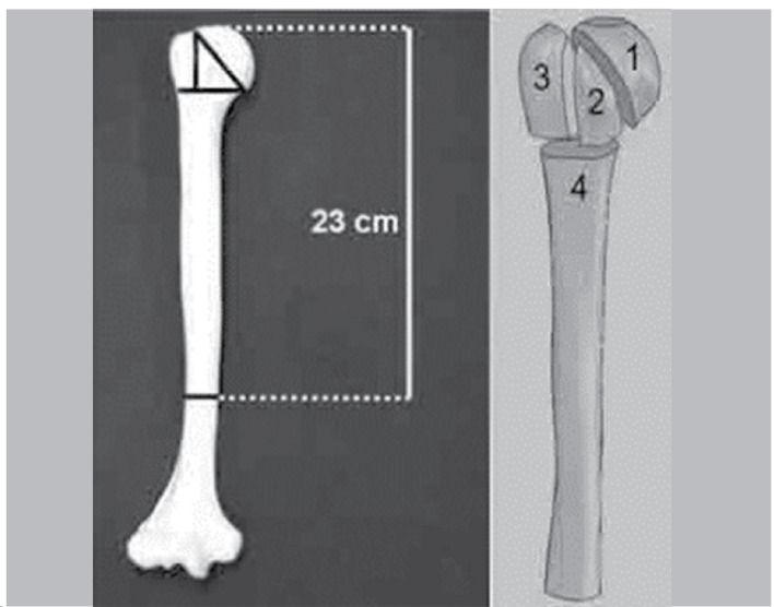
The first step of the investigation was to design a close to real model of the shoulder joint. A plastic human scapula and humeri (Nacional Ossos(r), Jaú, Brazil*), currently used for osteosynthesis drills, were used. Several clay molds were made using the plastic scapula as model and then used to produce aluminum scapulae in a local foundry. After finishing and polishing, three 4.5 mm diameter holes were drilled into the aluminum scapulae, on the supraespinatus fossa, infraespinatus and subscapulary fossae and in the lower face of the glenoid, later used for the fixation of leather straps mimicking the rotator cuff tendons and the axillary capsular recess (Figure 1). The synthetic humeri had their distal end removed 23 cm below the humeral head in order to facilitate fixation into the universal testing machine; the four-part fractures were prepared by making appropriate osteotomies at the anatomical neck, surgical neck and along the bicipital groove (Figure 2). The necessary number of 25mm wide straps of four different lengths were cut out from a 1mm-thick calf leather skin normally used for upholstery purposes. Ten 100mm-long straps were collected at random and checked for thickness regularity with a precision pachymeter and analyzed for mechanical behavior until rupture with the universal testing machine (EMIC(r), model DL10000, São José dos Pinhais, PR, Brazil**) linked to a computer equiped with specific software (Tesc(r), EMIC, São José dos Pinhais/PR, Brazil**), which allows the acquisition of data referring to time, load and deformation that automatically calculates the mechanical properties of the material, including maximum load and deformation (elongation), relative rigidity and tenacity. Thickness was found to be quite uniform (~1mm), but mechanical properties varied within a considerable wide range (Table 1), although without significant difference (p>0.05), making therefore the leather straps adequate for the purposes of the investigation. Thirteen centimeter long straps were used to mimic the 10cm infraespinatus tendon for both the supraespinatus and subscapularis tendons, and 5cm long straps for the axillary capsular recess. The straps were fixed into the greater and lesser tuberosities of the previously prepared bones model with a self-curing cyanoacrylate-ester glue, so as to mimic the insertions of the corresponding tendons, as mentioned above. The opposite end of the leather straps was fixed into the aluminum scapula with bolts and nuts, directly into the previously drilled holes, as mentioned above.
Figure 1. The anterior face of the aluminum scapula, showing the hole for fixation of both "subscapularis" and "infraspinatus" leather straps that mimic the tendons.
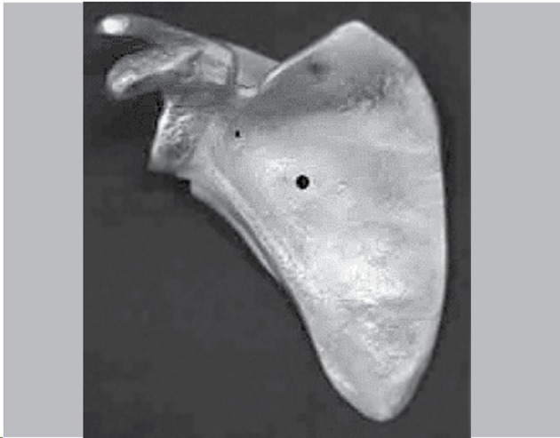
Figure 2. Diagram depicting the resection osteotomy of the distal end of the humeri (a) and the fragments of the four-part fracture (b): the head dome (1), lesser tuberosity (2), greater tuberosity (3) and diaphysis (4).
The fractures were assembled according to four different fixation techniques, as follows:
Group 1 (n=20): Intact humerus control models.
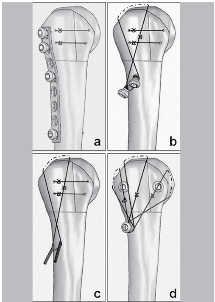
Group 2 (n=20): Fixation with an 8-hole 3.5mm DCP steel plate (Synthes(r), Limeira, Brazil), combined with two transosseous sutures between the lesser and greater tuberosities with caliber 5 braided polyester sutures. Two 40mm-long 3.5mm screws (holes 1 and 2) were inserted into the head dome and two other 25mm-long screws (holes 4 and 8) were fixed into the diaphyseal fragment (Figure 3a).
Figure 3. The four different assemblies for fixation of the four-part fractures: two transosseous sutures between the lesser and greater tuberosities and the 3.5 mm DCP plaque with the proximal screws directed to the head dome (a); two transosseous sutures between the lesser and greater tuberosities, two 4.5 mm cortical screws toward the head dome and one cerclage around the "supraespinatus" (b); two transosseous sutures between the lesser and greater tuberosities, two Kirschner wires toward the head dome and one cerclage around the "supraespinatus" (c); and two transosseous sutures between the lesser and greater tuberosities, two 3.5 mm cortical screws toward the head dome and one cerclages for each "tendon"(d).
Group 3 (n=20): Fixation with two 4.5 mm in diameter (50 and 55 mm-long) cortical screws from the diaphyseal fragment toward the humeral head dome, combined with two transosseous sutures between the greater and lesser tuberosity and one figure-of-eight cerclage between the screw heads, below, and the leather strap mimicking the supraespinatus tendon, above, with caliber 5 braided polyester sutures (Figure 3b).
Group 4 (n=20): Fixation with two 2 mm-thick Kirschner wires, introduced from the diaphyseal fragment toward the head dome, with two transosseous sutures between the lesser and greater tuberosities and one figure-of-eight cerclage between the wires, below, and the leather strap mimicking the supraespinatus tendon, above, with caliber 5 braided polyester sutures (Figure 3c).
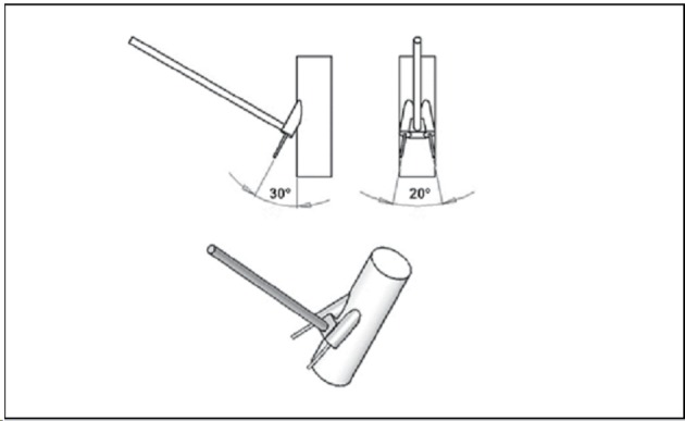
Group 5 (n=20): Fixation of both the greater and lesser tuberosity onto the head dome with two (one for each) 45 mm-long 3.5 mm cortical screws, combined with three figure-of-eight cerclages between the supraespinatus, infraespinatus and subscapularis straps and a third screw with a washer transversely inserted into the diaphysis, with caliber 5 braided polyester (Figure 3d). A special guide was designed to facilitate the introduction of both 4.5 mm screws and 2mm Kirschner wires, from the lateral aspect of the diaphyseal fragment into the head dome, in Groups 3 and 4, respectively (Figure 4).
Figure 4. Details of the guide used for insertion of the 4.5 mm cortical screws and Kirschner wires.
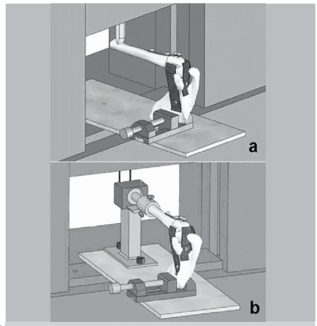
The bending and torsion essays were carried out with the same universal testing machine mentioned above, using ten assemblies for each essay, in all groups. The bending essays were planned to simulate resisted shoulder abduction and were performed with the aluminum scapula fixed onto a vise with the humerus on the horizontal position, the load being vertically applied to the humeral shaft from above at 200mm from the center of the humeral head (Figure 5a). A 5 N pre-load force was applied for 30 sec for system accommodation and the actual load was then continuously applied at the rate of 20mm/min until failure, usually a sudden fall in the applied load. Each assembly was analyzed once and the analyzed property was the relative rigidity (N/mm). An average value was calculated for the ten individual values in each group and used for comparisons. The torsion essays were planned to simulate resisted shoulder internal rotation and were also performed with the scapula fixed into the vise, but with the humeral shaft fixed into a rotary accessory connected to the load cell through a chain (Figure 5b). A 30 N pre-load was applied for 30 sec for system accommodation and the actual load was continuously applied at the rate of 20 mm/min rate, until failure, also a sudden fall in the applied load. The property analyzed was the relative torsion rigidity (N/º) and an average was calculated for the ten individual values in each group and used for comparisons.
Figure 5. Assemblies prepared for the bending (a) and torsion (b) tests. The aluminum scapula is fixed into a vise with the humeral diaphysis in the horizontal position, the load being applied at its opposite end in both cases.
Statistical analysis was carried out using the GraphPad Prism(r) version 5.0 software for analysis of data normality. One-way analysis of variance (ANOVA) and Tukey's post-test were then used for the parametric data, while the Kruskal-Wallis test and Dunn's post-test were used for data with a non-parametric behavior, at the 5% level of significance (p<0.05).
RESULTS
General findings: both for the bending and torsion tests, the load increased more or less uniformly during the elastic phase, then entering in the plastic phase and rapidly fail, usually with a sudden fall in the value of the applied burden, for the majority of the assembling. The failure occurred in the site of fixation, never in the leather strips or in its joint with the scapula or the humerus. However, in four assembling of Group 3 the failure was due to an oblique fracture towards the inside and up from the the most distal screw holes, probably due to concentration of stress in this site.
Biomechanical properties of the assemblies
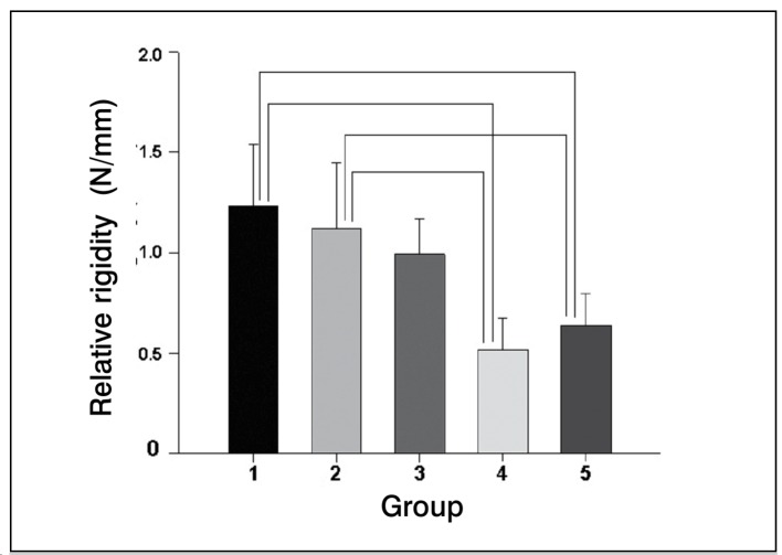
Bending essays: the average relative rigidity was 1.23 N/mm (range: 0.89 - 1.86 N/mm) in Group 1 (intact humeri), 1.12 N/mm (range: 0.59 - 1.65 N/mm) in Group 2 (plate), 0.95 N/mm (range: 0.64 - 1.31 N/mm) in Group 3 (4.5 mm cortical screws + cerclage), 0.51 N/mm (range: 0.31 - 0.68 N/mm) in Group 4 (K-wires + cerclage), and 0.64 N/mm (range: 0.40 - 0.86 N/mm) in Group 5 (3.5mm + cerclage) (Figure 6). Differences were significant (p<0.05) between Groups 1x4, 1x5, 2x4 and 2x5, but not (p>0.05) between Groups 1x2, 1x3, 2x3, 3x4, 3x5 and 4x5 (Figure 6).
Figure 6. Average values of relative rigidity of the bending essays, according to each group. Statistical differences (p<0.05) are indicated by the connecting lines. The higher performance of Groups 2 and 3 over Groups 4 and 5 is evident.
Torsion essays
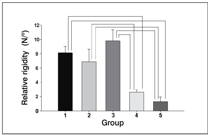
The average relative torsion rigidity was 8.11 N/º (range: 6.85 - 17.44 N/º) in Group 1; 6.89 N/º (range: 4.38 - 9.43 N/º) in Group 2; 9.79 N/º (range: 7.23 - 11.97 N/º) in Group 3; 2.63 N/º (range: 2.1 - 3.12 N/º) in Group 4; and 1.31 N/º (range: 0.6 - 2.49 N/º) in Group 5 (Fig 7). Differences were significant (p<0.05) between groups 1x4; 1x5; 2x4; 2x5; 3x4 and 3x5, but not (p>0.05) between Groups 1x2; 1x3; 2x3 and 4x5 (Figure 7).
Figure 7. Average values of relative rigidity of the torsion essays, according to each group. Statistical differences (p<0.05) are indicated by the connecting lines. The superior performance of Group 3 over Group 2 is evident, with Groups 4 and 5 presenting a much lower performance.
DISCUSSION
Whether or not associated with dislocation of the gleno-humeral joint, the four-part fractures of the proximal end of the humerus represent a very complex situation to deal with, from diagnosis to treatment. Surgical treatment is usually indicated, based in different arguments, particularly the need for an anatomical reduction of the articular fracture or to check whether the humeral head dome is somehow attached to soft tissues, which might work as a pathway for blood irrigation; the entirely loose head being considered unviable. Rigid fixation with a fixed-angle plate for a viable and primary arthroplastic replacement of an unviable head is among the current treatment indications, but definitive results and comparisons with other methods are not yet available. Actually, fixed-angle plate fixation is a relatively new resource with many theoretical advantages over other plates and primary arthroplastic replacement, but both are relatively expensive implants, not always available at small centers, a problem which must be properly addressed, particularly in developing countries. Furthermore, the implantation of a fixed-angle plate requires specific training and surgical expertise, otherwise it may not work as planned, sometimes evolving with loosening and loss of reduction. 8 Since functional results are not as good for fractures as for degenerative osteoarthritis, an arthroplastic replacement should only be indicated when the humeral head dome is completely disconnected from the surrounding soft tissues or reduction is impossible or very unsatisfactory. The indication for an arthroplastic replacement is also acceptable for elderly patients with a restricted activity demand and short life expectancy.
Because of the drawbacks mentioned above, techniques for limited open reduction and minimal internal fixation were devised and studied almost 20 years ago. Such techniques are lately undergoing revival, perhaps to become one of the first choices for younger patients or elderly active individuals, based on the observation that their functional results are usually better than those obtained with arthroplastic replacement. Avascular necrosis of the humeral head dome may always occur, but it is usually partial and paucisymptomatic, not to mention the possibility that spontaneous revascularization might occur later, or that the humeral head will be replaced in a second stage. 3 , 4 , 7
Minimal fixation of four-part fractures of the proximal end of the humerus can be achieved with different constructs using screws of different types and diameters, K-wires and metal or synthetic thread cerclage, separately or in combination. 1 , 6 , 7 , 10 , 11
No real fracture can be exactly reproduced in a single biomechanical study like ours and the results of such studies cannot be taken as the definite truth. However, provided the study is carefully planned and carried out, it may contribute to the knowledge on the subject and assist the surgeon in decision making between different techniques, until proper clinical randomized trials clarify the facts. In the particular case of the four-part fracture of the proximal end of the humerus, there seems to be no agreement about the most convenient type of minimal fixation, also because not all types have been comparatively studied, either clinically or by means of biomechanical studies. It is in this slot that our study fits.
The scientific literature is virtually devoid of publications on biomechanical models for the study of complex unstable fractures like ours. However, despite some controversies, the adaptation of a model for simpler stable fractures is a possibility. 12 , 13 Therefore, our model was developed according to the principle of indirect traction application onto the bone fragments through elastic reins, so as to provide the maximum elastic fixation of the greater and lesser tuberosities onto the aluminum scapula and humeral shaft, while the humeral head dome was fixed as rigidly as possible onto the proximal end of the humeral shaft. 9 , 15 , 16
The leather straps used to mimic the rotator cuff tendons gave the model a closer-to-real character, since the leather used was about 5 times as elastic as the untouched bone model, elasticity which permitted an elongation of about 70% of its original length before rupture, which was a sudden event at the end of the elastic phase. During the essays with the assemblies, it was noticed that the straps allowed a certain degree of accommodation, with the humeral shaft showing a slight adduction, followed by a period of stabilization before failure, during which the applied load concentrated at the fracture site. It seemed to us that such a behavior was quite similar to the real situation, in which the rotator cuff tendons stabilize the humeral head against the glenoid before the abduction movement begins and imposes the weight of the entire upper limb on the shoulder by the action of gravity, as simulated by the application of the load on the distal portion of the humeral shaft in the bending essays. Similarly, the torsion loads also tightened the anterior leather strap to a point at which it stopped permitting the external rotation of the proximal end of the humerus and the stress concentrated at the fracture site. That is why the model was designed to have a long lever arm, with both bending and torsion stresses being applied at 20 cm from the rotation center of the humeral head, which increases momentum, particularly for bending stresses. Polyurethane bones were used because they are a quite close reproduction of the human humeri, with all of the superficial anatomical details and even the internal structure, such as the typical cancellous bone of the proximal end and the transition between this and the cortical bone of the shaft. Furthermore, the plastic bones are made according to an industrial process and their mechanical properties tend to vary less than they would for natural human bones, usually obtained from autopsies or mortuaries and suffering the influence of a wide age range, body constitution, different health conditions and lifestyle, and so on. On the other hand, the synthetic bones do not present the same viscoelastic properties as the natural bones, thus inducing somewhat different mechanical properties and behavior. 16 Load protocols were applied according to previous studies, 15 , 17 with both the abduction-against-resistance and torsion essays being carried out similarly to the configurations previously proposed. 13 , 18 The four fixation techniques tested are among the ones described in the literature, being currently used in clinical practice. 10 , 19 , 20
Instead of ordinary rigidity, we decided to measure relative rigidity, which is based on the equalization of the load versus deformation graph, which is automatically calculated by the software; in fact, the graph normally obtained is quite irregular, with slight sudden ups and downs, while the equalized graph is rigorously linear, since the software calculates the average between points situated above and below a regular graph line. Furthermore, the relative rigidity closely reflects the other mechanical properties of the construct (maximum load and deformation, rigidity and tenacity), thus facilitating group comparison.
The results obtained here indicate that the studied groups can be divided into two sectors according to performance: Groups 2 and 3 in one, with a higher resistance to both bending and rotational stress, and Groups 4 and 5 in the other, with a slightly lower resistance. The DCP plaque (Group 2) presented a superior performance over the three other techniques in the bending essays, although without significant differences with the 4.5 mm screws combined with a eight shaped cerclage around the rotator cuff tendons (Group 3). The situation changed in the torsion essays, with the assemblies of Group 3 presenting a higher performance, but also without a significant difference compared to those of Group 2. Both techniques presented a similar performance to the intact humeri of Group 1, thus demonstrating that their resistance is adequate to clinical application for the fixation of the four-part fractures of the proximal end of the humerus, although those from Group 3 should perhaps be preferred, since it is less aggressive than that of Group 2. However, it should be emphasized that a diaphyseal fracture occurred in four constructs of Group 3, beginning at the point of entry of the most distal screw, certainly because of stress concentration at this point, a feature not observed with Groups 2 and 4 assemblies. Actually, the 3.5 mm DCP plaque of Group 2 probably absorbs and distributes tensions along the diaphysis, thus preventing stress concentration at one specific point, while the Kirschner wires of Group 4, introduced according to the same configuration as that of Group 3, are of a much smaller diameter than the 4.5 mm screws, therefore not favoring stress concentration at the entry holes.
On the other hand, our general results also indicated that the assemblies of Groups 4 and 5 supported bending and torsion loads to a level that indicates that both can well be used without great fear that they would quickly fail, provided protective and restraining measures are taken during the first three or four postoperative weeks. 15 , 17 In this respect, it must be considered that our model was relatively stable, since the flat distal surface of the two extra-articular proximal fragments sat on the flat osteotomy surface of the distal fragment, both transverse to the long axis of the humeral shaft, thus resulting in a considerable resistance against longitudinal compression forces. In the real situation in humans, there usually are some comminuted fragments around the main fracture lines, with contingent loss of the medial buttress due to impaction, in which case anatomical reduction is virtually impossible and stability is certainly less than in our constructs. Comparison with the results of other investigations is difficult, since different types of fractures, implants, experimental models, load application and definition of failure were used and considered. 22 At least twice as high rigidity as observed here was measured in two-part fractures of the proximal end of the humerus fixed with four 3 mm-thick self-tapping threaded Shanz-type pins in four different configurations; however, besides the different fracture type, far more stable than the four-part fracture, the assemblies were tested in the upright position, with an eccentric load being applied from the head down. 22 On the other hand, results similar to ours have been reported, with plaques showing a superior biomechanical behavior compared to K-wires and tension bands used separately or in combination. 11
Actually, according to the literature, the plaques seem to be uniformly more rigid than any other type of fixation, 23 supporting loads well above the natural demands, and studies comparing different types of plaques emphasize rigidity, as if this were the most important feature to be pursued. 9 , 12 , 19 However, other studies have emphasized the need for a balance between rigidity, to promote stability, and elasticity, to prevent loosening, together with the reduction of the posteromedial portion of the fracture line. 19 , 24 , 25
Besides the mechanical properties of the fixation system, other factors can also influence the final result of a surgical treatment. Patient's profile, correct diagnosis, fracture type and personality, bone stock and inherent vascular impairment, associated lesions, personal experience, quality of reduction, implant choice and rehabilitation program should always be the surgeon's concern and responsibility.
CONCLUSION
The four techniques are capable of supporting physiological loads and can be used in clinical situations in humans, but the 3.5 mm DCP plaque fixation and the 4.5 mm screws combined with a cerclage were the most relatively resistant assemblies. The biomechanical model used was adequate for the purposes of the study and led to reliable and reproducible results.
Footnotes
Research performed at the Bioengineer Laboratory, Department of Biomechanics, Medicine and Rehabilitation of the Locomotor System, Faculdade de Medicina de Ribeirão Preto, Universidade de São Paulo - Ribeirão Preto, SP, Brazil.
(www.ossos.com.br)
(www.universaltestingmachines.net)
Citation: Graça E, Okubo R, Shimano AC, Mazzer N, Barbieri CH. Biomechanics of four techniques for fixation of the four-part humeral head fracture. Acta Ortop Bras. [online]. 2013;21(1):34-39. Available from URL: http://www.scielo.br/aob.
REFERENCES
- 1.Gerber C, Werner CM, Vienne P. Internal fixation of complex fractures of the proximal humerus. J Bone Joint Surg Br. 2004;86(6):848–855. doi: 10.1302/0301-620x.86b6.14577. [DOI] [PubMed] [Google Scholar]
- 2.Nordqvist A, Petersson CJ. Incidence and causes of shoulder girdle injuries in an urban population. J Shoulder Elbow Surg. 1995;4(2):107–112. doi: 10.1016/s1058-2746(05)80063-1. [DOI] [PubMed] [Google Scholar]
- 3.Bigliani LU, Flatow EL, Pollock RG. Fractures of the proximal humerus. In: Rockwood CA, Green DP, Bucholz RW, Heckman JD, editors. Rockwood and Green's Fractures in Adults. 4. Philadelphia: Lippincott-Raven Publishers; 1996. pp. 1055–1107. [Google Scholar]
- 4.Zyto K, Ahrengart L, Sperber A, Törnkvist H. Treatment of displaced proximal humeral fractures in elderly patients. J Bone Joint Surg Br. 1997;79(3):412–417. doi: 10.1302/0301-620x.79b3.7419. [DOI] [PubMed] [Google Scholar]
- 5.Helmy N, Hintermann B. New trends in the treatment of proximal humerus fractures. Clin Orthop Relat Res. 2006;442:100–108. doi: 10.1097/01.blo.0000194674.56764.c0. [DOI] [PubMed] [Google Scholar]
- 6.Wijgman AJ, Roolker W, Patt TW, Raaymakers EL, Marti RK. Open reduction and internal fixation of three and four-part fractures of the proximal part of the humerus. J Bone Joint Surg Am. 2002;84(11):1919–1925. [PubMed] [Google Scholar]
- 7.Iannotti JP, Ramsey ML, Williams GR Jr, Warner JJ. Nonprosthetic management of proximal humeral fractures. Instr Course Lect. 2004;53:403–416. [PubMed] [Google Scholar]
- 8.Thanasas C, Kontakis G, Angoules A, Limb D, Giannoudis P. Treatment of proximal humerus fractures with locking platesa systematic review. J Shoulder Elbow Surg. 2009;18(6):837–844. doi: 10.1016/j.jse.2009.06.004. [DOI] [PubMed] [Google Scholar]
- 9.Chudik SC, Weinhold P, Dahners LE. Fixed-angle plate fixation in simulated fractures of the proximal humerusa biomechanical study of a new device. J Shoulder Elbow Surg. 2003;12(6):578–588. doi: 10.1016/s1058-2746(03)00217-9. [DOI] [PubMed] [Google Scholar]
- 10.Jakob RP, Miniaci A, Anson PS, Jaberg H, Osterwalder A, Ganz R. Four-part valgus impacted fractures of the proximal humerus. J Bone Joint Surg Br. 1991;73(2):295–298. doi: 10.1302/0301-620X.73B2.2005159. [DOI] [PubMed] [Google Scholar]
- 11.Koval KJ, Blair B, Takei R, Kummer FJ, Zuckerman JD. Surgical neck fractures of the proximal humerusa laboratory evaluation of ten fixation techniques. J Trauma. 1996;40(5):778–783. doi: 10.1097/00005373-199605000-00017. [DOI] [PubMed] [Google Scholar]
- 12.Seide K, Triebe J, Faschingbauer M, Schulz AP, Püschel K, Mehrtens G, Jürgens Ch. Clin Biomech. 2. Vol. 22. Bristol: 2007. Locked vs. unlocked plate osteosynthesis of the proximal humerusa biomechanical study; pp. 176–182. [DOI] [PubMed] [Google Scholar]
- 13.Siffri PC, Peindl RD, Coley ER, Norton J, Connor PM, Kellam JF. Biomechanical analysis of blade plate versus locking plate fixation for a proximal humerus fracturecomparison using cadaveric and synthetic humeri. J Orthop Trauma. 2006;20(8):547–554. doi: 10.1097/01.bot.0000244997.52751.58. [DOI] [PubMed] [Google Scholar]
- 14.Bono CM, Renard R, Levine RG, Levy AS. Effect of displacement of fractures of the greater tuberosity on the mechanics of the shoulder. J Bone Joint Surg Br. 2001;83(7):1056–1062. doi: 10.1302/0301-620x.83b7.10516. [DOI] [PubMed] [Google Scholar]
- 15.Wuelker N, Wirth CJ, Plitz W, Roetman B. A dynamic shoulder modelreliability testing and muscle force study. J Biomech. 1995;28(5):489–499. doi: 10.1016/0021-9290(94)e0006-o. [DOI] [PubMed] [Google Scholar]
- 16.Cristofolini L, Viceconti M, Cappello A, Toni A. Mechanical validation of whole bone composite femur models. J Biomech. 1996;29(4):525–535. doi: 10.1016/0021-9290(95)00084-4. [DOI] [PubMed] [Google Scholar]
- 17.Poppen NK, Walker PS. Forces at the glenohumeral joint in abduction. Clin Orthop Relat Res. 1978;135:165–170. [PubMed] [Google Scholar]
- 18.Carrera EF, Nicolao FA, Netto NA, Carvalho RL, Dos Reis FB, Giordani EJ. A mechanical comparison between conventional and modified angular plates for proximal humeral fractures. J Shoulder Elbow Surg. 2008;17(4):631–636. doi: 10.1016/j.jse.2008.02.003. [DOI] [PubMed] [Google Scholar]
- 19.Szyszkowitz R.Humerus proximalRüedi TP, Murphy WM.AO Principles of fracture management 2ndStuttgart: Georg Thieme Verlag; New York: Thieme New York; 2000271–289. [Google Scholar]
- 20.Tile M. Schatzker J, Tile M. The rationale of operative fracture care. 2. Berlin: Springer Verlag; 1996. Fractures of the proximal humerus; pp. 51–82. [Google Scholar]
- 21.Lill H, Hepp P, Korner J, Kassi JP, Verheyden AP, Josten C, et al. Proximal humeral fractureshow stiff should an implant be? A comparative mechanical study with new implants in human specimens. Arch Orthop Trauma Surg. 2003;123(2-3):74–81. doi: 10.1007/s00402-002-0465-9. [DOI] [PubMed] [Google Scholar]
- 22.Durigan A Jr, Barbieri CH, Mazzer N, Shimano AC. Two-part surgical neck fractures of the humerusMechanical analysis of the fixation with four Shanztype threaded wires in four different assemblies using the femur of swine as a model. J Shoulder Elbow Surg. 2005;14:96–102. doi: 10.1016/j.jse.2004.04.013. [DOI] [PubMed] [Google Scholar]
- 23.Sanders BS, Bullington AB, McGillivary GR, Hutton WC. Biomechanical evaluation of locked plating in proximal humeral fractures. J Shoulder Elbow Surg. 2007;16(2):229–234. doi: 10.1016/j.jse.2006.03.013. [DOI] [PubMed] [Google Scholar]
- 24.Gardner MJ, Weil Y, Barker JU, Kelly BT, Helfet DL, Lorich DG. The importance of medial support in locked plating of proximal humerus fractures. J Orthop Trauma. 2007;21(3):185–191. doi: 10.1097/BOT.0b013e3180333094. [DOI] [PubMed] [Google Scholar]
- 25.Liew AS, Johnson JA, Patterson SD, King GJ, Chess DG. Effect of screw placement on fixation in the humeral head. J Shoulder Elbow Surg. 2000;9(5):423–426. doi: 10.1067/mse.2000.107089. [DOI] [PubMed] [Google Scholar]


