

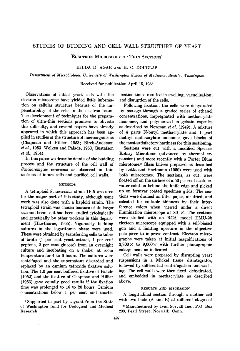
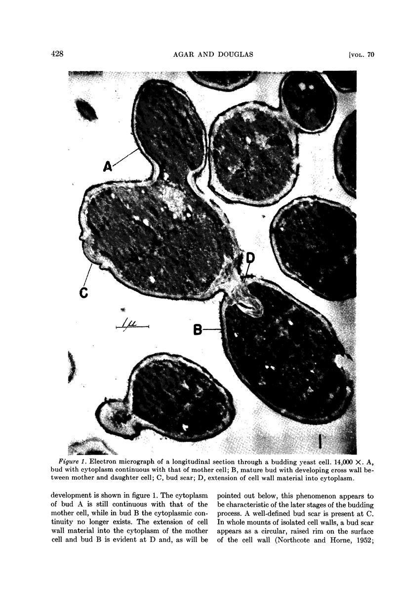
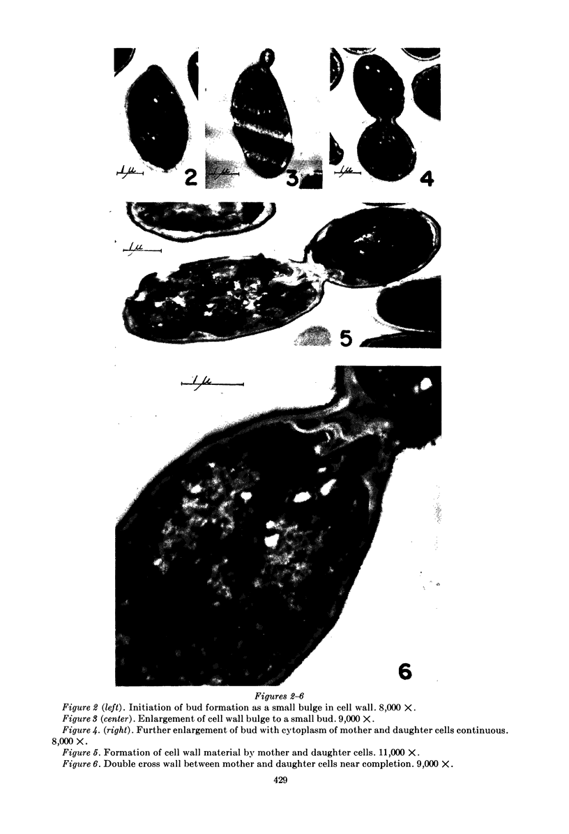
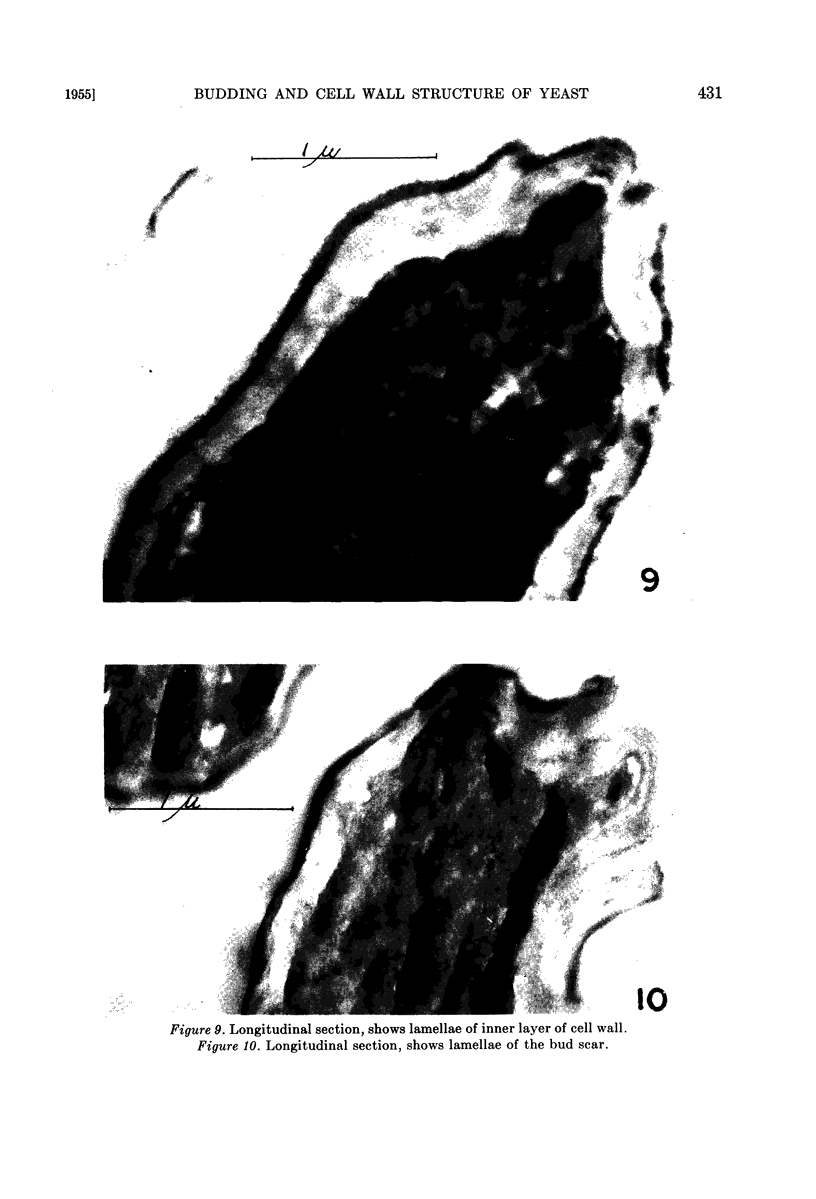
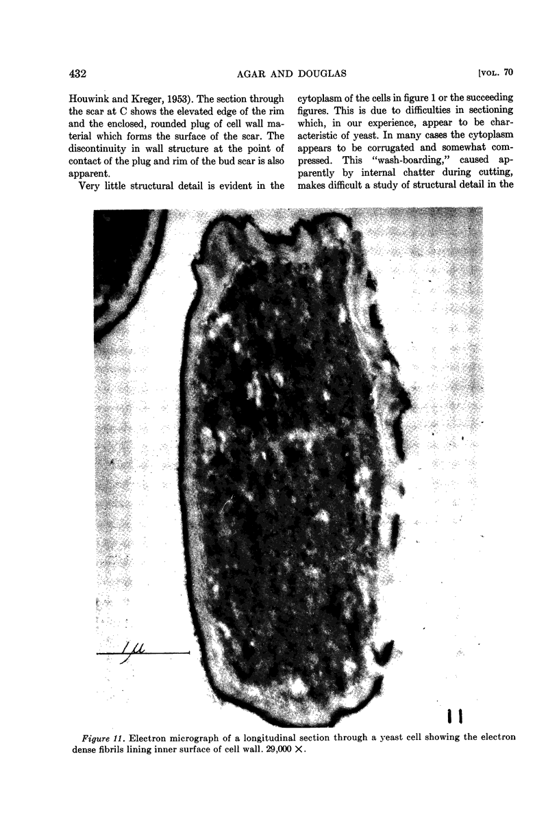
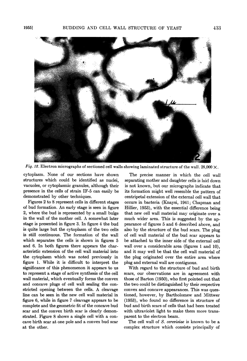
Full text
PDF

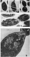
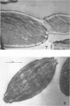
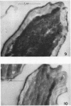
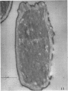
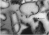
Images in this article
Selected References
These references are in PubMed. This may not be the complete list of references from this article.
- BARTHOLOMEW J. W., MITTWER T. Demonstration of yeast bud scars with the electron microscope. J Bacteriol. 1953 Mar;65(3):272–275. doi: 10.1128/jb.65.3.272-275.1953. [DOI] [PMC free article] [PubMed] [Google Scholar]
- BARTON A. A. Some aspects of cell division in saccharomyces cerevisiae. J Gen Microbiol. 1950 Jan;4(1):84–86. doi: 10.1099/00221287-4-1-84. [DOI] [PubMed] [Google Scholar]
- CHAPMAN G. B., HILLIER J. Electron microscopy of ultra-thin sections of bacteria I. Cellular division in Bacillus cereus. J Bacteriol. 1953 Sep;66(3):362–373. doi: 10.1128/jb.66.3.362-373.1953. [DOI] [PMC free article] [PubMed] [Google Scholar]
- GUSTAFSON P. V., AGAR H. D., CRAMER D. I. An electron microscope study of Toxoplasma. Am J Trop Med Hyg. 1954 Nov;3(6):1008–1022. [PubMed] [Google Scholar]
- HOUWINK A. L., KREGER D. R. Observations on the cell wall of yeasts; an electron microscope and x-ray diffraction study. Antonie Van Leeuwenhoek. 1953;19(1):1–24. doi: 10.1007/BF02594830. [DOI] [PubMed] [Google Scholar]
- Knaysi G. Observations on the Cell Division of Some Yeasts and Bacteria. J Bacteriol. 1941 Feb;41(2):141–153. doi: 10.1128/jb.41.2.141-153.1941. [DOI] [PMC free article] [PubMed] [Google Scholar]
- LATTA H., HARTMANN J. F. Use of a glass edge in thin sectioning for electron microscopy. Proc Soc Exp Biol Med. 1950 Jun;74(2):436–439. doi: 10.3181/00379727-74-17931. [DOI] [PubMed] [Google Scholar]
- NORTHCOTE D. H., HORNE R. W. The chemical composition and structure of the yeast cell wall. Biochem J. 1952 May;51(2):232–236. doi: 10.1042/bj0510232. [DOI] [PMC free article] [PubMed] [Google Scholar]
- PALADE G. E. A study of fixation for electron microscopy. J Exp Med. 1952 Mar;95(3):285–298. doi: 10.1084/jem.95.3.285. [DOI] [PMC free article] [PubMed] [Google Scholar]
- WOLKEN J. J., PALADE G. E. An electron microscope study of two flagellates, chloroplast structure and variation. Ann N Y Acad Sci. 1953 Oct 14;56(5):873–889. doi: 10.1111/j.1749-6632.1953.tb30266.x. [DOI] [PubMed] [Google Scholar]