

Introduction
Two commonly encountered alveolar defects are dehiscence and fenestration .1 An alveolar dehiscence denotes a lack of the facial or lingual alveolar cortical plate resulting in a denuded root surface, while an alveolar fenestration is a circumscribed defect of the cortical plate which exposes the underlying root surface, but does not involve the alveolar margin of the bone.2
Fenestration is an isolated area in which the tooth root is denuded of bone and the root surface is covered only by periosteum and overlying gingiva. Mucosal fenestration is a clinical entity in which the overlying gingiva or mucosa is also denuded thus the root is exposed to the oral cavity. It seems to have a multifactoral origin with relation to decreased thickness of the alveolar housing, labioversion of the tooth in the dental arch, contour of the root apex, occlusal factors, orthodontic tooth movement, periodontal and endodontic pathology and aberrant frenal attachment .3
It has been seen to be commonly associated with the anterior region of the arch, especially incisors. Mucosal fenestrations have been reported in literature but are far less prevalent as compared to normal fenestration.4 Several treatment modalities have been proposed in the literature. These include root planning along with chlorhexidene mouth rinsing, full thickness mucogingival flap with primary closure, pedicle flap surgery, free gingival grafting and guided tissue regeneration.5,6
The following case report describes a rare situation where a mucosal fenestration developed in the upper left central incisor due to chronic periapical inflammation which was successfully treated with combination of bone grafting and free mucosal graft.
Case report
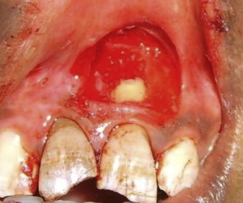
A 20-year-old female patient presented with a complaint of discolored upper front teeth. Patient gave a history of sustaining a fall 5 years ago. On examination, upper right and left central incisors were discolored and also non-vital on pulp testing and radiograph showed periapical radiolucency in both the central incisors. Clinically, upper left incisor had mucosal fenestration measuring approximately 3 mm for which the patient underwent root canal treatment for both the central incisors. However, even after successful endodontic treatment and apicectomy, the fenestration persisted (Fig 1). The patient was then taken for periodontal surgical therapy and a free mucosal graft was planned.
Fig. 1.

Mucosal fenestration seen in relation to upper left central incisor.
Region in relation to 11, 12, 21 & 22 was anesthetized using 2% lignocaine. (LIGNOX 2%A, INDOCO REMEDIES LTD). A partial thickness flap was raised with a no.15 blade and the recipient site was prepared 5 mm apical to fenestration (Fig. 2). Upon preparation of recipient bed periapical bone loss was evident, which was filled with bonegraft (PERIOGLAS, NovaBone products, LLC) to aid in bone regeneration (Fig. 3). A tin foil was placed on the recipient site and a template was prepared which was then placed overlying the palatal aspect in relation to upper first and second premolar. An incision was made all round the template to a depth of 2 mm. The incision outline was kept l mm larger than the outline of the tin foil to accommodate graft shrinkage (Fig. 4).
Fig. 2.

Recipient site prepared around the fenestration area.
Fig. 3.
Bone graft placed in recipient site bone defect.
Fig. 4.

The donor site marked on the palate.
Small tissue pliers were used to lift the graft's edge and the graft was separated along the outline. The undersurface of the graft was trimmed to remove the overhanging tissues and the harvested graft was placed onto gauze soaked in normal saline solution (Fig. 5). The graft was compressed and held in position for few minutes to reduce the dead space and immobilized with 4-0 black silk sutures. The suturing technique of Holbrook and Oschenbein was followed to hold the graft in place (Fig. 6).
Fig. 5.

Free gingival graft harvested from the palate.
Fig. 6.

Free gingival graft placed on the recipient bed and sutured.
The donor and recipient sites were protected with a periodontal pack. (Coe-Pack, GC America) Patient was prescribed antibiotics, amoxycillin 500 mg (Cap IDIMOX™, IDPL) one capsule three times a day for 5 days and analgesics, ibuprofen and paracetamol combination (Tab COMBIFLAM™, Sanofi Aventis Pharma) one tablet three times a day for 3 days. The patient was put on chlorhexidine mouthwash 0.2% (Hexidine, ICPA Health products) for 2 weeks. After which, post-operative instructions were given. The 1 week post-operative follow up visit showed successful healing and the 3 month visit showed the area of grafting having complete coverage of the mucosal fenestration in relation to upper left central incisor (Fig. 7).
Fig. 7.
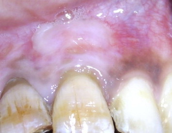
3 month follow up with complete coverage of the defect.
After complete healing at the end of 3 months, both central incisors were provided with porcelain fused to metal (PFM) crowns with excellent prognosis. The 6 month post-operative visit showed complete coverage of the mucosal fenestration (Fig. 8).
Fig. 8.

6 month follow up with complete coverage of the defect.
Discussion
Cases of mucosal fenestration have been earlier reported in history.1,2 Most common occurrences have been reported in maxillary and mandibular incisors. The exact etiology is not known but review of literature suggest that abnormally labioversed root tips, very thin labial plates and the presence of chronic periapical inflammation may be the probable cause.1,2 It has also been reported that exposed root tips favor the accumulation of plaque and calculus which prevents the reformation of mucosal covering.3 Various treatment modalities advocated in the literature for the management of mucosal fenestrations are lateral pedicle flap, Guided Tissue Regeneration (GTR) and apicoectomy combined with endodontic treatment.7,8
The present case report is amongst the first few to utilize free gingival graft in mucosal fenestration coverage. For the correction of periapical bony defect as well as the mucosal defect, it is necessary to create an environment for successful periapical healing, which was obtained in the case reported by conventional root canal treatment and apicectomy. One year follow up showed successful outcome of the surgical procedure.
Conclusion
Mucosal fenestrations and dehiscence are rare entities but whenever present, pose a difficult situation for the clinician. Large fenestration defects pose further challenge resulting in poor prognosis. There have been various non surgical and surgical procedures that have been documented for treatment. The present case report describes a rare situation where a mucosal fenestration developed in the upper left central incisor due to chronic periapical inflammation which was successfully treated with combination of bone grafting and free mucosal graft, showcasing the procedure as a viable treatment option in such cases.
Conflicts of interest
All authors have none to declare.
References
- 1.Elliot J.R., Bowers G.M. Alveolar dehiscence and fenestration. Periodontics. 1963;1:245–248. [Google Scholar]
- 2.Edel A. Alveolar bone fenestrations and dehiscence in dry Bedouin jaws. J Clin Periodontol. 1981;8:491–499. doi: 10.1111/j.1600-051x.1981.tb00898.x. [DOI] [PubMed] [Google Scholar]
- 3.Chen G., Fang C.T., Tong C. The management of mucosal fenestration: a report of two cases. Int Endod J. 2009;42(2):156–164. doi: 10.1111/j.1365-2591.2008.01463.x. [DOI] [PubMed] [Google Scholar]
- 4.Ju Y.R., Tsai H.Y., Wu Y.J. Surgical intervention of mucosal fenestration in a maxillary premolar: a case report. Quintessence Int. 2004;35:125–128. [PubMed] [Google Scholar]
- 5.Ling L.J. The treatment of fenestrated root: case reports. J Dent Sci. 1989;9:137–140. [Google Scholar]
- 6.Yang Z.P. Treatment of labial fenestration of maxillary central incisor. Endod Dent Traumatol. 1996;12:104–108. doi: 10.1111/j.1600-9657.1996.tb00106.x. [DOI] [PubMed] [Google Scholar]
- 7.Dawes W.L., Barnes I.E. The surgical treatment of fenestrated buccal roots of upper molars. Int Endod J. 1983;16:82–86. doi: 10.1111/j.1365-2591.1983.tb01301.x. [DOI] [PubMed] [Google Scholar]
- 8.von Arx T., Cochran D.L. Rationale for the application of the GTR principle using a barrier membrane in endodontic surgery: a proposal of classification and literature review. Int J Periodontics Restorative Dent. 2001;21:127–139. [PubMed] [Google Scholar]