

Abstract
Sarcoidosis is a systemic granulomatous disease that most commonly involves the lung and thoracic lymph nodes. However, any organ can be affected. Osseous sarcoidosis has been reported in 3–13% of the cases. The skeletal involvement on radiographs is usually seen late in the course of the disease and is rarely the initial manifestation. We report a case of sarcoidosis revealed by a lytic lesion of the phalanx.
Background
Sarcoidosis is a multisystemic inflammatory disease characterised by non-caseating granuloma and multinucleated giant cells. These granulomas can affect any tissue in the body. Bone involvement is common, but rarely it is the initial manifestation of sarcoidosis.1 Bones are usually involved in chronic disease or multivisceral sarcoidosis. Lesions are mainly seen in the small bones of the hands and feet. The clinical presentation is non-specific.2 Thus the diagnosis is often delayed in patients who have isolated bone lesions. We report a case of phalangeal osteolytic lesion leading to a diagnosis of sarcoidosis.
Case presentation
A 65-year-old woman, without any medical history, was hospitalised with a 2-year history of pain in her right hand. She noticed a progressive painful swelling of the right middle finger. She was a housewife and denied any trauma or injury. She was able to carry out her daily activities.
Physical examination showed a tender swelling in the middle phalanx between the proximal and distal interphalangeal joints. The passive and active range of motion of all joints was normal. There was no evidence of arthritis or sensory abnormality. A skin examination revealed a purple indurated plaque on the nose. There were no other cutaneous lesions. Cardiovascular, pulmonary and abdominal examinations were normal. No lymphadenopathy was found. The rest of the physical examination was normal.
Investigations
The blood investigations were normal. A radiograph of the hand showed mixed sclerotic and osteolytic abnormalities in the middle phalanx of the middle finger (figure 1). The radiographs of the remaining skeleton were normal. Thoracic radiography showed bilateral hilar masses. A thoracoabdominal CT scan revealed hilar lymphadenopathy. Small nodules (5–7 mm) were seen in the peribronchovascular regions and in the subpleural region. There was no evidence of other organ involvement. Skin biopsies of the cutaneous lesion in the nose showed a non-necrositing granuloma containing giant cells, consistent with sarcoidosis. Acid-fast bacilli were negative in the sputum and skin biopsies. The tuberculin skin test was negative. There was no elevation of the serum ACE. Pulmonary function testing was within normal limits. A final diagnosis of generalised sarcoidosis with bone involvement was made.
Figure 1.
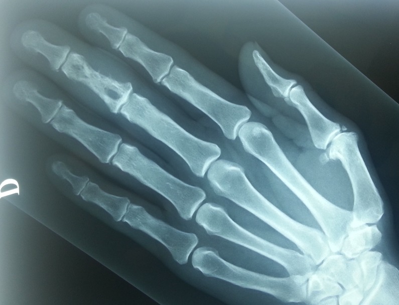
X-ray of the right hand showing lytic lesion involving the middle phalanx and swelling of the middle finger.
Treatment
The patient was treated with hydroxychloroquine 400 mg/day.
Outcome and follow-up
The patient was followed for 1 year. Her pain gradually decreased. There was no progression or involvement of other bones or new skin lesions. There has been no evidence of the systemic progression of disease.
Discussion
In 1928, Jungling3 described the characteristic bone involvement in sarcoidosis as ‘osteitis tuberculosa multiplex cystic’. The incidence of skeletal lesions is reported between 3% and 13% of cases, depending on radiological or clinical criteria.4 It occurs most often in patients known to have the chronic form of sarcoidosis with multivisceral manifestations.5 It is usually associated with skin manifestations, most frequently a lupus pernio, similar to the lesion on our patient's nose.
Radiographic diagnosis of osseous sarcoidosis is usually a late and asymptomatic manifestation of the disease. However, it can be the initial presentation of sarcoidosis in less than 1% of the cases.6 The clinical signs are variable and unspecific. Localised pain, swelling and redness are the main local symptoms and can be the first manifestation of the disease. Soft tissue swelling and thickening of the fingers or toes can also reveal phalangeal involvement.7 Bone lesions are recognised radiologically. MRI will show much more extensive and earlier osseous abnormalities.8 There is a predilection abnormalities of the metacarpals, metatarsals and phalanges of the hands and feet. They can be either small lytic lacunae in epiphyses or larger lytic areas involving medulla and cortex. The same lesions may occur in any bone in the body. Much less commonly, osteosclerotic lesions may be visualised.9 Extensive lytic lesions can progress leading to severe bone destruction and pathological fractures.10 In rare cases, mutilation and autoamputation of fingers, due to destruction of the terminal phalanges, has been reported.11
When isolated, osseous sarcoidosis often presents a diagnostic dilemma. The differential diagnosis includes enchondroma, subchondral cyst secondary to osteoarthritis, metastatic disease, hyperparathyroidism, epidermoid inclusion cyst, fibrous dysplasia and infection. Blood tests are not specific. When no other evidence of sarcoidosis is present, diagnosis may be confirmed by a bone biopsy, which shows the non-caseating granuloma and multinucleated giant cells.
The outcome of osseous sarcoidosis is usually good. However, management of this bone involvement is still controversial. Spontaneous resolution can occur within 2 years. Medical therapy is indicated in symptomatic lesions. Non-steroidal anti-inflammatory, corticosteroids and hydroxychloroquine can help to control pain and swelling. Methotrexate can be used in patients with poor response to corticosteroids or recurrent disease.5 Surgical excision has been reported in cases with fractures and bone destructions. In our case, the pain had improved with medical therapy.
Learning points.
Multiple organs may be involved in sarcoidosis. However, osseous sarcoidosis is rarely the presenting feature of the disease.
Although infection and malignancy are the classical causes of osteolytic lesions, sarcoidosis must be considered as a cause of lytic bony lesions.
Footnotes
Competing interests: None.
Patient consent: Obtained.
Provenance and peer review: Not commissioned; externally peer reviewed.
References
- 1.Smith K, Fort JG. Phalangeal osseous sarcoidosis. Arthritis Rheum 1998;41:176–9 [DOI] [PubMed] [Google Scholar]
- 2.Thelier N, Assous N, Job-Deslandre C, et al. Osteoarticular involvement in a series of 100 patients with sarcoidosis referred to rheumatology departments. J Rheumatol 2008;35:1622–8 [PubMed] [Google Scholar]
- 3.Jungling O. Uber ostitis tuberculosa multiplex cystoides. Bruns Klin Chirurg 1928;143:401 [Google Scholar]
- 4.Wilcoz A, Bharadwaj P, Sharma OP. Bone sarcoidosis. Curr Opin Rheumatol 2000;12:321–30 [DOI] [PubMed] [Google Scholar]
- 5.Zisman DA, Shorr AF, Lynch JP. Sarcoidosis involving the musculoskeletal system. Semin Respir Crit Care Med 2002;23:555–70 [DOI] [PubMed] [Google Scholar]
- 6.West SG, Kotzin BL. Sarcoidosis. In: Hochberg MC, Silman AJ, Smolen JS, Weinblatt ME, Weisman MH. eds. Rheumatology. 3rd edn Edinburgh: Mosby Elsevier, 2003:1735–52 [Google Scholar]
- 7.Shorr AF, Murphy FT, Kelly WF. Osseus sarcoidosis: clinical, radiographic, and therapeutic observations. J Clin Rheumatol 1998:186–92 [DOI] [PubMed] [Google Scholar]
- 8.Moore SL, Teirstein A, Golimbu C. MRI of sarcoidosis patients with musculoskeletal symptoms. AJR Am J Roentgenol 2005;185:154–9 [DOI] [PubMed] [Google Scholar]
- 9.Vagal A, Shipley R, Meyer C. Radiological manifestations of sarcoidosis. Clin Dermatol 2007;25:312–25 [DOI] [PubMed] [Google Scholar]
- 10.Ugwonali O, Parisien M, Nickerson KG, et al. Osseous sarcoidosis of the hand: pathologic analysis and review of the literature. J Hand Surg Am 2005;30:854–8 [DOI] [PubMed] [Google Scholar]
- 11.Cetinkaya R, Kavak A, Parlak AH, et al. Can sarcoidosis cause autoamputation of a finger phalanx. J Hand Surg Br 2006;31:413–15 [DOI] [PubMed] [Google Scholar]