Abstract
Although there are encouraging reports showing the use of dexamethasone implant (Ozurdex) in uveitis in adults, the literature is scanty regarding its benefits and side effects in children. A 12-year-old boy presented with intermediate uveitis with disc oedema. He had 20/20 visual acuity and intraocular pressure (IOP) of 18 mm Hg in both eyes. He was treated with intravitreal Ozurdex in his left eye (LE) due to progressive worsening of uveitis and disc oedema. He developed increased IOP (31 mm Hg) that could not be controlled on maximal antiglaucoma medications and required the removal of the Ozurdex implant at 2.5 months. His IOP remained persistently high leading to increased cup disc ratio necessitating glaucoma filtration surgery (GFS). At 9 months of post-GFS follow-up, IOP was 12 mm Hg in LE without any medication. Though dexamethasone implant is being increasingly used in children with uveitis, its potential risk factors such as intractable glaucoma should be considered.
Background
Intermediate uveitis (IU) is the second most common form of uveitis in childhood.1 In a graded approach, the treatment of idiopathic IU consists of local steroids followed by systemic steroids. Local treatment consists of subtenon or an intravitreal injection of long-acting steroids. Intravitreal injection of steroids such as triamcinolone is associated with a limited duration of action, raised intraocular pressure (IOP) and cataract.2 Systemic corticosteroids are known to cause adverse effects such as interference with growth, development of cushingoid habitus, etc especially in growing children.3 Dexamethasone implant (Ozurdex, Allergan Inc, Irvine, California, USA) has been shown to be beneficial and takes care of the above issues like limited duration of action and raised IOP especially in adults.4 Recently, Taylor et al5 in their study of dexamethasone implants in paediatric uveitis reported raised IOP-necessitating treatment. We report a case of intractable glaucoma necessitating dexamethasone implant (Ozurdex) removal and glaucoma surgery in a child with uveitis.
Case presentation
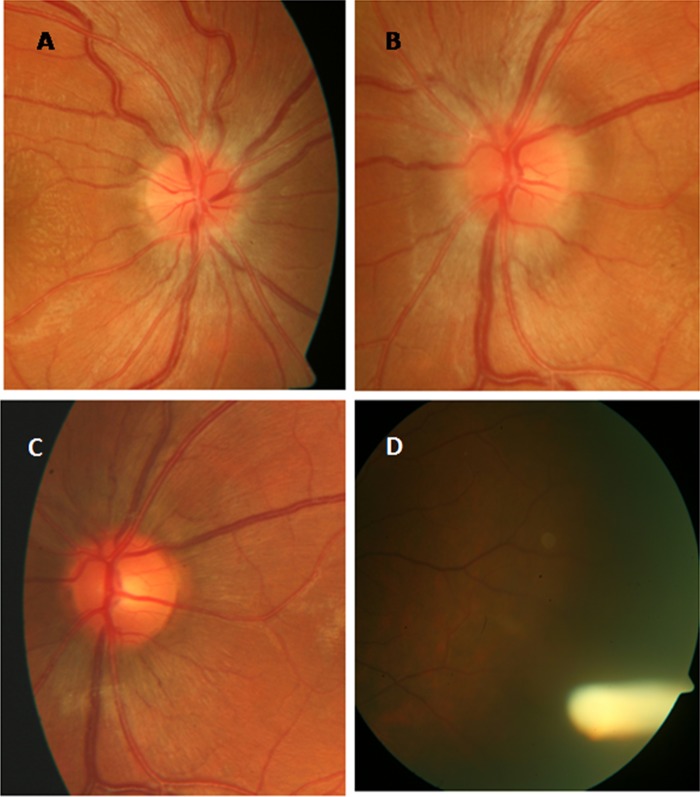
A 12-year-old boy was referred to the uveitis clinic with a diagnosis of bilateral IU. He was being treated with topical steroids without any relief of symptoms for the last 3 months elsewhere. On examination, his best-corrected visual acuity (BCVA) was 20/20 in both eyes (BE). His IOP was 18 mm Hg in BE. His central corneal thickness was 532 μ in the right eye (RE) and 528 μ in the left (LE). His anterior segment examination showed 1+ cells, and 1+ flare in BE. The posterior segment examination showed 1+ cellular reaction and multiple snowballs in vitreous and disc oedema in BE (figure 1A,B). He did not have any macular oedema. Fundus fluorescein angiography and optical coherence tomography of BE did not reveal any other positive findings except for disc staining. He was labelled as idiopathic IU and advised to continue on topical 0.1% β-methasone two hourly and twice daily homatropine 2% in BE. He was referred to a paediatrician to rule out any systemic association. Two weeks later, a progressive worsening of disc swelling was noted and his parents were counselled for treatment with systemic steroids or Ozurdex implant in BE. They agreed and consented for Ozurdex in the LE first after being explained about its benefits and complications. Intravitreal Ozurdex was inserted under all aseptic conditions in LE. On the first postoperative day, IOP was 31 mm Hg in LE. IOP in RE at this visit was 18 mm Hg. He was started on topical timolol maleate 0.5% in LE. We deferred the plan to implant Ozurdex in RE due to raised IOP in LE. IOP in LE at 2 weeks was 32 mm Hg and dorzolamide hydrochloride 2%, and brimonidine 0.2% was added. In the meantime, his systemic examination by a paediatrician revealed positive antinuclear antibodies, with no other systemic abnormality. He was started on oral steroids (1 mg/kg body weight) by the paediatrician along with the tablet azathioprine 50 mg once a day and the tablet hydrochloroquine 200 mg once a day. The tablet azathioprine was hiked to 125 mg over the next couple of weeks.
Figure 1.
Fundus photograph of the right and left eye showing optic disc oedema (A and B), the left eye showing resolution of the disc oedema at 4 weeks (C), and the left eye showing Ozurdex implant at 4 weeks (D).
Inflammation due to IU subsided with the Ozurdex implant within the next 4 weeks (figure 1C,D), but his IOP in LE was persistently high, that is, 30 mm Hg with healthy disc (cup disc ratio (CDR) 0.2) on maximal medical therapy (topical dorzolamide hydrochloride 2%, timolol maleate 0.5%, brimonidine 0.2%, tab acetazolamide 250 mg three times a day and syrup glycerol 30 mL three times a day). At this visit, IOP in RE was 32 mm Hg with healthy disc (CDR 0.2). He was started on topical dorzolamide hydrochloride 2%, timolol maleate 0.5% and brimonidine tartrate 0.2% in RE also. Oral and topical steroids were tapered off completely over 2 months and he was continued on oral azathioprine and hydrochloroquine.
Treatment
Two months after the insertion of the implant, despite maximal medical therapy, his IOP remained high and the disc started showing glaucomatous changes in LE. We planned to surgically remove the dexamethasone implant from the vitreous cavity by 23-gauge transconjunctival sutureless vitrectomy (TSV) at 2.5 months. The Ozurdex implant was removed with 23-gauge vitreous cutter. One week postoperatively, his BCVA was 20/20 in RE and 20/30 in LE, IOP was 20 mm Hg on topical brinzolamide 1% and oral acetazolamide 250 mg three times a day in LE. RE had IOP of 16 mm Hg on treatment.
Two months after the removal of the implant, IOP in LE was 36 mm Hg on maximum medical therapy. Disc showed the effect of raised IOP and increased CDR with diffuse loss of neuroretinal rim. Glaucoma filtration surgery (GFS) with 2% mitomycin-C (MMC) was carried out in LE. Four weeks post-GFS surgery, all antiglaucoma medications were stopped in LE.
Outcome and follow-up
Nine months after GFS, the patient had BCVA of 20/20 in BE with quiescent anterior and posterior segments. IOP was 12 mm Hg at last follow-up on dorzolamide and timolol combination with healthy disc in RE and 12 mm Hg in LE without any antiglaucoma medication with 0.4 CDR.
Discussion
Our case highlights the adverse effect of dexamethasone implants in the paediatric age group that necessitated removal of the steroid implant and glaucoma surgery.
The general consensus is to treat IU, inspite of the patient presenting with vision better than 6/9. There is always a risk of increase in inflammation progressively, if we do not treat. Our patient was already on a topical treatment for the last 3 months without any improvement before consulting with us. Local therapy in the form of intravitreal dexamethasone implant was given to avoid the systemic effects of oral steroids.
Various studies in adults have shown raised IOP as an adverse effect.4 6 7 In a study using flucinolone acetonide implant for the treatment of uveitis,7 32% of the cases required surgical intervention at 3 years due to raised IOP. In dexamethasone implant trial for the treatment of uveitis,4 raised IOP was controlled medically, while none of the cases required surgical intervention. There was no paediatric patient in these studies. Taylor et al5 has reported the successful use of Ozurdex in paediatric patients with intermediate or posterior uveitis. Authors5 reported rise in IOP in 4 of the 13 eyes, which necessitated treatment, 2 with topical medication, 1 with systemic acetazolamide and 1 with glaucoma drainage surgery. None of their children required removal of the implant. The literature on steroid responsiveness in children documents variable effects on IOP.8 9 In our patient, oral steroids were not given for bilateral disease to avoid systemic side effects initially. The paediatrician started him on oral steroids later because systemic immunosuppression would have taken atleast couple of weeks for its action to appear. IOP raised on day 1 was unusual in our case. We speculate that this patient might have been a steroid responder as he was already exposed to topical steroids, and intravitreal Ozurdex acted like a final insult, which lead to an immediate hike in IOP in only that eye. Later, when systemic steroids were added, it happened for the RE as well leading to an increase in IOP.
The removal of the Ozurdex implant is reported in aphakic10 eyes. In these eyes, there was an anterior migration of the Ozurdex implant into the anterior chamber that resulted in the decompensation of the corneal endothelium. About 1–5% of patients with steroid-induced iatrogenic glaucoma need surgery to normalise their IOP.11 In our case, the decision to perform TSV and implant removal first before GFS was the treating physician's personal choice. Apart from the other advantages of TSV, it preserves the conjunctiva for any future surgeries. Later, GFS with MMC was carried out to normalise IOP.
Although recently there have been encouraging reports showing the use of dexamethasone implants in uveitis, there is limited literature regarding its benefits and side effects in children, and the potential risk factors such as intractable IOP rise should always be considered. The removal of the Ozurdex implant alone from the vitreous cavity may not result in the complete reversal of steroid-induced IOP rise in these patients.
Learning points.
While implanting Ozurdex implant, the possibility of intractable intraocular pressure (IOP) rise should be kept in mind especially in children.
If intractable IOP rise occurs, then the Ozurdex implant can be removed by 23G transconjunctival sutureless vitrectomy. It will help to preserve the conjunctiva for future use.
Implant removal from the vitreous cavity may not result in a complete reversal of steroid-induced IOP rise.
Acknowledgments
The authors would like to acknowledged Professor Amod Gupta for his advice and support.
Footnotes
Contributors: NK contributed to conception and design, acquisition and interpretation of the data, drafting the manuscript and final approval. SP contributed in acquisition of photographs, drafting the manuscript and final approval. SK contributed in revising the manuscript critically and final approval. RS contributed to conception and design, revising the manuscript critically and final approval.
Competing interests: None.
Patient consent: Obtained.
Provenance and peer review: Not commissioned; externally peer reviewed.
References
- 1.Nagpal A, Leigh JF, Acharya NR. Epidemiology of uveitis in children. Int Ophthalmol Clin 2008;48:1–7 [DOI] [PubMed] [Google Scholar]
- 2.Taylor SR, Isa H, Joshi L, et al. New developments in corticosteroid therapy for uveitis. Ophthalmologica 2010;224(Suppl 1):46–53 [DOI] [PubMed] [Google Scholar]
- 3.McDonough AK, Curtis JR, Saag KG. The epidemiology of glucocorticoid-associated adverse events. Curr Opin Rheumatol 2008;20:131–7 [DOI] [PubMed] [Google Scholar]
- 4.Lowder C, Belfort R, Jr, Lightman S, et al. Ozurdex HURON Study Group Dexamethasone intravitreal implant for noninfectious intermediate or posterior uveitis. Arch Ophthalmol 2011;129:545–53 [DOI] [PubMed] [Google Scholar]
- 5.Taylor SRJ, Tomkins-Netzer O, Joshi L, et al. Dexamethasone implant in paediatric uveitis. Ophthalmology 2012;119:2412-2412.e2. [DOI] [PubMed] [Google Scholar]
- 6.Callanan DG, Jaffe GJ, Martin DF, et al. Treatment of posterior uveitis with a flucinolone acetonide implant: three-year clinical trial results. Arch Ophthalmol 2008;126:1191–201 [DOI] [PubMed] [Google Scholar]
- 7.Goldstein DA, Godfrey DG, Hall A, et al. Intraocular pressure in patients with uveitis treated with flucinolone acetonide implants. Arch Ophthalomol 2007;125:1478–85 [DOI] [PubMed] [Google Scholar]
- 8.Jones R, III, Rhee DJ. Corticosteroid induced hypertension and glaucoma—a brief review and update of the literature. Curr Opin Ophthalmol 2006;17:163–7 [DOI] [PubMed] [Google Scholar]
- 9.Kwok AK, Lam DS, Ng FS, et al. Ocular hypertensive response to topical steroids in children. Ophthalmology 1997;104:2112–16 [DOI] [PubMed] [Google Scholar]
- 10.Bansal R, Bansal P, Kulkarni P, et al. Wandering Ozurdex® implant. J Ophthal Inflamm Infect 2012;2:1–5 [DOI] [PMC free article] [PubMed] [Google Scholar]
- 11.Razeghinejad MR, Hatz LJ. Steroid-induced iatrogenic glaucoma. Ophthalmic Res 2012;47:66–80 [DOI] [PubMed] [Google Scholar]


