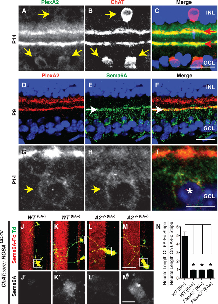
Fig. 1. Sema6Ais expressed in On, but not Off, SACs in vivo, and SACs lacking Sema6A avoid exogenous Sema6A in vitro.
(A to C) Mouse P14 retina sections immunostained with antibodies directed against PlexA2 (green) and ChAT (red) reveal colocalization of PlexA2 and ChAT immunoreactivity in cell bodies (yellow arrows) and in dendritic processes (red arrows). (D to I) Postnatal retina sections immunostained with antibodies to PlexA2 (red) and antibodies to Sema6A (green) show that PlexA2 and Sema6A exhibit mostly complementary protein distributions in the inner plexiformlayer [white arrows in (E) and (F)] but that they are coexpressed in On SACs [yellow arrow heads in (G) and (H), white asterisk in (I)]. (J to N) Genetically labeled, dissociated SACs from wild-type (WT) retinas are divided into two populations in vitro: Sema6A− (J and J′) and Sema6A+ (K and K′). Neurites extending from WT Sema6A− SACs avoid exogenous Sema6AFc stripes (J), whereas WT Sema6A+ SACs do not (K). PlexA2−/− SACs, both Sema6A+ and Sema6A−, extend freely over Sema6A-Fc+ stripes [(L) and (M)]. The ratio of SAC neurite length off of Sema6AFc stripes to neurite length on Sema6A-Fc stripes is shown in (N) (n ≥ 15 SACs per genotype). Error bars, mean ± SEM. *P < 0.01. Scale bars, 20 µm in (C) for (A) to (C), 20 µm in (F) for (D) to (F), 10 µm in (I) for (G) to (I), 40 µm in (M) for (J) to (M), and 10 µm in (M′) for (J′) to (M′).

