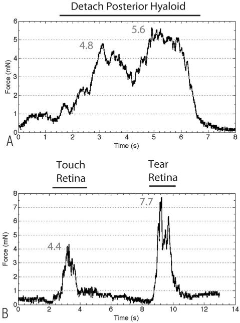
Fig. 3.
A. Representative tracing of the raw forces measured by the force-sensing micropick during posterior hyaloid detachment. A gradual increase in force was seen, with a general plateau phase, followed by a rapid decrease when the hyaloid was separated from the optic nerve. B. Representative tracing of the forces measured during creation of a retinal tear. From 2 seconds to 4 seconds, a maximum force of 4.4 mN was generated when the instrument was touching the retina. From 8 seconds to 11 seconds, when a retinal tear was created, there was a rapid increase in force, a short plateau phase, and a rapid decrease in force when the retina was torn.

