
FIGURE 5.
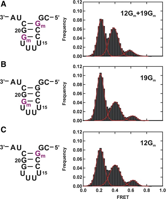
Charaterization of individual modifications. (A) Schematic structure of the U2 stem I containing 2′-O-methylguanosines (Gm) at positions 12 and 19 (left) and corresponding FRET histogram at 40 mM Mg2+ (right). (B) Schematic structure of the U2 stem I containing Gm at position 19 (left) and corresponding FRET histogram at 40 mM Mg2+ (right). (C) Schematic structure of the U2 stem I containing Gm at position 12 (left) and corresponding FRET histogram at 40 mM Mg2+ (right). All modified bases are shown in purple.