Abstract
Artifacts present in computed tomography (CT) image often degrade the image quality and ultimately, the diagnostic outcome. Ring artifact in trans-axial image is caused by either miscalibrated or defective detector element of detector row, which is often categorized as scanner based artifact. A ring artifact detected on trans-axial CT image of positron emission tomography/computed tomography (PET/CT), was caused by contamination of CT tube aperture by droplet of injectable contrast medium. This artifact was corrected by removal of contrast droplet from CT tube aperture. The ring artifact is a very common artifact, commonly cited in the literature. Our case puts forward an uncommon cause of this artifact and its method of correction, which also, has no mention in the existing literature.
Keywords: Artifact, computed tomography tube, computed tomography tube aperture, mylar window, trans-axial
INTRODUCTION
Image quality of computed tomography (CT) is entirely dependent on the performance characteristic of the scanner. Acceptance test is of utmost importance and should be performed, at installation and after any major service, before its clinical use.[1] Artifacts that appear on CT images are mainly caused by metallic implant, patient motion, improper patient preparation and improper calibration of equipment.[2,3] Even in proper calibrated system, artifacts can appear because of improper scan protocol selection and can be overcome by proper selection of protocol.[2] All the above mentioned artifacts are commonly identified and can be corrected. Most of these artifacts are due to patient or scanner related factors. We through our case want to document an artifact that was caused due to contamination of CT tube aperture by a small droplet of contrast medium.
CASE REPORT
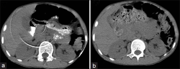
We encountered an unusual scenario in our busy positron emission tomography (PET)/CT department. In the afternoon, after half of the scheduled patients were successfully scanned, when all of a sudden, there was appearance of a ring artifact on CT trans-axial image [Figure 1a-arrow], in the CT component of PET/CT images acquired on our PET/CT scanner, Discovery ST, GE Medical Systems, USA. We immediately informed this occurrence to service engineer. Service engineer suspected that there might be defective detector element. While recalling the events that had occurred during that day, since we started acquiring scans, it struck to us that a contrast had splashed on CT scanner during acquisition of a scan due to a defective infusion tube during contrast infusion. Such events had occurred in the past and are not unusual in imaging departments. However, appearance of ring artifact, even when the scanner had passed daily CT quality control test in the morning, at the start, raised doubts. We had a strong suspicion that contrast might have trickled down inside the gantry because Mylar window was having a small slit, at its center. Service engineer opened the gantry and we inspected the detector ring and CT tube aperture for any contrast contamination. We found a small droplet of contrast on the CT tube aperture. We cleaned the CT tube aperture. We cleaned the CT tube aperture and repeated the scan. In repeat scan CT trans-axial slices was free of ring artifact [Figure 1b].
Figure 1.

Clinical image (a) Trans-axial image showing a ring artifact (arrows), (b) trans-axial image showing no artifact
DISCUSSION
Ring artifact in trans-axial CT scan is well documented over all these years since new generation CT scanners came into practice. The ring artifact can appear in trans-axial images of multi detector computer tomography because of two reasons.[4,5,6] A defective detector element or set of detector elements can cause ring artifact in trans-axial CT image. This artifact can be eliminated by replacement of CT detector module with defective element followed by CT number calibration. This artifact can be corrected by recognizing the faulty element or group of elements and generating correction for them in sinogram.[5,6] In very few circumstances, replacement of CT detector module might require to eliminate the artifact when defective detector element are significant in one module. Sometimes, improper calibration of scanner, when wrong CT number is assigned during calibration for an individual or set of detector elements, can lead to appearance of ring artifact on trans-axial image. There are methods described in the literature to remove ring artifact from the CT trans-axial image.[5,6] The resolution of ring artifact caused by above-mentioned reasons requires large amount of time, technical effort and capital investment. The ring artifact described in our case report also seemed to be originated from either of the above reason. However, our technical alertness helped us pick up the exact cause of artifact, which was very rare and could have been easily missed, if a log of daily accidents would not have been maintained. It thereby saved reasonable amount of time, technical effort and capital investment. Though ring artifact and its cause have extensively described in the literature in the past, we put forward a unique cause of this artifact, which needs to be kept in mind, as such scenarios often occur in a PET/CT department performing contrast enhanced studies.
CONCLUSION
The ring artifact seen in trans-axial image was caused by contrast contamination of CT tube aperture. Proper fitting of Mylar window and avoidance of contrast leakage can avoid this occurrence. And if ring artifact appears in the CT image this cause needs to be ruled out.
Footnotes
Source of Support: Nil.
Conflict of Interest: None declared.
REFERENCES
- 1.Seeram E. Computed Tomography: Physical Principles, Clinical Applications and Quality Control. 2nd ed. Philadelphia, PA: Saunders; 2012. Image quality; pp. 174–99. [Google Scholar]
- 2.Wilting JE, Timmer J. Artefacts in spiral-CT images and their relation to pitch and subject morphology. Eur Radiol. 1999;9:316–22. doi: 10.1007/s003300050673. [DOI] [PubMed] [Google Scholar]
- 3.Barrett JF, Keat N. Artifacts in CT: Recognition and avoidance. Radiographics. 2004;24:1679–91. doi: 10.1148/rg.246045065. [DOI] [PubMed] [Google Scholar]
- 4.Hsieh J. Computed Tomography: Principles, Design, Artifacts and Recent Advances. Bellingham, Wash: SPIE Press; 2003. Image artifacts: Appearances, causes and corrections; pp. 167–240. [Google Scholar]
- 5.Abu Anas EM, Lee SY, Hasan MK. Removal of ring artifacts in CT imaging through detection and correction of stripes in the sinogram. Phys Med Biol. 2010;55:6911–30. doi: 10.1088/0031-9155/55/22/020. [DOI] [PubMed] [Google Scholar]
- 6.Anas EM, Lee SY, Hasan K. Classification of ring artifacts for their effective removal using type adaptive correction schemes. Comput Biol Med. 2011;41:390–401. doi: 10.1016/j.compbiomed.2011.03.018. [DOI] [PubMed] [Google Scholar]


