

Abstract
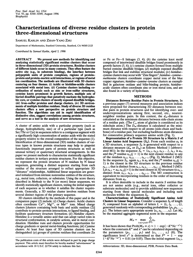
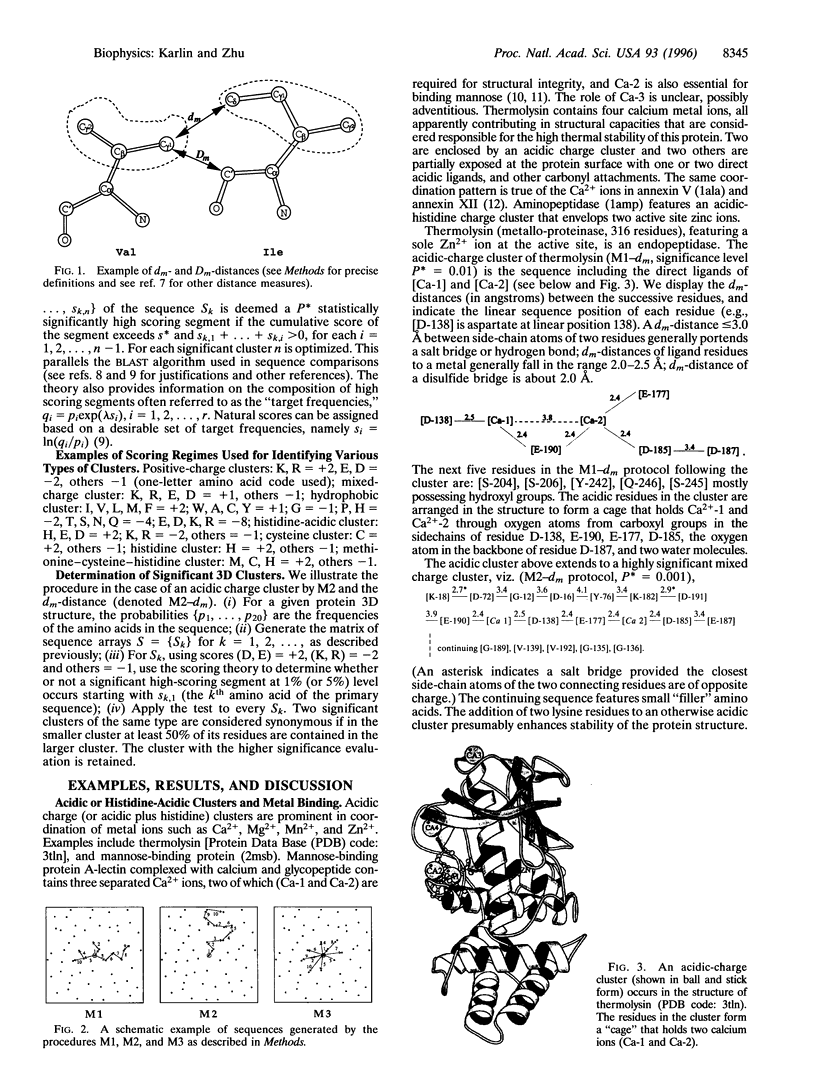
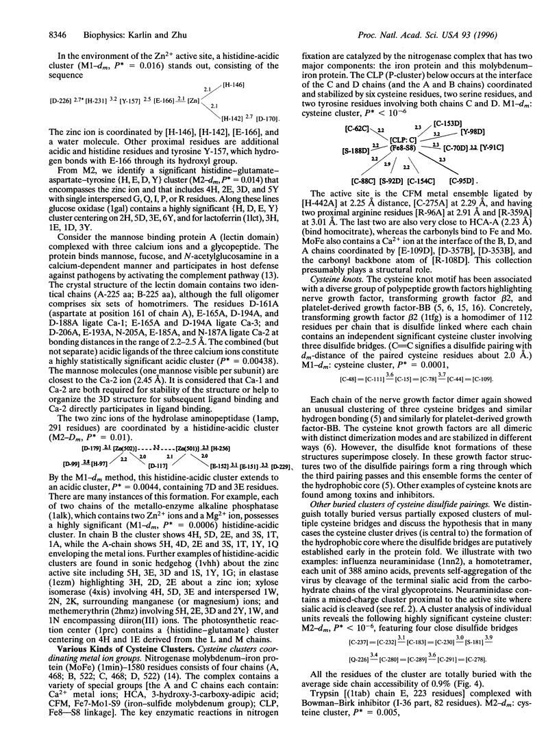
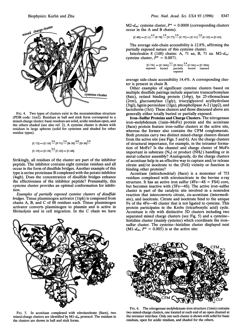
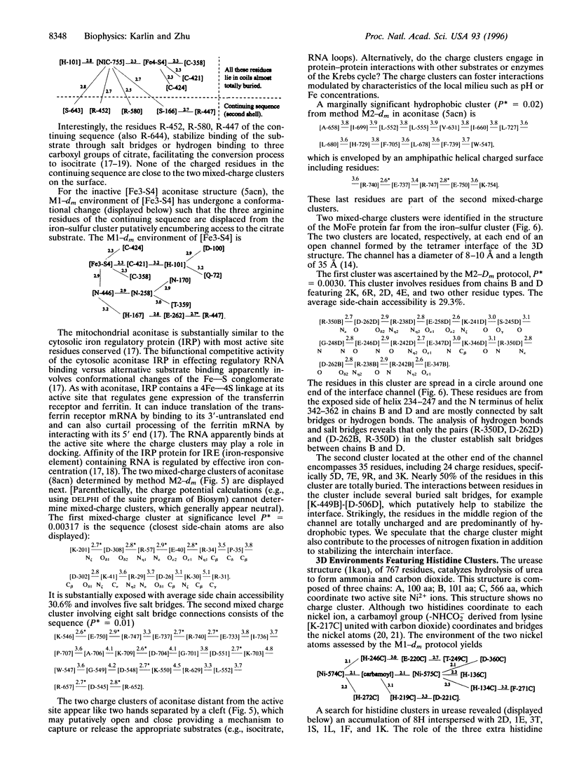
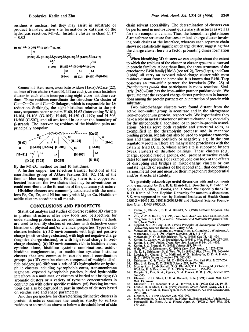
We present new methods for identifying and analyzing statistically significant residue clusters that occur in three-dimensional (3D) protein structures. Residue clusters of different kinds occur in many contexts. They often feature the active site (e.g., in substrate binding), the interface between polypeptide units of protein complexes, regions of protein-protein and protein-nucleic acid interactions, or regions of metal ion coordination. The methods are illustrated with 3D clusters centering on four themes. (i) Acidic or histidine-acidic clusters associated with metal ions. (ii) Cysteine clusters including coordination of metals such as zinc or iron-sulfur structures, cysteine knots prominent in growth factors, multiple sets of buried disulfide pairings that putatively nucleate the hydrophobic core, or cysteine clusters of mostly exposed disulfide bridges. (iii) Iron-sulfur proteins and charge clusters. (iv) 3D environments of multiple histidine residues. Study of diverse 3D residue clusters offers a new perspective on protein structure and function. The algorithms can aid in rapid identification of distinctive sites, suggest correlations among protein structures, and serve as a tool in the analysis of new structures.
Full text
PDF
Images in this article
Selected References
These references are in PubMed. This may not be the complete list of references from this article.
- Daopin S., Piez K. A., Ogawa Y., Davies D. R. Crystal structure of transforming growth factor-beta 2: an unusual fold for the superfamily. Science. 1992 Jul 17;257(5068):369–373. doi: 10.1126/science.1631557. [DOI] [PubMed] [Google Scholar]
- Drickamer K., Taylor M. E. Biology of animal lectins. Annu Rev Cell Biol. 1993;9:237–264. doi: 10.1146/annurev.cb.09.110193.001321. [DOI] [PubMed] [Google Scholar]
- Jabri E., Carr M. B., Hausinger R. P., Karplus P. A. The crystal structure of urease from Klebsiella aerogenes. Science. 1995 May 19;268(5213):998–1004. [PubMed] [Google Scholar]
- Karlin S., Blaisdell B. E., Brendel V. Identification of significant sequence patterns in proteins. Methods Enzymol. 1990;183:388–402. doi: 10.1016/0076-6879(90)83026-6. [DOI] [PubMed] [Google Scholar]
- Karlin S., Brendel V. Chance and statistical significance in protein and DNA sequence analysis. Science. 1992 Jul 3;257(5066):39–49. doi: 10.1126/science.1621093. [DOI] [PubMed] [Google Scholar]
- Karlin S. Statistical studies of biomolecular sequences: score-based methods. Philos Trans R Soc Lond B Biol Sci. 1994 Jun 29;344(1310):391–402. doi: 10.1098/rstb.1994.0078. [DOI] [PubMed] [Google Scholar]
- Karlin S., Zuker M., Brocchieri L. Measuring residue associations in protein structures. Possible implications for protein folding. J Mol Biol. 1994 Jun 3;239(2):227–248. doi: 10.1006/jmbi.1994.1365. [DOI] [PubMed] [Google Scholar]
- Kim J., Rees D. C. Structural models for the metal centers in the nitrogenase molybdenum-iron protein. Science. 1992 Sep 18;257(5077):1677–1682. doi: 10.1126/science.1529354. [DOI] [PubMed] [Google Scholar]
- Klausner R. D., Rouault T. A., Harford J. B. Regulating the fate of mRNA: the control of cellular iron metabolism. Cell. 1993 Jan 15;72(1):19–28. doi: 10.1016/0092-8674(93)90046-s. [DOI] [PubMed] [Google Scholar]
- Lauble H., Stout C. D. Steric and conformational features of the aconitase mechanism. Proteins. 1995 May;22(1):1–11. doi: 10.1002/prot.340220102. [DOI] [PubMed] [Google Scholar]
- Lippard S. J. At last--the crystal structure of urease. Science. 1995 May 19;268(5213):996–997. doi: 10.1126/science.7754394. [DOI] [PubMed] [Google Scholar]
- Luecke H., Chang B. T., Mailliard W. S., Schlaepfer D. D., Haigler H. T. Crystal structure of the annexin XII hexamer and implications for bilayer insertion. Nature. 1995 Nov 30;378(6556):512–515. doi: 10.1038/378512a0. [DOI] [PubMed] [Google Scholar]
- McDonald N. Q., Hendrickson W. A. A structural superfamily of growth factors containing a cystine knot motif. Cell. 1993 May 7;73(3):421–424. doi: 10.1016/0092-8674(93)90127-c. [DOI] [PubMed] [Google Scholar]
- McDonald N. Q., Lapatto R., Murray-Rust J., Gunning J., Wlodawer A., Blundell T. L. New protein fold revealed by a 2.3-A resolution crystal structure of nerve growth factor. Nature. 1991 Dec 5;354(6352):411–414. doi: 10.1038/354411a0. [DOI] [PubMed] [Google Scholar]
- Messerschmidt A., Ladenstein R., Huber R., Bolognesi M., Avigliano L., Petruzzelli R., Rossi A., Finazzi-Agró A. Refined crystal structure of ascorbate oxidase at 1.9 A resolution. J Mol Biol. 1992 Mar 5;224(1):179–205. doi: 10.1016/0022-2836(92)90583-6. [DOI] [PubMed] [Google Scholar]
- Murray-Rust J., McDonald N. Q., Blundell T. L., Hosang M., Oefner C., Winkler F., Bradshaw R. A. Topological similarities in TGF-beta 2, PDGF-BB and NGF define a superfamily of polypeptide growth factors. Structure. 1993 Oct 15;1(2):153–159. doi: 10.1016/0969-2126(93)90029-g. [DOI] [PubMed] [Google Scholar]
- Ng K. K., Drickamer K., Weis W. I. Structural analysis of monosaccharide recognition by rat liver mannose-binding protein. J Biol Chem. 1996 Jan 12;271(2):663–674. doi: 10.1074/jbc.271.2.663. [DOI] [PubMed] [Google Scholar]
- Weis W. I., Drickamer K. Trimeric structure of a C-type mannose-binding protein. Structure. 1994 Dec 15;2(12):1227–1240. doi: 10.1016/S0969-2126(94)00124-3. [DOI] [PubMed] [Google Scholar]
- Zhu Z. Y., Karlin S. Clusters of charged residues in protein three-dimensional structures. Proc Natl Acad Sci U S A. 1996 Aug 6;93(16):8350–8355. doi: 10.1073/pnas.93.16.8350. [DOI] [PMC free article] [PubMed] [Google Scholar]