Abstract
Methamphetamine and other drugs activate a small proportion of all neurons in the brain. We previously developed a FACS-based method to characterize molecular alterations induced selectively in activated neurons that express the neural activity marker Fos. However, this method requires pooling samples from many rats. We now describe a modified FACS-based method to characterize molecular alterations in Fos-expressing dorsal striatal neurons from a single rat using a multiplex pre-amplification strategy. Fos and NeuN (a neuronal marker) immunohistochemistry indicate that 6–7% of dorsal striatum neurons were activated 90 min after acute methamphetamine injections (5 mg/kg, i.p) while less than 1% of neurons were activated by saline injections. We used FACS to separate NeuN-labeled neurons into Fos-positive and Fos-negative neurons and assessed mRNA expression using RT-qPCR from as little as 5 Fos-positive neurons. Methamphetamine induced 3–20-fold increases of immediate early genes arc, homer-2, c-fos, fosB and its isoforms (ΔfosB and a novel isoform ΔfosB-2) in Fos-positive but not Fos-negative neurons. IEG mRNA induction was 10-fold lower or absent when assessed in unsorted samples from single dorsal striatum homogenates. Our modified method makes it feasible to study unique molecular alterations in neurons activated by drugs or drug-associated cues in complex addiction models.
Keywords: flow cytometry, immediate early genes, gene expression
Introduction
A main premise of numerous drug addiction studies over the last several decades has been that chronic drug exposure causes long-lasting neuroadaptations within components of the mesocorticolimbic dopamine reward circuit and other brain circuits, leading to compulsive drug use and long-term relapse vulnerability (Nestler 2002, White & Kalivas 1998, Wolf et al. 2004, Shaham & Hope 2005). However, this premise is based on studies limited to measuring drug-induced molecular changes in homogenates of brain regions, or in neurons selected independently of their activation state during drug taking or seeking. The distinction between drug-induced molecular neuroadaptations in activated versus non-activated neurons is important, because many studies using in vivo electrophysiology (Carelli & Wightman 2004, Kiyatkin & Rebec 1997, Chang et al. 1998) and immediate early gene expression (Neisewander et al. 2000, Hamlin et al. 2007, Hope et al. 2006) methods have shown that both drugs and drug-associated cues activate only a small proportion of neurons in a given brain area (Cruz et al. 2013). Selective inactivation of only the behaviorally activated neurons attenuates subsequent drug-taking behaviors and conditioned drug effects in a context-specific manner, which indicates that these neurons play a causal role in encoding these learned behaviors (Bossert et al. 2011, Koya et al. 2009, Fanous et al. 2012).
Based on the above considerations, we have begun to develop novel procedures to characterize unique synaptic physiology and gene expression patterns in activated Fos-expressing neurons that encode drug- and food-seeking behaviors and other learned behaviors (Cifani et al. 2012, Fanous et al. 2013, Guez-Barber et al. 2011, Cruz et al. 2013). Fos is the protein product of the immediate early gene c-fos, which is rapidly induced in response to unconditioned and conditioned stimuli and has been used in many studies as a cellular marker of neuronal activity (Morgan & Curran 1991, Curran & Morgan 1995). One of our new procedures includes a method for dissociating intact cell bodies from adult rat brains and using FACS to sort these cells using commercially available antibodies against the nuclear neuronal markers NeuN (Mullen et al. 1992) and Fos (Guez-Barber et al. 2011, Guez-Barber et al. 2012, Fanous et al. 2013). We used this method to study unique molecular alterations in behaviorally relevant Fos-expressing neurons of striatum and prefrontal cortex, using RT-qPCR (reverse transcriptase–quantitative real time polymerase chain reaction) assays (Fanous et al. 2012, Guez-Barber et al. 2011, Guez-Barber et al. 2012). However, our original FACS procedure requires pooling 6–10 rat striatum or prefrontal cortex tissues, which is not feasible for many studies on mechanisms of drug reward and relapse that require surgery and many weeks of labor-intensive behavioral training.
Here we describe an improved FACS procedure to characterize molecular alterations in behaviorally activated Fos-expressing dorsal striatal neurons from a single rat using multiplex pre-amplification strategy.
Methods
Subjects
We used a total of 70 male Sprague-Dawley rats (Charles Rivers) that weighed 300–350 g in our experiments. The rats were group-housed (two per cage) and maintained in the animal facility under a reverse 12:12 h light/dark cycle (lights off at 8 am) with food and water freely available. Procedures followed the guidelines outlined in the Guide for the Care and Use of Laboratory Animals (8th edition; http://grants.nih.gov/grants/olaw/Guide-for-the-Care-and-Use-of-Laboratory-Animals.pdf) and were approved by the local Animal Care and Use Committee.
Drug injection procedure
Rats were handled for 2 days and then received saline injections (1 ml/kg, i.p.) once daily for 3 more days prior to testing for the purpose of habituating them to the injection procedure. For each experiment, we used either saline (1 ml/kg, i.p.) or (+)-methamphetamine-HCl (NIDA drug supply; 5 mg/kg, i.p). In Exp. 1 (immunohistochemistry), 4 groups of rats (n=6 per group) were injected with saline or methamphetamine in either their home cage or a novel environment (Med Associates self-administration chambers). Rats were anesthetized 90 min later and perfused for immunohistochemistry. In Exp. 2 (RT-qPCR of FACS-sorted neurons), two groups of rats (n=6 per group) were injected with either saline or methamphetamine. Rats were decapitated 90 min later, and dorsal striatum was dissected (see below). In Exp. 3 (RT-qPCR of dorsal striatum homogenates) two groups of rats (n=3 per group) were injected with either saline or methamphetamine. Rats were decapitated 90 min later, and dorsal striatum of each hemisphere was dissected and processed independently. In Exp. 4 (Fos and FosB time course), we used flow cytometry to assess the time course of Fos− and FosB-labeled neurons in three groups of rats (n=8 per group) that were injected with either saline or methamphetamine. Rats were decapitated 90 min (saline group and one methamphetamine group) and 180 min (another methamphetamine group) after the injections, and dorsal striatum was dissected.
Experiment 1. Fos immunohistochemistry and Fos+NeuN double-labeling
Fos immunohistochemistry
The Fos immunohistochemistry procedure is based on our previous reports (Bossert et al. 2011, Bossert et al. 2012, Nair et al. 2011, Cifani et al. 2012). Ninety min after injections of methamphetamine or saline, rats were deeply anaesthetized with isoflurane for approximately 80 sec and perfused transcardially with 100 ml of 10 mM phosphate-buffered saline (PBS) followed by 400 ml of 4% paraformaldehyde in 0.1M sodium phosphate buffer (pH 7.4). Brains were removed and post-fixed in 4% paraformaldehyde for 2 h before transfer to 30% sucrose in 10 mM sodium phosphate buffer (pH 7.4) for 48 hours at 4°C. Brains were subsequently frozen in powdered dry ice and stored at −80°C until sectioning. Coronal sections (40 µm) containing dorsal striatum (approximately +0.6 to 1.8 mm from Bregma) were cut using a cryostat (Leica Microsystems Inc.) at −20°C, and collected in PBS for immunohistochemical processing within two weeks or collected in cryoprotectant (20% glycerol and 2% dimethylsulfoxide in 0.1 M sodium phosphate buffer, pH 7.4) for longer-term storage at −80°C.
Free-floating sections were rinsed (3 times for 10 min each) in PBS, incubated for 1 h in 3% normal goat serum (NGS) in PBS with 0.25% Triton X-100 (PBST), and incubated overnight at 4°C with rabbit anti-Fos primary antibody (sc-52, Santa Cruz Biotechnology) diluted 1:4000 in 3% NGS in PBST. Sections were then rinsed in PBS and incubated for 2 h with biotinylated anti-rabbit IgG secondary antibody (BA-1000, Vector Labs) diluted 1:600 in 1% NGS in PBST. Sections were again rinsed in PBS and incubated in avidin-biotin-peroxidase complex (ABC Elite kit, PK-6100, Vector Labs) in PBST for 1 h. Sections were then rinsed in PBS and developed using 0.012% peroxide hydrogen and 0.35 mg/ml 3,3'-diaminobenzidine (DAB). Sections were rinsed in PBS, mounted onto chromalum/gelatin-coated slides, and air-dried. Slides were dehydrated through a graded series of alcohol concentrations (30, 60, 90, 95, 100, 100% ethanol), cleared with Citrasolv (Fisher Scientific, Hampton, NH), and coverslipped with Permount (Fisher Scientific).
Bright-field images of immunoreactive (IR) cells in dorsomedial and dorsolateral striatum were digitally captured using an EXi Aqua camera (Biovision Inc.) using a 2.5X (PLAN NEOFLUAR, N.A = 0.075) objective attached to a Zeiss Axioskop 2. Labeled Fos-IR nuclei were quantified and averaged from 2 sections in left and right hemispheres for each rat in the 4 groups (Saline-Home Cage, Saline-Novel Cage, Methamphetamine-Home Cage, Methamphetamine-Novel Cage; n=6 per group). The number of Fos-IR nuclei in these images were quantified using iVision software (version 4.0.15 for Mac; Biovision Inc.) in a blind manner by QRL, XH and JMB (inter-rater reliability r= 0.96, p<0.05). Images in Fig. 2D were captured using a 5× objective (Zeiss PLAN NEOFLUAR, N.A = 0.15).
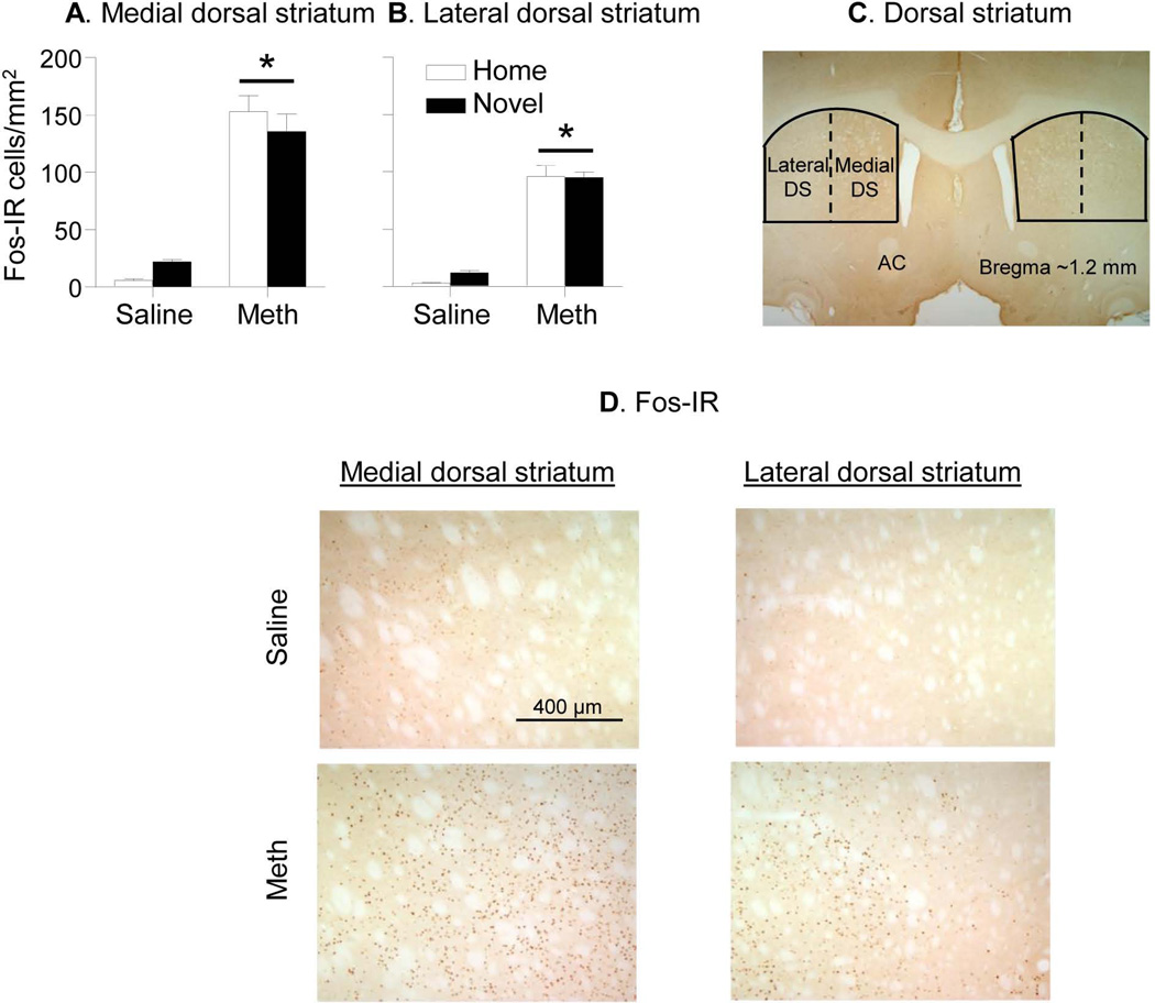
Figure 2. Fos labeling in medial and lateral dorsal striatum of rats injected in Home cage or Novel cage.
(A,B) Number of Fos-IR nuclei per mm2 in medial and lateral dorsal striatum 90 min after Saline or Methamphetamine (Meth) injections. (C) Areas where Fos-IR was quantified. Image was captured using 1.25× objective. (D) Representative images of Fos-IR (dark brown) in the medial (left) and lateral (right) dorsal striatum. Images were captured using 5× objective. Abbreviations: AC: anterior commissure; DS: dorsal striatum. * Different from the Saline condition, p<0.05, n=6 per experimental condition.
Double labelling of Fos and NeuN immunohistochemistry
A double-labeling assay for Fos and NeuN, a marker for neuronal-specific nuclear membrane protein (Mullen et al. 1992), was used to determine the proportion of neurons expressing Fos in dorsal striatum. Brain sections from 12 rats from Exp. 1 (4 groups; n=3 per group) were thawed and washed (3 times for 10 min each) in Tris-buffered saline (TBS; 0.025 M Tris-HCl, 0.5 M NaCl, pH 7.5) and incubated for 20 min in TBS with 0.25% Triton X-100 (TBST). Sections were washed in TBS and incubated for 48 h at 4°C with the rabbit anti-Fos primary antibody (1:500 dilution of sc-52, Santa Cruz Biotechnology) and mouse anti-NeuN primary antibody (1:2,000 dilution of MAB377, Millipore) in TBST. Sections were again washed in TBS and incubated for 1 h with the secondary antibodies Alexa 488-labeled donkey anti-rabbit and Alexa 568-labeled goat anti-mouse (1:200 dilutions in TBST for each antibody, Invitrogen, Life Technologies). Finally, sections were washed in TBS, mounted on chromalum/gelatin-coated slides, air-dried, and coverslipped with Vectashield fluorescent mounting medium (H-1400, Vector Laboratories).
Fluorescent images of dorsal striatum were captured with Qimaging Exi Aqua camera (Biovision) attached to a Zeiss AXIO Imager M2 microscope using a 10× objective. Sampled areas for lateral and medial dorsal striatum were about 1 mm2. The number of Fos-labeled, NeuN-labeled, and double-labeled immunoreactive nuclei in these images were manually counted using iVision (version 4.0.15 for Mac; Biovision) in a blind manner by QRL and JMB. For Fig. 2D, multiple z-stack (390 nm) images were captured using a 40× objective (Zeiss PLAN-APOCHROMAT, N.A.=1.3) with oil immersion. These images were deconvolved with Huygens software (v3.7, Scientific Volume Imaging), pseudocolored (NeuN green and Fos red) and joined using iVision software.
Double-labeling of Fos and Arc immunohistochemistry
The Fos+Arc double-labeling immunohistochemistry procedure is based on our previous report (Guez-Barber et al. 2011). We used coronal sections obtained from three rats that were injected with methamphetamine in Exp. 1. Sections were washed three times in Tris-buffered saline (TBS) and permeabilized for 30 min in TBS with 0.2% Triton X-100. Sections were washed again in TBS and incubated in primary antibodies diluted in TBS with 0.3% Triton X-100 for 24 hours on a shaker at 4°C. We used a rabbit polyclonal against c-Fos antibody (1:500 dilution, Cat# sc-52, Santa Cruz Biotechnology, Santa Cruz, CA) and a mouse monoclonal Arc antibody (1:100 dilution, Cat# sc-17839, Santa Cruz Biotechnology). Sections were washed three times in TBS and incubated in secondary antibodies diluted in TBS for 1.5 hours on a shaker at room temperature. We used Alexa Fluor 488-labeled donkey anti-rabbit antibody (1:200 dilution, Cat# A21206, Invitrogen) and Alexa Fluor 568-labeled goat anti-mouse antibody (1:200 dilution, Cat# A11004, Invitrogen). Sections were washed in TBS, mounted onto chrom-alum coated slides, and coverslipped with VectaShield hard-set mounting media.
Fluorescent images of dorsal striatum were captured with Qimaging Exi Aqua camera (Biovision) attached to a Zeiss AXIO Imager M2 microscope using a 20× objective. Sampled areas were 0.25 mm2. The number of Fos-labeled, Arc-labeled, and double-labeled immunoreactive nuclei in these images were manually counted using iVision (version 4.0.15 for Mac; Biovision) in a blind manner by FJR. For Fig. 7, multiple z-stack (352 nm) images were captured using a 40× objective (Zeiss PLAN-APOCHROMAT, N.A.=1.3) with oil immersion. These images were deconvolved with Huygens software (v3.7, Scientific Volume Imaging), pseudocolored (Fos green and Arc red) and joined using iVision software.
Figure 7. Fos+Arc double-labeling dorsal striatum after acute methamphetamine injections.
Immunohistochemistry demonstrated that Arc protein was co-expressed in 65 ± 1% of all Fos-positive cells whereas Fos protein was detected in 80 ± 4% of all Arc-positive cells (n=3). Images for counting labeled cells from 4–6 hemispheres per rat were captured using 20× objective. Representative images were captured using 40× objective with oil immersion and deconvolved using Huygens software. Single Fos-positive nuclei (green, filled arrowhead), single Arc-positive cells (red, empty arrowhead), or double-labeled cells (arrow) are indicated in the images.
Experiment 2: FACS and RT-qPCR quantification of gene expression in Fos-positive and Fos-negative neurons
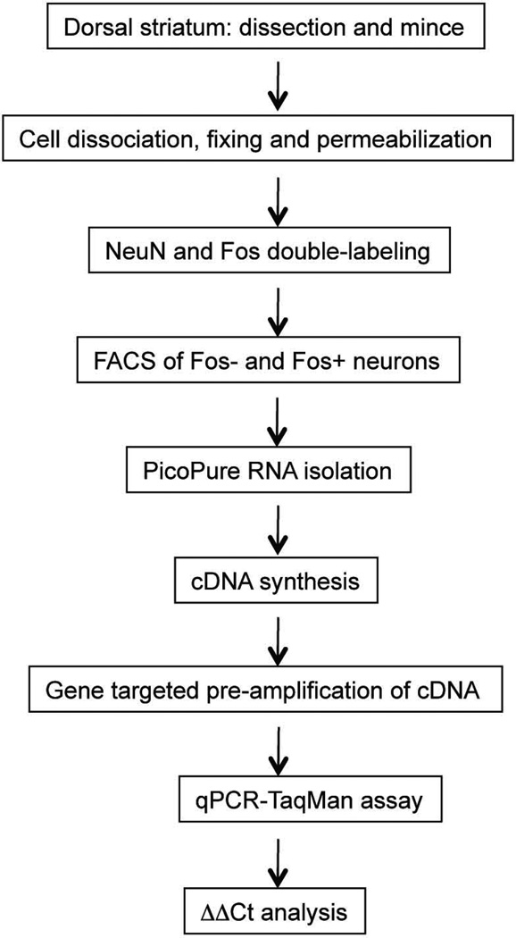
The FACS-PreAmplification-qPCR method is based on our previous method (Fanous et al. 2012, Guez-Barber et al. 2011, Guez-Barber et al. 2012) with modifications for neuronal dissociation and pre-amplification procedures, as described in Fig. 1 workflow chart. In our previous studies, we used a Percoll purification step to get rid of the large amount of debris from our larger samples of pooled tissue so that the debris did not increase FACS time. The smaller samples from smaller brain areas from single rats make this step unnecessary in the current protocol.
Figure 1. FACS-Preamplification-qPCR workflow chart.
Cell dissociation
Rats were decapitated 90 min after saline or methamphetamine injections and their dorsal striatum was extracted within 2 min. Dorsal striatum was dissected from 2 mm thick coronal sections and minced with razor blades on an ice-cold glass plate (dried before each mincing) and placed in 1 ml of Hibernate A (Cat# HA-lf; Brain Bits). The tissues were enzymatically digested in 1 ml of Accutase (4°C) per dorsal striatum (Cat# SCR005; Millipore) with end-over-end mixing for 30 min at 4°C. Tissue was centrifuged for 2 min at 425 × g and the pellet re-suspended in 0.6 ml of ice-cold Hibernate A. The cells were dissociated in three steps by trituration (up and down 10×) with fire-polished glass Pasteur pipettes of successively smaller diameters (1.3, 0.8, and 0.4 mm). After each trituration step, the tubes containing cells were briefly placed on ice for 2 min to sediment undissociated tissue and the supernatants were collected and transferred to another tube sitting on ice. Each cloudy supernatant containing dissociated cells was combined with the previous supernatant for a total volume of 1.8 ml that was then filtered through 100 µm and then 40 µm cell strainers (Falcon brand, BD Biosciences) that were pre-wetted with PBS. Two aliquots of 650 µl per filtered sample were used to fix cells.
Immunolabeling and FACS
Dissociated cells were fixed and permeabilized by adding an equal volume of ice-cold ethanol to the above cell suspension for a final concentration of 50% ethanol and kept on ice for 15 min with occasional shaking. Cells were centrifuged for 4 min at 1,000 × g and resuspended in 0.7 ml of cold PBS. Cells were incubated with primary antibodies including biotinylated mouse anti-NeuN antibody (1:1000 dilution, Cat# MAB377B, Millipore) and rabbit anti-c-Fos antibody (1:1000 dilution of sc-52, Santa Cruz Biotechnology). The cell suspension was rotated end-over-end in primary antibodies for 30 min at 4°C and then washed by adding 0.8 ml of cold PBS with centrifugation for 3 min at 425 × g. The pellet was resuspended in 0.7 ml of cold PBS and incubated with streptavidin conjugated to R-phycoerythrin (R-PE Streptavidin, 1:1000 dilution, Cat# SA1004-1, Invitrogen, Life Technologies) for neuronal-labeling and the donkey anti-rabbit IgG antibody conjugated to Alexa Fluor 488 (1:1000 dilution, Cat# A-21206, Invitrogen, Life Technologies) for Fos-labeling, with rotation end-over-end for 15 min in darkness (tubes were wrapped in aluminum foil). Cells were washed first by adding 0.8 ml of cold PBS with centrifugation for 3 min at 425 × g followed by the second wash with 1 ml of cold PBS. The pellet was resuspended in 0.5 ml of cold PBS for FACSAria (BD Biosciences) sorting. Control samples were analyzed first to determine optimal criteria for sorting test samples. Specifically, fixed cells stained with DAPI and without antibodies were used to set the light scatter gate. Fixed cells treated with fluorescent streptavidin or secondary antibody, but without primary antibody, were then used to set thresholds for endogenous tissue fluorescence and nonspecific binding by fluorescent streptavidin or secondary antibody. Next, control samples single-labeled with primary and either R-PE Streptavidin or secondary antibody conjugated with Alexa Fluor 488 for each protein (NeuN or Fos, respectively) were used to compensate for fluorescent overlap in neighboring channels. Finally, test samples labeled with fluorescent markers were analyzed and sorted. Sorted cells were collected into low-binding tubes (Cat# 022431081, Eppendorf) with 0.1 ml of cold PBS in each tube.
RNA isolation and cDNA synthesis
PicoPure RNA isolation kit (Arcturus Bioscience, Inc.) was used to isolate RNA. The FACS-sorted neurons were centrifuged at 4,500 × g at 4°C for 10 min. The pellets were suspended in 50 µl of the PicoPure RNA extraction buffer by pipetting up and down 10 times and then incubated at 42°C for 30 min. The suspension was centrifuged at 1,000 × g at 4°C for 2 min and the supernatant was collected in an RNase-free tube (for long-term storage at −80°C or immediately processed for RNA isolation). Column filtration, washing, and elution of RNA from the columns are described in the Protocol for Use with CapSure Macro Laser Capture Microscope (LCM) Caps as described in section C of the PicoPure RNA isolation protocol. RNA integrity numbers were measured using Agilent RNA 600 Pico kit (Agilent Technologies) and found to be ~3–4 for the FACS samples. Single strand cDNAs were synthesized with the Superscript III first strand cDNA synthesis kit according to the manufacturer’s protocol (Invitrogen, Life Technologies).
Pre-amplification and RT-qPCR
TaqMan PreAmp Master Mix Kit was used for cDNA preamplification (Cat# 4391128, Applied Biosystems, Life Technologies) using pooled primer mixes of 20 × dilution of TaqMan Gene Expression Assay and 80 nM of customer-designed primer sets (Table 1). cDNAs were pre-amplified in an ABI 9700 Thermal Cycler using the program: 95°C hold for 10 min and then 14 cycles of denaturation at 90°C for 15 seconds and annealing and extension at 60°C for 4 min. The pre-amplification PCR products were immediately diluted 5 times with molecular biology-grade water (Cat# 2500010, 5-Prime) and stored at −20°C or immediately processed for qPCR. Duplex qPCR assays were performed on technical duplicates using a Fam-labeled probe for each target gene (Table 1) and a Vic-labeled probe for the endogenous control gene (gapdh), along with TaqMan® Advanced Fast PCR Master Mix (Cat# 4444963, Life Technologies). To avoid amplification of any genomic DNA contamination, Primer Express 3.0 (Applied Biosystems) was used to select primers and probes for pre-amplification and RT-qPCR that amplified across exon-exon junctions for each target gene. RT-qPCR reactions were run in a 7500 Fast TaqMan instrument using the program: 95°C hold for 20 seconds followed by 40 cycles of denaturation at 95°C for 3 seconds and annealing and extension at 60°C for 30 sec. Calculations of relative expression from Ct data were carried out according to User Bulletin #2 for ABI Prism 7900 Sequence Detection System. For each target gene, the average ΔCt value from the Saline-Fos-negative samples was subtracted from ΔCt = Ct (target gene) − Ct (gapdh) for each sample to obtain ΔΔCt for each sample. Relative expression = 2e(−ΔΔCt) was then calculated and interpreted as fold-difference over Saline-Fos-negative expression levels. Uniformity of the pre-amplification was verified by comparing cDNA templates from the pre-amplified and unamplified dorsal striatum samples. ΔΔCt values were within range of ±1.5 for all target genes between the pre-amplified and unamplified samples.
Table 1.
List of qPCR probes and primers
| Gene | TaqMan probe | Forward primer | Reverse primer |
|---|---|---|---|
| c-fos | Rn00487426_g1* | ||
| fosB (total) | CGAGAAGAAACACTTACC | CCAGAGCCAGGCCTAGAAGA | CTGCGAACCCTTCGTTTTTC |
| ΔfosB | AGAGGAAAAGGCAGAGCT | CGAGAAGAAACAGAAACGGATCA | CCAGGCGTTCCTTCTCTTTTT |
| ΔfosB-2 | CCCTTCCCCGTTGTT | TGGCCGAGTGAAGTTCAAGTC | CGGGCAGGTGAGGACAAA |
| arc | Rn00571208_g1* | ||
| homer 2 | Rn00584015_m1* | ||
| drd1 | Rn00432253_m1* | ||
| drd2 | Rn00561126_m1* | ||
| darpp-32 | TCGCCAGAGGAGGAG | TTCCGGGTCTCAGAGCATTC | TGCCCCTCTCCTGAGGTTCT |
| Δdarpp | CCCTGTGAAATAAAG | TGAGCCCCGGACTTTTCA | GCCGGATCCTGCTTTCAAT |
| ampa1 | Rn00709588_m1* | ||
| cb1r | TGAGAAGGGGTTCC | GTGCCGAGGGAGCTTCTG | GACTCAAGGTGACTGAGAAAGA |
| gapdh | CTCATGACCACAGTCCA | GACAACTTTGGCATCGTGGAA | CACAGTCTTCTGAGTGGCAGTGA |
| iba-1 | Rn00574125_g1* | ||
| neun | CACTCCAACAGCGTGAC | GGCCCCTGGCAGAAAGTAG | TTCCCCCTGGTCCTTCTGA |
| gfap | Rn00566603_m1* |
Taqman catalog number for ordering from Applied Biosystems.
Experiment 3: RT-qPCR of dorsal striatum homogenates
The procedure was similar to that described in our previous publication (Theberge et al. 2012). Briefly, dorsal striatum was dissected from 2 mm thick coronal sections and total RNA was extracted from two hemisphere (~0.5 mg) per sample using TRIzol Reagent protocol (Invitrogen, Life Technologies). The RNA integrity numbers were 7–8 using Agilent RNA 600 Nano kit (Agilent Technologies). Single strand cDNAs were synthesized with the Superscript III first strand cDNA synthesis kit according to the manufacturer’s protocol (Invitrogen, Life Technologies). The same duplex qPCR assays were performed on technical duplicates using a Fam-labeled probe for each target gene (Table 1) and a Vic-labeled probe for the endogenous control gene (Gapdh). Relative expression = 2e(−ΔΔCt) was calculated as above and interpreted as fold-difference over Saline expression levels.
Experiment 4: Time course for Fos and FosB
Three groups of rats (n=8 per group) were injected with either saline (1 ml/kg) or methamphetamine (5 mg/kg) and were decapitated 90 or 180 min later, and dorsal striatum was dissected. Cells were dissociated as described above and incubated with primary antibodies including biotinylated mouse anti-NeuN antibody (1:1000 dilution, Cat# MAB377B, Millipore), rabbit anti-Fos antibody (1:1000 dilution of sc-52, Santa Cruz Biotechnology) or goat anti-FosB antibody (1:500 dilution of sc-48-G, Santa Cruz Biotechnology). The cells were than labeled with streptavidin conjugated to R-phycoerythrin (R-PE Streptavidin, 1:1000 dilution, Cat# SA1004-1, Invitrogen, Life Technologies), the donkey anti-rabbit IgG antibody conjugated to Alexa Fluor 647 (1:300 dilution, Cat# A-31573, Invitrogen, Life Technologies) and donkey anti-goat IgG antibody conjugated to Alexa Fluor 647 (1:500 dilution, Cat# A-21447, Invitrogen, Life Technologies) for neurons, Fos and FosB, respectively. Numbers of Fos and FosB-immunoreactive cells were assessed using flow cytometry as described above and in the Results section.
Statistical analyses
In Exp. 1, we analyzed the Fos and NeuN immunoreactivity data using mixed ANOVA (SPSS version 20, GLM procedure or GraphPad software) with the between-subjects factors of Context (home, novel) and Drug (saline, methamphetamine), and the within-subjects factor of Dorsal Striatum Area (lateral, medial). In Exp. 2, we analyzed cell numbers data for FACS-sorted NeuN-positive, Fos-positive and Fos-negative neurons using the between-subjects factor of Drug (saline, methamphetamine). We analyzed relative gene expression data from the FACS-sorted samples using the between-subjects factor of Drug (saline, methamphetamine) and the within-subjects factor of neuron type (Fos-positive, Fos-negative). In Exp. 3, we analyzed relative gene expression data using Student’s t-test for the between-subjects factor of Drug (saline, methamphetamine). We used Fisher PLSD post-hoc tests where appropriate. In Exp. 4, we analyzed the time courses for Fos− and FosB-labeled dorsal striatal neurons using one-way ANOVA using 3 groups: saline-90 min, methamphetamine-90 min, and methamphetamine-180 min. We used Fisher PLSD post-hoc tests where appropriate. Significance was p<0.05.
Results
Exp. 1: Fos immunohistochemistry and Fos+NeuN double-labeling
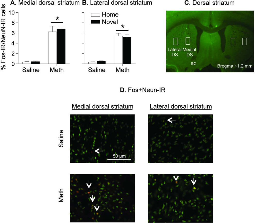
In an initial experiment, we determined the pattern of Fos expression in medial and lateral dorsal striatum, using traditional Fos and Fos+NeuN double labeling immunohistochemistry in rats injected with saline or methamphetamine in the home cage or novel cage. This was done in order to estimate the proportion of Fos-positive neurons in dorsal striatum before proceeding to the FACS method, and for determining whether the context of drug exposure would modulate striatal Fos expression, as we have previously shown in studies using cocaine (Mattson et al. 2008, Hope et al. 2006). A single injection of 5 mg/kg methamphetamine significantly increased Fos-IR in both medial and lateral dorsal striatum and this effect was independent of the context of drug injections (home or novel). These injections also had a stronger effect on Fos-IR in medial than in lateral dorsal striatum (Fig. 2). The statistical analysis indicates significant main effects of Drug (F1,20=297.7, p<0.001) and Dorsal Striatum Area (F1,20=25.9, p<0.001), and an interaction between these two factors (F1,20=15.7, p<0.001); the effect of Context or interactions between Context and the other factors were not significant (p> 0.05). In the Fos+NeuN double-labeling experiment, 5–6% of dorsal striatum neurons (NeuN) were Fos-positive after methamphetamine injections, while less than 0.5% of these neurons were Fos-positive after saline injections (Fig. 3). The context of methamphetamine injections (home, novel) had no effect on the number of Fos+NeuN double-labeling cells in medial or lateral dorsal striatum.
Figure 3. Fos+NeuN double-labeling in medial and lateral dorsal striatum.
(A,B) Percent Fos-IR cells out of all NeuN-IR cells (per mm2, mean±sem) in medial and lateral dorsal striatum 90 min after Saline or Methamphetamine (Meth) injections. (C) Rectangular areas where Fos+NeuN was quantified. Image was captured using 1.25× objective. (D) Representative images of Fos-IR (red), NeuN-IR (green) and double-labeled cells (orange) in the medial (left) and lateral (right) dorsal striatum (n=3 per experimental condition). Images were captured using 40× objective with oil immersion and deconvolved using Huygens software. * Different from the Saline condition, p<0.05, n=3 per experimental condition.
Experiment 2: FACS and RT-qPCR quantification of gene expression in Fos-positive and Fos-negative neurons
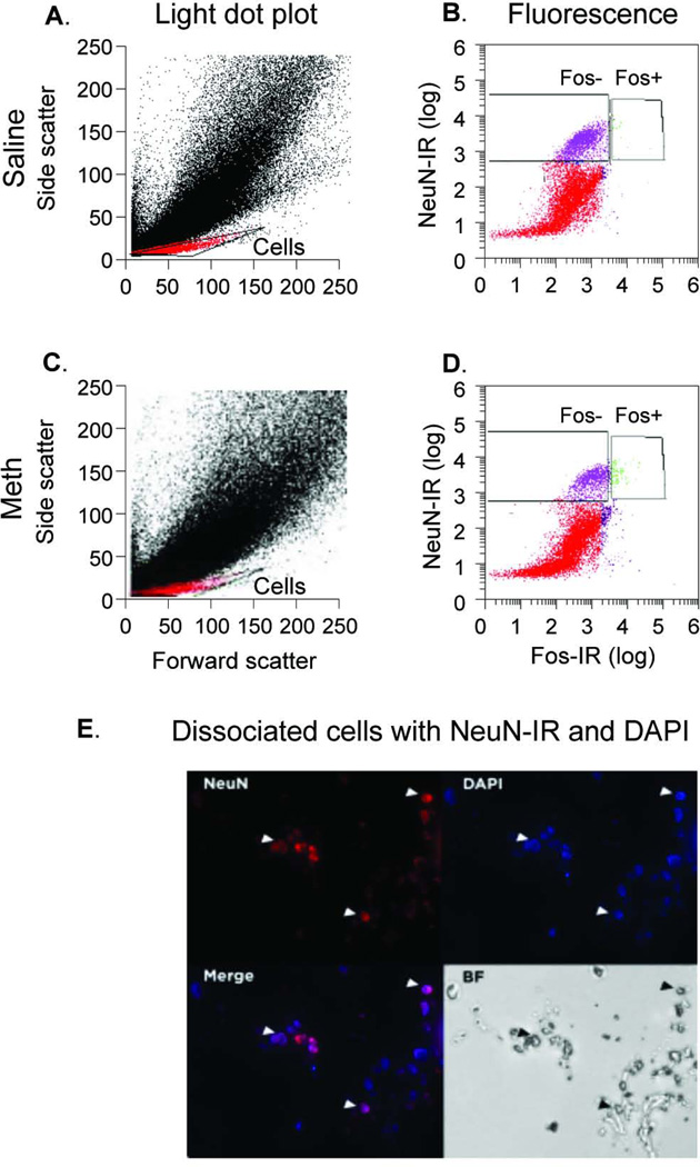
Since we found that the environmental context did not modulate methamphetamine-induced Fos expression in dorsal striatum, we used rats injected only in their home cage for our FACS and RT-qPCR experiments that assessed gene expression in Fos-positive and Fos-negative neurons. In Fig. 4, the Light scattergrams on the left indicate characteristics such as size in the x-axis (forward scatter, FSC) and granularity in the y-axis (side-scatter, SSC). The events (red and purple dots) within the ‘Cells’ gate were selected based on our previous studies (Guez-Barber et al. 2011) and shown in the current study to contain mostly cells: DAPI staining labeled >95% of events within this gate. We used microscopy to confirm that the majority of NeuN-positive events were DAPI positive after sorting (Fig. 4E). The fluorescence scattergrams on the right indicate fluorescent characteristics of cells with their degree of NeuN labeling (R-phycoerythrin) on the y-axis and Fos labeling (Alexa 488) on the x-axis. Cells with high NeuN labeling were considered neurons (purple and green dots). Approximately 2000–7000 neurons were obtained from 2 mg of dorsal striatum. Neurons comprised 30% of all cells selected in the light scatter gate for both saline- and methamphetamine-injected rats. Of these neurons, high levels of Fos labeling were considered Fos-positive neurons (green dots) while the remainder were considered Fos-negative (purple dots); these neurons are indicated within rectangular gates. The mean ratio of Fos+/Fos− neurons was 57/7035 (0.9%, range 5–148 neurons were Fos+) in saline-injected rats and 115/1940 (5.7%, range 9–147 were Fos+) in methamphetamine-injected rats. The proportion of Fos-positive neurons from the methamphetamine-injected rats was significantly higher than the proportion of Fos-positive neurons from the saline-injected rats (F1,10=208.1, p<0.0001). These percentages of Fos-positive neurons were similar to those observed in our Fos+NeuN immunohistochemistry assays in Exp. 1.
Figure 4. FACS analyses of neurons from dorsal striatum.
(A,C) Forward scatter (X axis, cell size) and Side scatter (Y axis, granularity) linear plots in which the cell population (Cells) was gated for further analysis with fluorescence. (B,D) Fluorescence from cells double-labeled for NeuN (R-phycoerythrin on Y axis) and Fos (Alexa 488 on X axis) logarithmic plots (a log10 scale). Fos-negative and Fos-positive neurons (left and right gates, respectively) on the X axis were represented by purple (left gate, Fos-negative) and green (right gate, Fos-positive) dots. (E) DAPI labeling of FACS-isolated neurons. Dissociated NeuN-labeled neurons were centrifuged briefly to concentrate them and spread on a microscope slide and labeled with DAPI. Bottom right-hand panel indicates bright field (BF) labeling of cells that are now visibly clumped due to centrifugation. Other panels indicate NeuN, DAPI, and merged labeling of these same cells.
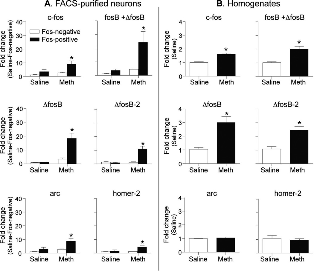
Methamphetamine induced large increases (3–20 fold higher than saline) of mRNA expression of the immediate early genes (IEGs) arc, homer-2, c-fos, and fosB in Fos-positive neurons but not in Fos-negative neurons (Fig. 5A); these increases included total levels of all fosB mRNA isoforms (total fosB+ΔfosB), as well as two spliced isoforms of fosB mRNA (ΔfosB and ΔfosB-2) that were identified by amplification across the splice junction sites. Details of the statistical analyses are shown in Table 2. In contrast, methamphetamine did not increase mRNA expression levels of several other genes, including dopamine D1 and D2 receptors, CB1 receptors, DARPP-32, a truncated version of DARPP-32, MeCP2, and GluA1 (see Table 2).
Figure 5. IEG expression in FACS-purified Fos-negative and Fos-positive neurons and in homogenates from dorsal striatum.
Exp. 2: Left panels (A) show IEG expression (0–40 fold in Y axis) in Fos-negative and Fos-positive neurons from dorsal striatum of rats injected with Saline or Methamphetamine (Meth) (n=5–6 per group). Exp. 3: Right panels (B) show IEG expression (0–4 fold in Y axis) in homogenates from dorsal striatum of rats injected with Saline or Methamphetamine (Meth) (n=3 per group). All genes are normalized to the endogenous control gene gapdh. * Different from the Saline group, p<0.05.
Table 2.
Results of the statistical analyses of Experiment 2: FACS and RT-qPCR quantification of gene expression in Fos-positive and Fos-negative neurons (n=5–6 per group).
| Main effect of Drug | Main effect of Fos cell type |
Interaction | |
|---|---|---|---|
| Immediate early genes | |||
| c-fos | F(1,9)=4.8, p=0.055 | F(1,9)=9.4, p=0.014 | F(1,9)=2.1, p=0.186 |
| fosB (total) | F(1,9)=7.6, p=0.022 | F(1,9)=5.9, p=0.037 | F(1,9)=3.6, p=0.090 |
| ΔfosB | F(1,10)=21.0, p=0.001 | F(1,10)=20.6, p=0.001 | F(1,10)=23.9, p=0.001 |
| ΔfosB-2 | F(1,10)=19.0, p=0.001 | F(1,10)=23.6, p=0.001 | F(1,10)=22.6, p=0.001 |
| arc | F(1,9)=6.0, p=0.037 | F(1,9)=14.5, p=0.004 | F(1,9)=3.9, p=0.081 |
| homer-2 | F(1,9)=4.7, p=0.059 | F(1,9)=5.1, p=0.050 | F(1,9)=3.1, p=0.113 |
| Other genes | |||
| drd1 | F(1,10)=0.0, p=0.990 | F(1,10)=3.1, p=0.108 | F(1,10)=0, p=0.870 |
| drd2 | F(1,10)=2.1, p=0.175 | F(1,10)=0.8, p=0.388 | F(1,10)=0.1, p=0.808 |
| darpp-32 | F(1,10)=0.1, p=0.730 | F(1,10)=0.5, p=0.496 | F(1,10)=0.1, p=0.830 |
| Δdarpp-32 | F(1,10)=2.5, p=0.145 | F(1,10)=0.0, p=0.925 | F(1,10)=3.9, p=0.075 |
| cb1r | F(1,10)=1.1, p=0.330 | F(1,10)=0.0, p=0.942 | F(1,10)=0.1, p=0.829 |
| ampa1 | F(1,10)=1.2, p=0.300 | F(1,10)=0.1, p=0.768 | F(1,10)=1.0, p=0.345 |
Experiment 3: RT-qPCR of dorsal striatum homogenates
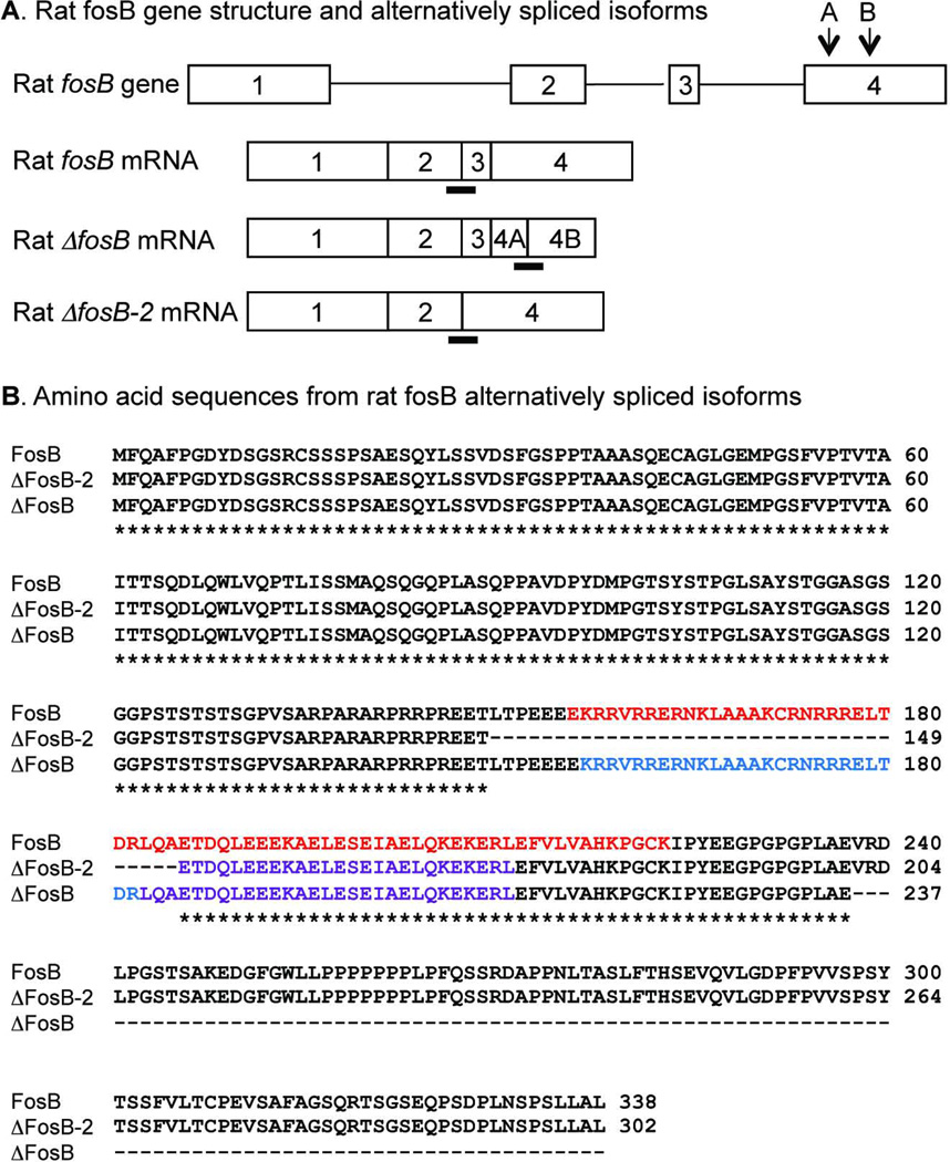
To demonstrate the different magnitudes of gene expression in homogenates relative to expression selectively in behaviorally activated Fos-expressing neurons, we also assessed gene expression in homogenates of dorsal striatum neurons in rats injected with saline or methamphetamine in their home cages. Methamphetamine induced smaller increases or no alteration (1–3 fold higher than saline) of mRNA expression of the IEGs c-fos, (total fosB+ΔfosB), and the distinct splice isoforms ΔfosB and ΔfosB-2 (Fig. 5B) in dorsal striatum homogenates from a single hemisphere. Details of the statistical analyses are shown in Table 3. In contrast, methamphetamine did not increase mRNA expression levels of the IEGs arc and homer-2 (Fig. 5B), or expression levels of the other genes, including dopamine D1 and D2 receptors, CB1 receptors, DARPP-32, a truncated version of DARPP-32, and mGluR1 (see Table 3). The gene structure, TaqMan probe positions for fosB (total and splice isoforms), and amino acid sequences are shown in Fig. 6A and B.
Table 3.
Results of the statistical analyses of Experiment 3: qPCR of dorsal striatum homogenates (n=3 per group)
| Main effect of Drug | |
|---|---|
| Immediate early genes | |
| c-fos | t(4)=6.8, p=0.002 |
| fosB (total) | t(4)=3.3, p=0.029 |
| ΔfosB | t(4)=3.6, p=0.022 |
| ΔfosB-2 | t(4)=3.3, p=0.030 |
| arc | t(4)=0.6, p=0.584 |
| homer-2 | t(4)=0.2, p=0.825 |
| Other genes | |
| drd1 | t(4)=1.3, p=0.249 |
| drd2 | t(4)=1.5, p=0.209 |
| darpp-32 | t(4)=1.2, p=0.308 |
| Δdarpp–32 | t(4)=1.7, p=0.160 |
| cb1r | t(4)=0.1, p=0.896 |
| ampa1 | t(4)=0.9, p=0.430 |
Figure 6. Rat fosB gene structure and its alternatively spliced isoforms.
(A) The open horizontal bars represent exons while the solid lines represent introns. Within exon 4 there are two intra-exonal splicing sites (4A and 4B), indicated by arrows above the exon. The alternatively spliced isoforms are shown under the gene structure. A TaqMan probe was designed to detect both full-length fosB and ΔfosB combined by hybridizing across the junction between exons 2 and 3. Another TaqMan probe was designed to detect only the ΔfosB isoform by hybridizing across the junction within exon parts 4A and 4B. Another Taqman probe was designed to detect only the ΔfosB-2 isoform by hybridizing across the junction between exon 2 and 4. The short black horizontal bars under the exon junctions represent the TaqMan probes. (B) Comparison of amino acid sequences for FosB, ΔFosB, and ΔFosB-2. Asterisks represent identical amino acids and the dashed lines deletions. Red font represents basic leucine zipper domain (155–218 AA) domain including the basic motif (157–182 AA, blue) and leucine-zipper motif (183–211AA, purple). ΔFosB-2 lacks the basic motif (150–185 AA).
Double-labeling of Fos and Arc protein
We performed immunohistochemistry to assess whether protein expression of Fos and Arc correlates with mRNA expression results from our FACS experiment. Immunohistochemistry revealed that Arc protein was co-expressed in 65 ± 1% of all Fos-positive cells whereas Fos protein was detected in 80 ± 4% of all Arc-positive cells (Fig. 7).
Experiment 4: Time course of Fos and FosB expression
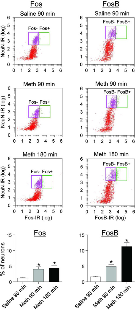
We performed FACS of dorsal striatum in rats injected with either saline (90 min time point) and or methamphetamine (90 or 180 time points). We found time-dependent increases for FosB but not Fos expression (Fig. 8). The one-way ANOVA demonstrated a significant group effect for both FosB (F2,21=56.8, p<0.01) and Fos (F2,21=56.8, p<0.01). Post-hoc group differences are indicated in Figure 8.
Figure 8. Time course for number of Fos and FosB expression in dorsal striatum after acute methamphetamine injections.
Fluorescence from cells double-labeled for NeuN (R-phycoerythrin on Y axis) and either Fos or FosB (Alexa 647 on X axis) logarithmic plots (a log10 scale). Fos/FosB-negative and Fos/FosB-positive neurons (left and right gates, respectively) on the X axis were represented by purple (left gate, Fos/FosB-negative) and green (right gate, Fos/FosB-positive) dots, respectively. The Y axes of the bar graphs on the bottom represent percentages of Fos-positive or FosB-positive neurons from all NeuN-labeled neurons while the X axis indicates time points following acute saline or methamphetamine injections (n=7–8 per group). * Different from the Saline group, p<0.05.
Discussion
The main goal of our report was to describe a more sensitive FACS-based method to study unique gene expression alterations in activated Fos-expressing neurons from a single brain area of a single rat. As mentioned in the Introduction, our original FACS method requires pooling up to 10 rat striatum or prefrontal cortex tissues (Fanous et al. 2013, Guez-Barber et al. 2011, Guez-Barber et al. 2012), and therefore may not be feasible for long-term studies on mechanisms of drug reward and relapse, as assessed in animal models of drug addiction (Badiani et al. 2011, Bossert et al. 2013). Here, we improved the existing FACS method (Guez-Barber et al. 2012) by introducing two main modifications. First, we have used Arcturus PicoPure RNA Kit for Laser Captured Cells to isolate RNA from less than 100 Fos-positive neurons. Second, we used Applied Biosystems TaqMan PreAmp technology to pre-amplify target genes, using cDNA templates reverse transcribed from the isolated RNAs. These modifications allowed us to reliably measure gene expression changes in a limited number (at least 5) of Fos-expressing neurons from dorsal striatum of rats acutely injected with saline or methamphetamine.
Our modified FACS method reliably separates activated Fos-positive neurons from the majority of Fos-negative neurons. First, the proportion of Fos-positive neurons (~5–6%) was similar to the proportion detected by more traditional NeuN+Fos double-labeling immunohistochemistry (Fig. 3–4). Second, as in our previous studies (Fanous et al. 2013, Guez-Barber et al. 2011, Guez-Barber et al. 2012), the Fos-positive neurons co-express other immediate early genes (Fig. 5, left column), including arc, c-fos, fosB, and homer2. These IEGs are established markers of neuronal activation (Guzowski et al. 1999, Nakabeppu & Nathans 1991, Worley et al. 2007, Hope et al. 1994b, Cruz et al. 2013). On the other hand, expression levels of several receptor genes and DARPP-32 were not different between Fos-positive and Fos-negative neurons. These genes are expressed at relatively high levels in the striatum and their expression was not expected to be altered within 90 min by neural activity in Fos-positive neurons (Table 2). Finally, our improved FACS method was 10-fold or more sensitive than homogenate preparations for detecting IEG alterations that were induced selectively in Fos-positive neurons (Fig. 5B). Unique gene expression alterations in Fos-positive neurons would be diluted by the relative lack of gene expression in Fos-negative neurons that comprise the majority of neurons in brain homogenate preparations. Positive detection of increased arc and homer-2 expression in FACS-purified Fos-positive neurons without any alterations detected in homogenates exemplifies the advantage of measuring gene expression selectively in Fos-expressing neurons and how potentially relevant gene expression information can be lost when using homogenate preparations.
In our FACS time course experiment the numbers of methamphetamine-induced Fos-labeled neurons were similar at the 90 and 180 min time points while FosB (including all FosB isoforms) expression was substantially higher after 180 min than after 90 min (Fig. 8). This corresponds with previous time course experiments that found FosB expression is delayed and more prolonged than for Fos expression (Hope et al. 1994a, Chen et al. 1995, Chen et al. 1997). This study demonstrates that our modified FACS procedure can also reliably identify FosB-positive neurons, and that this immediately early gene can be used to study molecular characteristics of behaviorally-relevant activated neurons.
Methodological considerations
FACS was first used in adult brain to purify striatal neurons expressing GFP driven by the promoters for D1 and D2 receptors in mice (Lobo et al. 2006, Lobo 2009). In our original FACS method (Fanous et al. 2012, Guez-Barber et al. 2011, Guez-Barber et al. 2012), we used commercially available antibodies to identify neurons expressing Fos and other protein markers from both wild-type and transgenic animals. In our procedure we have to dissociate cells, permeabilize their cell membranes with ethanol to label intracellular proteins such as the nuclear antigens NeuN and Fos, and pass them through a FACS machine before extracting RNA. Thus the integrity (RIN: 3–4) of RNA extracted from Fos-positive neurons after FACS was lower than the integrity (RIN: 7–8) of RNA extracted directly from homogenates. To compensate for lower RNA integrity, we used PCR-amplicons that were less than 100 bp and thus less affected by limited RNA degradation. One caveat is that IEG expression is dynamic following psychostimulant injections. Thus different fold levels of IEG expression will likely be observed at earlier or later time points than the single 90-minute time point that we used.
In preliminary experiments, we tested different candidate reference genes, including gapdh, β-actin, pde10A, and gorasp, by assessing their expression in Fos-positive and Fos-negative neurons from rats injected with saline or methamphetamine. Gapdh expression was the least variable between these groups and thus selected as our endogenous reference gene (data not shown). We used different reference genes (gorasp and pde10A) in our previous FACS studies (Fanous et al. 2013, Guez-Barber et al. 2011, Guez-Barber et al. 2012), because we were also examining gene expression in different cell types in which gapdh expression was greater between cell types than differences of gorasp and pde10A expression. The validity of our modified FACS technique to measure gene expression levels in selected neurons was verified by assessing NeuN-positive FACS-sorted cells for gene expression of the cell-type markers, neuN for neurons, gfap for astrocytes, and iba-1 for microglia. NeuN mRNA levels were ~200-fold higher than gfap mRNA levels and 2000-fold higher than Iba-1 mRNA levels (data not shown). Finally, we also verified that the subtracted ΔCt values (ΔCt preamp − ΔCt cDNA) for 30 gene targets between pre-amplified and unamplified dorsal striatum cDNA samples were within a range of ±1.5. Our gene expression results for arc and c-fos mRNA in FACS-sorted Fos-positive and Fos-negative neurons were validated by showing that the majority (80 ± 4%) of Arc protein expression was found in Fos-positive neurons in immunohistochemically labeled sections obtained from methamphetamine-injected rats.
A novel spliced isoform of the fosB gene
We identified a novel spliced isoform of the fosB gene (ΔfosB-2) that deletes exon 3 encoding a 36 amino acid sequence normally involved in binding DNA, using the “Gnomon” NCBI eukaryotic gene prediction tool (http://www.ncbi.nlm.nih.gov/RefSeq/Gnomon-description.pdf). An in-frame deletion of exon 3 in ΔfosB-2 removes part of the positively charged DNA binding motif but keeps the N- and C-terminal transactivation domains and the leucine zipper motif necessary for dimerizing with Jun in the AP-1 transcription factor complex (Morgan & Curran 1991). In comparison, the previously described fosB spliced isoform ΔfosB is produced by alternative splicing within the last coding exon that truncates the C-terminal domain (101 amino acids) of FosB (Nakabeppu & Nathans 1991). The protein product of ΔfosB is often called deltaFosB and thought to be involved in altered responses to chronic exposure to drugs of abuse in different brain regions (Hope et al. 1994b, Nestler 2001, McClung et al. 2004, Kelz et al. 1999, Grueter et al. 2013, Perrotti et al. 2008, Zachariou et al. 2006). All fosB mRNA isoforms were increased in Fos-positive neurons activated by methamphetamine in dorsal striatum. The order of mRNA levels for FosB isoforms in dorsal striatum were fosB>ΔfosB>ΔfosB-2 (data not shown). The functional significance of ΔfosB-2 for transcription regulation, gene expression, and drug-related behaviors is a subject for future research.
Conclusions
We described a modified FACS method that makes it feasible to study unique gene expression alterations in neurons from a single rat that were selectively activated by drugs such as methamphetamine. This method could be used to study neurons activated by drug-associated contexts or cues in complex addiction models. Since drug- and context-activated Fos-expressing neurons are known to play a critical role in encoding learned associations between drugs and the environment where they were administered (Koya et al. 2009, Bossert et al. 2011, Fanous et al. 2012, Cruz et al. 2013), this procedure enables us and other investigators to study gene expression alterations induced selectively within neurons that underlie the formation and maintenance of these learned associations. This procedure can also be used to assess gene expression in interneuron populations that form a similarly small proportion of neurons in various brain areas.
Acknowledgments
This research was supported by the National Institute on Drug Abuse, Intramural Research Program. FJR was supported by an appointment to the NIDA Research Participation Program sponsored by the National Institutes of Health and administered by the Oak Ridge Institute for Science and Education and also by Becas-Chile Scholarship managed by CONICYT. QRL, YS and BH designed the experiments, performed statistical analyses and wrote the manuscript; QRL, FJR, SF, JMB, NJM, and XH performed experiments. We thank Dr. Joe Chrest and Dr. Raffaello Cimbro for excellent technical assistance with FACS. The Johns Hopkins FACS Core facility was supported by the National Institute of Arthritis and Musculoskeletal and Skin Diseases of the National Institute of Health under Award Number P30AR053503. We also thank Thi N. Khuc for help in handling rats and Dr. Fabio Cruz for advice on flow cytometry analysis.
Footnotes
The authors have no conflict of interests.
References
- Badiani A, Belin D, Epstein D, Calu D, Shaham Y. Opiate versus psychostimulant addiction: the differences do matter. Nat. Rev. Neurosci. 2011;12:685–700. doi: 10.1038/nrn3104. [DOI] [PMC free article] [PubMed] [Google Scholar]
- Bossert JM, Marchant NJ, Calu DJ, Shaham Y. The reinstatement model of drug relapse: recent neurobiological findings, emerging research topics, and translational research. Psychopharmacology (in press) 2013 doi: 10.1007/s00213-013-3120-y. [DOI] [PMC free article] [PubMed] [Google Scholar]
- Bossert JM, Stern AL, Theberge FR, Cifani C, Koya E, Hope BT, Shaham Y. Ventral medial prefrontal cortex neuronal ensembles mediate context-induced relapse to heroin. Nat Neurosci. 2011;14:420–422. doi: 10.1038/nn.2758. [DOI] [PMC free article] [PubMed] [Google Scholar]
- Bossert JM, Stern AL, Theberge FR, Marchant NJ, Wang HL, Morales M, Shaham Y. Role of projections from ventral medial prefrontal cortex to nucleus accumbens shell in context-induced reinstatement of heroin seeking. J. Neurosci. 2012;32:4982–4991. doi: 10.1523/JNEUROSCI.0005-12.2012. [DOI] [PMC free article] [PubMed] [Google Scholar]
- Carelli RM, Wightman RM. Functional microcircuitry in the accumbens underlying drug addiction: insights from real-time signaling during behavior. Current opinion in neurobiology. 2004;14:763–768. doi: 10.1016/j.conb.2004.10.001. [DOI] [PubMed] [Google Scholar]
- Chang JY, Janak PH, Woodward DJ. Comparison of mesocorticolimbic neuronal responses during cocaine and heroin self-administration in freely moving rats. J. Neurosci. 1998;18:3098–3115. doi: 10.1523/JNEUROSCI.18-08-03098.1998. [DOI] [PMC free article] [PubMed] [Google Scholar]
- Chen J, Kelz MB, Hope BT, Nakabeppu Y, Nestler EJ. Chronic Fos-related antigens: stable variants of deltaFosB induced in brain by chronic treatments. J Neurosci. 1997;17:4933–4941. doi: 10.1523/JNEUROSCI.17-13-04933.1997. [DOI] [PMC free article] [PubMed] [Google Scholar]
- Chen J, Nye HE, Kelz MB, Hiroi N, Nakabeppu Y, Hope BT, Nestler EJ. Regulation of delta FosB and FosB-like proteins by electroconvulsive seizure and cocaine treatments. Mol Pharmacol. 1995;48:880–889. [PubMed] [Google Scholar]
- Cifani C, Koya E, Navarre BM, et al. Medial prefrontal cortex neuronal activation and synaptic alterations after stress-induced reinstatement of palatable food seeking: a study using c-fos-GFP transgenic female rats. J. Neurosci. 2012;32:8480–8490. doi: 10.1523/JNEUROSCI.5895-11.2012. [DOI] [PMC free article] [PubMed] [Google Scholar]
- Cruz FC, Koya E, Guez-Barber DH, Bossert JM, Lupica CR, Shaham Y, Hope BT. New technologies for examining the role of neuronal ensembles in drug addiction and fear. Nat Rev Neurosci (under review) 2013 doi: 10.1038/nrn3597. [DOI] [PMC free article] [PubMed] [Google Scholar]
- Curran T, Morgan JI. Fos: an immediate-early transcription factor in neurons. J. Neurobiol. 1995;26:403–412. doi: 10.1002/neu.480260312. [DOI] [PubMed] [Google Scholar]
- Fanous S, Goldart EM, Theberge FR, Bossert JM, Shaham Y, Hope BT. Role of orbitofrontal cortex neuronal ensembles in the expression of incubation of heroin craving. J. Neurosci. 2012;32:11600–11609. doi: 10.1523/JNEUROSCI.1914-12.2012. [DOI] [PMC free article] [PubMed] [Google Scholar]
- Fanous S, Guez-Barber DH, Goldart EM, Schrama R, Theberge FR, Shaham Y, Hope BT. Unique gene alterations are induced in FACS-purified Fos-positive neurons activated during cue-induced relapse to heroin seeking. J Neurochem. 2013;124:100–108. doi: 10.1111/jnc.12074. [DOI] [PMC free article] [PubMed] [Google Scholar]
- Grueter BA, Robison AJ, Neve RL, Nestler EJ, Malenka RC. FosB differentially modulates nucleus accumbens direct and indirect pathway function. Proc Natl Acad Sci U S A. 2013;110:1923–1928. doi: 10.1073/pnas.1221742110. [DOI] [PMC free article] [PubMed] [Google Scholar]
- Guez-Barber D, Fanous S, Golden SA, et al. FACS identifies unique cocaine-induced gene regulation in selectively activated adult striatal neurons. J. Nneurosci. 2011;31:4251–4259. doi: 10.1523/JNEUROSCI.6195-10.2011. [DOI] [PMC free article] [PubMed] [Google Scholar]
- Guez-Barber D, Fanous S, Harvey BK, Zhang Y, Lehrmann E, Becker KG, Picciotto MR, Hope BT. FACS purification of immunolabeled cell types from adult rat brain. J. Neurosci. Methods. 2012;203:10–18. doi: 10.1016/j.jneumeth.2011.08.045. [DOI] [PMC free article] [PubMed] [Google Scholar]
- Guzowski JF, McNaughton BL, Barnes CA, Worley PF. Environment-specific expression of the immediate-early gene Arc in hippocampal neuronal ensembles. Nat Neurosci. 1999;2:1120–1124. doi: 10.1038/16046. [DOI] [PubMed] [Google Scholar]
- Hamlin AS, Newby J, McNally GP. The neural correlates and role of D1 dopamine receptors in renewal of extinguished alcohol-seeking. Neuroscience. 2007;146:525–536. doi: 10.1016/j.neuroscience.2007.01.063. [DOI] [PubMed] [Google Scholar]
- Hope BT, Kelz MB, Duman RS, Nestler EJ. Chronic electroconvulsive seizure (ECS) treatment results in expression of a long-lasting AP-1 complex in brain with altered composition and characteristics. J Neurosci. 1994a;14:4318–4328. doi: 10.1523/JNEUROSCI.14-07-04318.1994. [DOI] [PMC free article] [PubMed] [Google Scholar]
- Hope BT, Nye HE, Kelz MB, Self DW, Iadarola MJ, Nakabeppu Y, Duman RS, Nestler EJ. Induction of a long-lasting AP-1 complex composed of altered Fos-like proteins in brain by chronic cocaine and other chronic treatments. Neuron. 1994b;13:1235–1244. doi: 10.1016/0896-6273(94)90061-2. [DOI] [PubMed] [Google Scholar]
- Hope BT, Simmons DE, Mitchell TB, Kreuter JD, Mattson BJ. Cocaine-induced locomotor activity and Fos expression in nucleus accumbens are sensitized for 6 months after repeated cocaine administration outside the home cage. The European journal of neuroscience. 2006;24:867–875. doi: 10.1111/j.1460-9568.2006.04969.x. [DOI] [PubMed] [Google Scholar]
- Kelz MB, Chen J, Carlezon WA, Jr, et al. Expression of the transcription factor deltaFosB in the brain controls sensitivity to cocaine. Nature. 1999;401:272–276. doi: 10.1038/45790. [DOI] [PubMed] [Google Scholar]
- Kiyatkin EA, Rebec GV. Activity of presumed dopamine neurons in the ventral tegmental area during heroin self-administration. Neuroreport. 1997;8:2581–2585. doi: 10.1097/00001756-199707280-00032. [DOI] [PubMed] [Google Scholar]
- Koya E, Golden SA, Harvey BK, et al. Targeted disruption of cocaine-activated nucleus accumbens neurons prevents context-specific sensitization. Nat Neurosci. 2009;12:1069–1073. doi: 10.1038/nn.2364. [DOI] [PMC free article] [PubMed] [Google Scholar]
- Lobo MK. Molecular profiling of striatonigral and striatopallidal medium spiny neurons past, present, and future. Int Rev Neurobiol. 2009;89:1–35. doi: 10.1016/S0074-7742(09)89001-6. [DOI] [PubMed] [Google Scholar]
- Lobo MK, Karsten SL, Gray M, Geschwind DH, Yang XW. FACS-array profiling of striatal projection neuron subtypes in juvenile and adult mouse brains. Nat Neurosci. 2006;9:443–452. doi: 10.1038/nn1654. [DOI] [PubMed] [Google Scholar]
- Mattson BJ, Koya E, Simmons DE, Mitchell TB, Berkow A, Crombag HS, Hope BT. Context-specific sensitization of cocaine-induced locomotor activity and associated neuronal ensembles in rat nucleus accumbens. Eur J Neurosci. 2008;27:202–212. doi: 10.1111/j.1460-9568.2007.05984.x. [DOI] [PubMed] [Google Scholar]
- McClung CA, Ulery PG, Perrotti LI, Zachariou V, Berton O, Nestler EJ. DeltaFosB: a molecular switch for long-term adaptation in the brain. Brain research. Molecular brain research. 2004;132:146–154. doi: 10.1016/j.molbrainres.2004.05.014. [DOI] [PubMed] [Google Scholar]
- Morgan JI, Curran T. Stimulus-transcription coupling in the nervous system: involvement of the inducible proto-oncogenes fos and jun. Annu. Rev. Neurosci. 1991;14:421–451. doi: 10.1146/annurev.ne.14.030191.002225. [DOI] [PubMed] [Google Scholar]
- Mullen RJ, Buck CR, Smith AM. NeuN, a neuronal specific nuclear protein in vertebrates. Development. 1992;116:201–211. doi: 10.1242/dev.116.1.201. [DOI] [PubMed] [Google Scholar]
- Nair SG, Navarre BM, Cifani C, Pickens CL, Bossert JM, Shaham Y. Role of dorsal medial prefrontal cortex dopamine D1-family receptors in relapse to high-fat food seeking induced by the anxiogenic drug yohimbine. Neuropsychopharmacology. 2011;36:497–510. doi: 10.1038/npp.2010.181. [DOI] [PMC free article] [PubMed] [Google Scholar]
- Nakabeppu Y, Nathans D. A naturally occurring truncated form of FosB that inhibits Fos/Jun transcriptional activity. Cell. 1991;64:751–759. doi: 10.1016/0092-8674(91)90504-r. [DOI] [PubMed] [Google Scholar]
- Neisewander JL, Baker DA, Fuchs RA, Tran-Nguyen LT, Palmer A, Marshall JF. Fos protein expression and cocaine-seeking behavior in rats after exposure to a cocaine self-administration environment. J. Neurosci. 2000;20:798–805. doi: 10.1523/JNEUROSCI.20-02-00798.2000. [DOI] [PMC free article] [PubMed] [Google Scholar]
- Nestler EJ. Molecular basis of long-term plasticity underlying addiction. Nat. Rev. Neurosci. 2001;2:119–128. doi: 10.1038/35053570. [DOI] [PubMed] [Google Scholar]
- Nestler EJ. From neurobiology to treatment: progress against addiction. Nat Neurosci. 2002;5(Suppl):1076–1079. doi: 10.1038/nn945. [DOI] [PubMed] [Google Scholar]
- Perrotti LI, Weaver RR, Robison B, et al. Distinct patterns of DeltaFosB induction in brain by drugs of abuse. Synapse. 2008;62:358–369. doi: 10.1002/syn.20500. [DOI] [PMC free article] [PubMed] [Google Scholar]
- Shaham Y, Hope BT. The role of neuroadaptations in relapse to drug seeking. Nat. Neurosci. 2005;8:1437–1439. doi: 10.1038/nn1105-1437. [DOI] [PubMed] [Google Scholar]
- Theberge FR, Pickens CL, Goldart E, Fanous S, Hope BT, Liu QR, Shaham Y. Association of time-dependent changes in mu opioid receptor mRNA, but not BDNF, TrkB, or MeCP2 mRNA and protein expression in the rat nucleus accumbens with incubation of heroin craving. Psychopharmacology (Berl) 2012;224:559–571. doi: 10.1007/s00213-012-2784-z. [DOI] [PMC free article] [PubMed] [Google Scholar]
- White FJ, Kalivas PW. Neuroadaptations involved in amphetamine and cocaine addiction. Drug Alcohol Depend. 1998;51:141–153. doi: 10.1016/s0376-8716(98)00072-6. [DOI] [PubMed] [Google Scholar]
- Wolf ME, Sun X, Mangiavacchi S, Chao SZ. Psychomotor stimulants and neuronal plasticity. Neuropharmacology. 2004;471(suppl):61–79. doi: 10.1016/j.neuropharm.2004.07.006. [DOI] [PubMed] [Google Scholar]
- Worley PF, Zeng W, Huang G, Kim JY, Shin DM, Kim MS, Yuan JP, Kiselyov K, Muallem S. Homer proteins in Ca2+ signaling by excitable and non-excitable cells. Cell Calcium. 2007;42:363–371. doi: 10.1016/j.ceca.2007.05.007. [DOI] [PMC free article] [PubMed] [Google Scholar]
- Zachariou V, Bolanos CA, Selley DE, et al. An essential role for DeltaFosB in the nucleus accumbens in morphine action. Nat Neurosci. 2006;9:205–211. doi: 10.1038/nn1636. [DOI] [PubMed] [Google Scholar]


