Fig. 2.
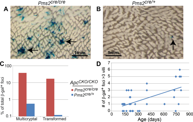
β-gal+ foci number, crypt fission and adenoma formation in Pms2 cre/cre and Pms2 cre/+ intestines. Wholemount images of the proximal small intestine from a Pms2 cre/cre (A) and Pms2 cre/+ (B) mouse. Arrows show multicryptal β-gal+ foci in (A) and a single β-gal+ focus in (B). Note the difference in both number and size of β-gal+ foci between Pms2 cre/cre and Pms2 cre/+ intestines. (C) Pms2 cre/cre; Apc CKO/CKO intestines have a higher percentage of normal-appearing multicryptal β-gal+ foci and transformed β-gal+ foci compared with Pms2 cre/+; Apc CKO/CKO intestines. The percentage of transformed β-gal+ foci was determined by scoring frozen sections counterstained with nuclear fast red. (D) The number of β-gal+ foci containing more than three villi results in a linear regression of y = 0.004x + 0.2, which is a statistically significant increase with age (P < 0.001). The number of β-gal+ foci containing more than three villi was determined via wholemount.

