Abstract
The basolateral amygdala (BLA) plays an integral role in the etiology of anxiety disorders and alcoholism. Although much is known about the intrinsic circuitry that governs BLA excitability, our understanding of the neuromodulators that control BLA excitation is incomplete. In many brain regions, adenosine (ADO) regulates neuronal excitability, primarily via A1 receptor inhibition of glutamate release, and basal adenosinergic tone is high enough to tonically inhibit neuronal excitation. Although ADO signaling modulates many anxiety- and alcohol-related behaviors, little is known about ADO regulation of BLA neurotransmission. To that end, we used patch clamp methods in rodent brain slices to characterize adenosinergic modulation of excitatory neurotransmission onto BLA pyramidal cells. ADO significantly inhibited EPSCs evoked by stimulation of either medial or external glutamatergic inputs into the BLA. This effect was mimicked by an A1, but not by an A2a, agonist. Paired-pulse ratio and miniature EPSC experiments revealed that A1 receptors reside at a presynaptic locus on BLA glutamatergic synapses. Moreover, bath application of an A1 receptor antagonist significantly enhanced EPSCs, providing evidence of tonic adenosinergic tone at BLA glutamatergic synapses. In addition, tonic ADO was regulated by adenosine kinase, but not adenosine deaminase. Finally, activation of A1 receptors had no direct effects on the intrinsic excitability of BLA pyramidal cells. Collectively, these data suggest that tonic A1 receptor signaling may play an important role in regulating BLA excitability and suggest a possible neurobiological substrate through which ADO may contribute to the pathophysiology of anxiety disorders and alcohol addiction.
Keywords: Adenosine, Basolateral Amygdala, EPSCs, Cyclopentyladenosine
1. Introduction
The basolateral amygdala (BLA) represents a key hub for consolidating and relaying sensory information within the CNS and a growing amount of evidence suggests that dysregulation in this brain region contributes to the pathophysiology of anxiety disorders (Etkin et al., 2009; Mahan and Ressler, 2012). Afferent projection neurons originating in the thalamus and cortex synapse onto excitatory BLA projection neurons after entering the BLA along distinct anatomical pathways (Aggleton et al., 1980; LeDoux et al., 1991). Glutamatergic projection neurons within the BLA consolidate these signals and project onto downstream extended amygdala structures thereby driving the physiological manifestations of anxiety-like behaviors (Tye et al., 2011) Notably, pharmacological agents that decrease excitability of BLA projection neurons, by increasing GABAergic inhibition or decreasing glutamatergic transmission, are efficacious at attenuating phenotypic expressions of anxiety (Millan, 2003). Additionally, there is now compelling evidence that stress and anxiety play a critical role in alcoholism (Koob, 2013; Silberman et al., 2009). Acute ethanol consumption can reduce anxiety-like behaviors while withdrawal from chronic ethanol can exacerbate anxiety (Koob, 2013; McCool et al., 2010). However, despite many recent advances, our understanding of the key neuromodulatory systems that regulate BLA excitability are not fully understood.
In recent years, the neuromodulator adenosine and its receptor system have emerged as key regulators of neuronal transmission (Boison, 2012; Dunwiddie and Masino, 2001). Particularly, adenosine A1 receptor-mediated inhibition of glutamate release has been demonstrated in multiple regions of the mammalian central nervous system (Choi et al., 2004; Courjaret et al., 2012; Hawryluk et al., 2012; Lupica et al., 1992; Patel et al., 2001). Adenosine A1 receptors are G protein-coupled receptors that are widely expressed throughout the CNS (Braas et al., 1986). Activation of these receptors liberates the Gi protein which inhibits the adenylyl cyclase signaling cascade (Londos et al., 1980). This inhibition leads to decreased Ca2+ entry at synaptic terminals, reducing transmitter release (Dunwiddie and Masino, 2001). Activation of A1 receptors may also increase K+ conductance and decrease voltage-gated calcium channel activity via the Go protein (Fredholm and Dunwiddie, 1988; McCool and Farroni, 2001)
Adenosine is present in the extracellular space of CNS tissue at a concentration thought to fluctuate between 20 – 300 nM (Dunwiddie and Masino, 2001). These concentrations are sufficient to activate high-affinity adenosine A1 receptors in some brain regions, an effect evidenced by enhancement of excitatory postsynaptic currents (EPSCs) following adenosine A1 receptor blockade (Choi et al., 2004; Dunwiddie and Diao, 1994). Thus, basal extracellular adenosine may serve as a tonic modulator of neuronal excitability (Greene et al., 1985). Not surprisingly, extracellular adenosine levels are dynamically regulated via a number of mechanisms. Adenosine is transported into the extracellular space through equilibrative nucleoside transporters located on the membrane of both neurons and glia (Dunwiddie and Masino, 2001), or produced from the dephosphorylation of ATP by ectonucleotidases (Dunwiddie et al., 1997; Dunwiddie and Hoffer, 1980). Clearance of extracellular adenosine levels is achieved by a combination of metabolism to inosine by adenosine deaminase, and by transport back through equilibrative transporters (ENT1) into neurons and glia (Arch and Newsholme, 1978). Once in the intracellular space, adenosine can be phosphorylated by adenosine kinase to form AMP (Lloyd and Fredholm, 1995).
An increasing amount of evidence suggests that adenosine receptor signaling modulates anxiety-like behaviors in rodents and humans. For instance, the anxiogenic activity of caffeine is generally attributed to its non selective blockade of adenosine receptors (Nehlig et al., 1992), while mice lacking A1 receptors display increased anxiety-like behavior (Giménez-Llort et al., 2002). Additionally, selective activation of A1 receptors decreases anxiety-like behavior in naïve mice (Jain et al., 1995), and also reduces ethanol withdrawal-associated anxiety (Prediger et al., 2006). There is also evidence suggesting that adenosine is critically involved in a range of ethanol-mediated behaviors and ethanol self-administration (Nam et al., 2011), effects possibly driven by ethanol blockade of ENT1, which raises extracellular adenosine levels (Nagy et al., 1990). Additional reports have indicated that adenosine receptor modulators influence ethanol’s ataxic (Dar, 1990), hypnotic (Choi et al., 2004), and anxiolytic effects (Prediger et al., 2004). Furthermore, adenosine receptors are expressed in the BLA (Braas et al., 1986), lending credence to the idea that the BLA adenosine system may contribute to the regulation of anxiety-like behaviors.
To examine the effects of adenosine signaling on BLA neurotransmission we implemented whole cell patch clamp electrophysiological methods to record from rat BLA pyramidal neurons. Our data reveal that activation of presynaptic adenosine A1 receptors decreases glutamate release onto BLA pyramidal neurons without direct effects on the intrinsic excitability of these cells. Additionally, we found evidence to support the hypothesis that basal adenosine levels in the BLA tonically regulate glutamate release onto BLA pyramidal neurons and that tonic levels of adenosine are regulated, in part, by adenosine kinase. Our findings establish a critical link between the adenosine system, known to be involved in the regulation of anxiety and addiction, and the BLA, a region that is thought to play an integral role in the etiology of both disease states.
2. Materials and Methods
2.1 Animals
Male Sprague-Dawley rats between the ages of 4-8 weeks were used for all experiments. Animals arrived from a commercial supplier (Harlan) at postnatal day 21 and were allowed to acclimate for 1 week. Animals were pair housed in a vivarium with a 12 hour light-dark cycle and had ad libitum access to food and water. All experiments were performed in accordance with the Wake Forest University Animal Care and Use Committee.
2.2 Electrophysiological Recordings
Transverse amygdala slices (400 μm) were prepared each recording day using a Leica VT1000S vibratome (Leica Microsystems Inc., Buffalo Grove, IL). Rats were anesthetized with halothane, decapitated and the brains were quickly isolated in ice cold artificial cerebral spinal fluid (aCSF) composed of (in mM): 124 NaCl, 3.3 KCl, 2.4 MgCl, 2.5 CaCl2, 1.2 KH2PO4, 10 D-glucose, and 25 NaHCO3, saturated with 95% O2 and 5% CO2. Slices were then maintained at ambient temperature for at least two hours in oxygenated aCSF. Amygdala slices were transferred to a recording chamber and superfused with oxygenated aCSF at a flow rate of 2 mL/min using a calibrated flow meter (Gilmont Instruments, Racine, WI). 2 – 3 cells were recorded from each animal and drug effects were consistent across subjects. Evoked AMPA receptor-mediated EPSCs were recorded using an internal solution containing 130 mM K-gluconate, 10 nM KCl, 1 mM EGTA, 100 μM CaCl2, 2 mM Mg- ATP, 200 μM Tris-guanosine, 5’-triphosphate, and 10 nM HEPES, pH adjusted with KOH, 275-280 mOsm. Miniature EPSCs were recorded using a similar internal solution, replacing equimolar Cs-gluconate for K-gluconate. For all AMPA EPSC recordings, 5 mM N-(2,6-dimethyl-phenylcarbamoyl-methyl)-triethylammonium chloride (QX-314) was included in the recording solution to block voltage-gated sodium channels. BLA pyramidal neurons were voltage-clamped at \m=-\65 to \m=-\70 mV for EPSCs experiments. Whole cell currents were acquired using an Axoclamp 2B amplifier, digitized (digidata 1321 A; Axon Instruments, Union City, CA), and analyzed online and offline using an IBM-compatible computer and pClamp 10.1 software (Axon Instruments).
For perforated patch-clamp recordings, gramicidin was diluted in dimethylsulfoxide (DMSO) to a stock concentration of 50 mg/ml. The stock solution was further diluted to a final concentration of 200 ug/ml in a patch-pipette solution containing (in mM): KCl 135, HEPES 10, MgCl2 2, Na2-EGTA 5, CaCl2 0.5, adjusted to 7.2 pH with KOH. The KCl-gramicidin solution was sonicated for 1-5 min at the beginning of each day and vortexed for 15-30 sec before filling each electrode. No filtering was applied. Each electrode was backfilled with gramicidin-free KCl in order to avoid interference of the antibiotic with seal formation, and the remainder of the electrode was filled with KCl-gramicidin. After forming a high-resistance seal (GOhm), the cell was held in current-clamp mode for 25-75 min until perforation occurred and access resistance stabilized. All cells were maintained at a membrane potential of -60mV with direct current injection. The rheobase was determined by applying a 30 ms current step, increasing from 0 by 20 pA per step, every 5 seconds until an action potential was generated. Action potential frequency was assessed by applying an 800 ms current step every 20 sec, ranging from 100 to 500 pA, in 50 pA increments. Perforated patch experiments were conducted in the presence of 50 μM APV, 20 μM bicuculline, and 20 μM DNQX.
To isolate postsynaptic AMPA currents, 100 μM AMPA was applied directly to the soma of BLA pyramidal neurons (20 psi, 250 msec) using a picospritzer III (General Valve, Fairfield, NJ). AMPA was applied every 3 minutes while whole cell currents were recorded. For these experiments a blocker cocktail of 500 nM Tetrodotoxin (TTX), 20 μM bicuculline, and 50 μM APV was used.
2.3 Pharmacological Isolation of Synaptic Currents
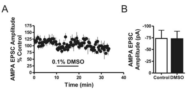
Filamented borosilicate glass capillary tubes (inner diameter 0.86 mm) were pulled using a horizontal pipette puller (P-97; Sutter instruments, Novato, CA) to prepare recording electrodes. AMPA-mediated EPSCs were pharmacologically isolated using 50 μM APV and 20 μM bicuculline to block NMDA and GABAa receptors, respectively. In most experiments, synaptic currents were evoked every 20 s by electrical stimulation (0.2 ms duration) using a concentric bipolar stimulation electrode (FHC, Bowdoinham, ME) placed along the external capsule to target cortical inputs, or along the medial border of the BLA to target thalamic inputs. For paired-pulse ratio experiments, paired EPSCs were evoked every 45 s with an interstimulus interval of 25 ms. In both of these experiments, stimulation intensity was adjusted to 10-20% of maximal currents (typically 80-150 pA). Spontaneous, action potential-independent miniature EPSCs (mEPSCs) were recorded in voltage-clamp mode from BLA pyramidal neurons in the presence of 500 nM TTX. mEPSCs were digitized at 5-10kHz in continuous 3 min epochs. Unless otherwise noted, all drugs were purchased from Sigma (St. Louis, MO). All adenosine receptor modulators were purchased from Tocris (Ellisville, MO). Bath applied drugs were prepared as 100 to 400-fold concentrates suspended in water or DMSO and applied directly into the perfusion chamber via calibrated syringe pumps (Razel Scientific Instruments, Stanford, CT). Final DMSO concentrations did not exceed 0.1%, a concentration that had no effect on EPSCs in control experiments (see Fig 3).
FIGURE 3. DMSO vehicle does not modulate BLA EPSCs.
(A) Time course illustrating the effect of bath applied 0.1% DMSO on BLA EPSCs (each point equals the mean ± SEM from 5 cells). Solid bar below points indicates the length of drug application. (B) Bar graph summarizing the effect of 0.1% DMSO bath applied to BLA pyramidal neurons on EPSCs. DMSO did not affect EPSC amplitude (paired t-test, p > 0.05, n = 5)
2.4 Statistics
Drug effects on EPSCs were quantified as percent change relative to the mean of baseline and washout values. Paired-pulse ratios were derived by quantifying the change in the ratio of the second peak relative to the first, during control and drug applications. Although fully separable EPSCs could not be evoked at this short interstimulus interval under our recording conditions, this protocol has proven to be a reliable index of presynaptic changes in empirical experiments in our lab and has been used in other recent studies where EPSCs did not fully decay to baseline (Pannasch et al., 2011). mEPSCs were first identified using Clampfit event detection software (pClamp 10.1) and then visually inspected to avoid inclusion of spurious events. mEPSCs in each epoch were then averaged and the amplitude of the averaged traces was calculated. Effects of CPA and DPCPX on mEPSCs were quantified as the absolute change in amplitude and frequency of miniature events relative to the mean of control values. For rheobase experiments using the perforated patch preparation, 3 trials were conducted and the average current needed to generate the first action potential was compared between baseline and 40 nM CPA. For action potential frequency experiments, 5 randomly selected sweeps were averaged at each current step and compared pre and post drug. Statistical analyses on drug effects were conducted using paired and upaired two-tailed t-tests, or a two-way analysis of variance followed by Bonferroni post hoc tests, where applicable, with a minimal level of significance of p < 0.05. Statistical analysis of mEPSC data was performed using a one-tailed Mann-Whitney U-test for group results, as these data did not follow a Gaussian distribution The minimal level of significance was set at p < 0.05, and confirmed by the Kolmorgov-Smirnov (K-S) test on individual cells, with a minimal level of significance of p < 0.01. All grouped results are reported as mean percent change from control ± SEM. In all experiments, control values are derived as the average of baseline and washout values.
3. Results
3.1 Effect of Adenosine Receptor Activation on AMPA Receptor-Mediated EPSCs
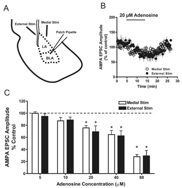
As mentioned above, previous reports have demonstrated that adenosine functions as a powerful modulator of neuronal excitability in multiple brain regions. Because the BLA plays a critical role in regulating anxiety-like behaviors, and recent evidence suggesting a role of the adenosine receptor system in modulating anxiety measures, we set out to characterize the role adenosine plays in regulating BLA excitability. Based on data from other brain regions, we hypothesized that adenosine would reduce AMPA receptor-mediated EPSCs onto BLA pyramidal neurons. To examine this hypothesis, we first tested the acute effect of exogenous adenosine on EPSCs evoked from either the medial or external inputs into the BLA (see FIG 1 A for schematic). At both inputs, significant effects of adenosine were observed at concentrations of 20 μM and higher (FIG 1 C). Statistical comparison of the data revealed a significant overall effect of adenosine (F = 26.97, p 0.0001) with no significant effect of stimulus location (F = 0.48, p > 0.05) and no significant stimulus-adenosine interaction (F = 0.23, p > 0.05). Inhibition of EPSCs was apparent within 5-10 min and reversed upon washout (FIG 1 B). No detectable changes were seen in either the holding current or input resistance (data not shown). We next set out to determine which adenosine receptor subtype mediates the reduction in EPSC amplitude. To date, four subtypes of adenosine receptors have been identified (Dunwiddie and Masino, 2001). Studies in other brain regions have demonstrated that activation of A1 receptors, coupled to Gi/Go, can inhibit release of glutamate (Brambilla et al., 2005), dopamine (Cechova et al., 2010), acetylcholine (Van Dort et al., 2009), and norepinephrine (Schütte et al., 2006). Conversely, activation of the A2A subtype, coupled to Gs, tends to facilitate release of these transmitters (Abbracchio et al., 2009; Rebola et al., 2003; Van Dort et al., 2009). We therefore hypothesized that adenosinergic inhibition of EPSCs recorded from BLA pyramidal neurons was due to activation of the A1 receptor subtype. To test this hypothesis, we bath applied the adenosine A1 receptor subtype specific agonist N6 - cyclopentyladenosine (CPA) while recording AMPA-mediated EPSCs in the BLA. At a concentration of 40 nM, CPA significantly inhibited both medially- (54.1% ± 7.2%, n = 11, p < 0.001) and externally- (40.4% ± 5.8%, n = 10, p < 0.01) evoked EPSCs (FIG 2). In a separate experiment, CPA mediated inhibition was blocked by the A1 receptor antagonist dipropylcyclopentylxanthine (DPCPX) when tested at both medial (−2.3% ± 6.18%, n = 4, p > 0.05), and external inputs (6.4% ± 17.7%, n = 4, p > 0.05) (FIG 2). To examine the role of A2a receptors in regulating glutamate transmission, we bath applied the A2a specific agonist, CGS-21680, while recording AMPA-mediated EPSCs from BLA pyramidal neurons. At a concentration sufficient to enhance release of various transmitters in other brain regions (Rebola et al., 2003; Shindou et al., 2001), we saw no significant effect on the amplitude of EPSCs recorded from BLA pyramidal neurons while stimulating either the medial (-4.1% ± 8.9%, n = 5, p > 0.05) or external input (4.3% ± 5.9%, n = 6, p > 0.05) (FIG 2). It should be noted that CPA, CGS, and DPCPX were solubilize in 0.1% DMSO, Therefore, we conducted a control experiment to examine possible effects of the DMSO vehicle on medial EPSCs recorded from BLA pyramidal neurons. Bath application of 0.1% DMSO had no significant effect on the amplitude of BLA EPSCs (0.4% ± 1.7%, n = 5, p > 0.05) (FIG 3 A & B).
FIGURE 1. Adenosine receptor activation inhibits EPSC amplitude.
(A) Schematic diagram illustrating the placement of stimulating and recording electrodes used to activate medial and external glutamatergic afferents and record EPSCs from BLA pyramidal neurons. (B) Time course illustrating the effect of 20 μM adenosine on BLA EPSCs (each point equals the mean ± SEM from5 cells). Solid bar above the points indicates the length of drug application (C) Bar graph summarizing the effect of adenosine (5-80 μM), on the amplitude of AMPA EPSCs evoked by either medial or external stimulation in the BLA. Dashed line depicts control levels (average of baseline and washout). 5-11 cells tested at each concentration. (*, p < 0.05; significant difference from control; paired t-test.)
FIGURE 2. A1 receptor activation inhibits BLA EPSC amplitude at medial and external inputs.
(A) Bar graph summarizing the effect of the A1 selective agonist CPA (40 nM) and the A2A selegtive agonist CGS on BLA EPSCs evoked at either medial or external synapses, as well as the blockade of CPA inhibition by the A1 receptor antagonist DPCPX (**, p < 0.01; ***, p < 0.001; significant difference from control (dashed line), paired t-test). Five to 11 cells recorded for each condition. Traces above the graph are representative of individual cells during each treatment (averages of 10-20 sweeps). Scale bars are 50 pA by 50 ms.
3.2 Synaptic Locus of Adenosine A1 Receptor Inhibition of BLA EPSCs
The next sets of experiments were designed to determine the synaptic locus underlying CPA inhibition of AMPA-mediated EPSCs. We first assessed the effect of 40 nM CPA on paired-pulse ratios. Typically, changes in paired pulse ratio are inversely correlated with changes in neurotransmitter release probability (Katz and Miledi, 1968). Therefore, if CPA inhibited evoked AMPA EPSCs through a decrease in glutamate release probability, this inhibition should be associated with an increase in paired-pulse ratio. Pairs of EPSCs were evoked at an inter-stimulus interval of 25 ms, and after an 8-10 min baseline, slices were treated with 40 nM CPA for 15 min. CPA inhibited the amplitude of both peaks and this inhibition was associated with an increased paired-pulse ratio at both medial (from 1.6 ± 0.1 to 2.0 ± 0.2, n = 5, p < 0.05) and external (from 1.6 ± 0.1 to 1.8 ± 0.1, n = 8, p < 0.05) BLA inputs (FIG 4). Together, these data suggest that A1 receptors are likely located presynaptically at glutamatergic synapses onto BLA pyramidal neurons and that activation of these receptors inhibits glutamate release probability.
FIGURE 4. CPA application increases the paired-pulse ratio of BLA EPSCs.
Bar graph summarizing the effect of 40 nM CPA on the ratio of paired EPSCs (25 ms interstimulus interval) evoked from either medial or external BLA inputs (*, p < 0.05; significant difference between control and CPA groups, paired t-test, n = 5 - 8 cells/group). Traces to the left of the graph are averages of 5 consecutive sweeps from representative cells illustrating the effect of CPA on the paired-pulse ratio of medial and externally derived EPSCs.
To further resolve the synaptic locus of BLA A1 receptors, we assessed the effect of 40 nM CPA on miniature EPSCs (mEPSCs). Like the paired-pulse ratio, mEPSC analyses can also be used to determine the synaptic locus of drug effects. According to the quantal hypothesis, changes in frequency of mEPSCs typically reflect alterations in presynaptic release while changes in amplitude are generally associated with postsynaptic changes (del Castillo and Katz, 1954; Fatt and Katz, 1952). We thus hypothesized that bath application of 40 nM CPA would significantly reduce mEPSC frequency with no effect on the amplitude of these events. Two 3 min baseline epochs of mEPSCs were recorded from BLA pyramidal neurons in the presence of 500 nM TTX to block voltage-gated sodium channels. Following a 15 min application of 40 nM CPA, two additional 3 min epochs of mEPSCs were recorded. In support of our hypothesis, CPA attenuated the mean frequency of mEPSC events onto BLA pyramidal neurons (in Hz, from 1.4 ± 0.3 to 1.0 ± 0.3, n = 7, p < 0.05) (FIG 5 A & C). To further evaluate these findings we performed a Kolmogorov-Smirnov (K-S) test on individual cells. Using this analysis, (5/7) cells showed a significant reduction in mEPSC frequency. When the K-S test was applied to individual runs to evaluate mEPSC amplitude, 5/7 cells showed a significant difference. However, there was not a significant difference in mean mEPSC amplitude across all cells (from -15.3 ± 1.7 to -16.2 ± 3.3, n = 7, p > 0.05) (FIG 5 A & B).
FIGURE 5. CPA decreases mEPSC frequency but not amplitude.
(A) Representative current traces of mEPSCs recorded from BLA pyramidal neurons at baseline and in the presence of 40 nM CPA. (B) The distribution of cumulative amplitudes for mEPSCs during baseline and 40 nM CPA conditions. Insert: Bar graph depicting mean amplitude of mEPSCs for all cells recorded (p > 0.05, Mann-Whitney U-test, n = 7). (C) The distribution of cumulative interevent intervals for mEPSCs under baseline and 40 nM CPA conditions. Insert: bar graph depicting mean frequency of mEPSCs under baseline and 40 nM CPA conditions (*, p < 0.05; significant difference from baseline frequency, Mann-Whitney U-test, n = 7).
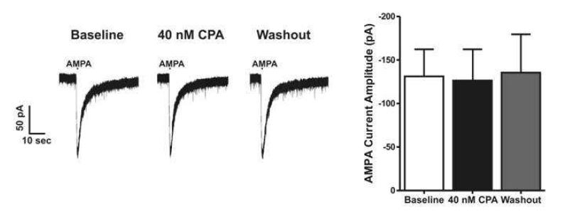
To further exclude postsynaptic mechanisms of A1 receptor-mediated inhibition, exogenous AMPA was applied directly to BLA pyramidal neurons by pressure ejection. BLA pyramidal neurons were patch clamped and a glass pipette containing 100 μM AMPA was positioned directly above the patched neuron using visual guidance. AMPA currents were evoked every 3 minutes and 40 nM CPA was bath applied for 18 – 22 minutes. In agreement with the PPR and mEPSC data, CPA had no effect on the amplitude of AMPA-evoked currents (\m=-\6.6% ± 14.6%, n = 5, p > 0.05) (FIG 6), further illustrating a presynaptic locus for adenosine A1 receptor in the BLA.
FIGURE 6. Currents evoked by exogenous AMPA application are not modulated by A1 receptor activation.
Bar graph illustrating summary data illustrating that bath application of the A1 agonist CPA (40 nM) had no effect on currents elicited by pressure application of 100 μM AMPA onto BLA pyramidal neurons (paired t-test, n = 5, p > 0.05 between control and CPA values). To the left of are graph are representative traces of AMPA-evoked currents during baseline, 40 nM CPA bath application, and CPA washout. Horizontal bar above traces indicates AMPA application.
3.3 Basal Adenosine Tone at Glutamatergic Synapses onto BLA Pyramidal neurons
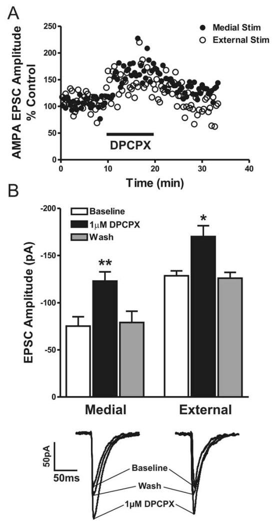
In several brain regions, basal adenosine levels fluctuate between 25-300 nM (Dunwiddie and Masino, 2001). While low, these concentrations are sufficient to activate high affinity A1 and A2a receptors. The stimulatory effects of methylxanthines, such as caffeine and theophylline, are attributed to antagonism of these receptors, and thus blockade of tonic adenosinergic inhibition (Fredholm, 1985; Nehlig et al., 1992). Fluctuations in adenosine tone have been shown to alter regional excitability and treatments that increase tonic adenosine show promise in the cessation of increased neuronal excitability associated with epilepsy (Boison, 2013; Masino et al., 2011). To that end, we sought to determine if basal adenosine levels in the BLA were high enough to regulate glutamatergic synaptic transmission in this brain region. To address this hypothesis, we first bath applied the A1 specific antagonist DPCPX while recording AMPA receptor-mediated EPSCs from BLA pyramidal neurons. 1 μM DPCPX significantly potentiated EPSCs at medial (75.0% ± 20.6%, n = 6, p < 0.01) and external inputs (32.3% ± 7.9%, n = 4, p < 0.05) (FIG 7). We also observed an increase in mEPSC frequency following DPCPX application (94.3% ± 32.7%, n = 5, p < 0.05) (5/5 cells; K-S test). In contrast, the amplitude of mEPSCs was not altered following 1 μM DPCPX application (3.9% ± 7.2%, n = 5, p > 0.05) (0/5 cells; K-S test), confirming our hypothesis that basal extracellular adenosine acts on presynaptic A1 receptors to tonically inhibit glutamate release in the BLA (FIG 8).
FIGURE 7. Evidence for adenosinergic tone in the BLA.
(A) Time course from representative cells indicating the effect of the A1 antagonist DPCPX (1 μM) on medially and externally evoked EPSCs in the BLA relative to control values. Solid bar below points indicates length of DPCPX application. (B) Bar graph depicting mean amplitude of BLA EPSCs at baseline, following bath application of 1 μM DPCPX, and during washout at medial and external inputs (*, p < 0.05; **, p < 0.01; paired t-test, n = 4 - 6 cells/group). Traces below the graph are averages of 10-20 consecutive sweeps from representative cells illustrating the effect of DPCPX on BLA EPSCs evoked from external and medial stimulation.
FIGURE 8. DPCPX increases the frequency of mEPSCs without changing mEPSC amplitude.

(A) Representative current traces of mEPSCs recorded from BLA pyramidal neurons at baseline and in the presence of 1 μM DPCPX. (B) The distribution of cumulative amplitudes of mEPSCs under baseline and 1 μM DPCPX conditions. Insert: Bar graph depicting mean amplitude of mEPSCs at baseline and following 1 μM DPCPX (p > 0.05, Mann Whitney U-test, n = 5). (C) The distribution of cumulative interevent intervals of mEPSCs under baseline and 1 μM DPCPX conditions. Insert: bar graph depicting mean frequency of mEPSCs at baseline and bath application of 1 μM DPCPX (*, p < 0.05, Mann-Whitney U-test, n = 5).
As mentioned above, extracellular adenosine is thought to be inactivated by either deamination to inosine by adenosine deaminase (ADA), or phosphorylated to adenosine monophosphate, an event that is catalyzed by adenosine kinase (Meghji and Newby, 1990). To that end, our next sets of experiments were designed to address if either of these catabolic mechanisms regulate basal adenosine levels in the BLA. As our previous experiments did not reflect differential effects of adenosine on the two main excitatory inputs into the BLA, we chose to examine only the medial input for these experiments. Bath application of the ADA inhibitor EHNA (20 μM) had no significant effect on EPSC amplitude (0.4% ± 11.9%, n = 8, p > 0.05) (FIG 9 A & C). Conversely, when ADK was inhibited with 5-Iodotubercidin (5-ID), significant inhibition of EPSCs was observed (50.3% ± 10.8%, n = 5, p < 0.05) (FIG 9 B & C).
FIGURE 9. Inhibition of adenosine kinase suppresses EPSC amplitude.

(A) Time course of BLA EPSCs demonstrating that inhibition of adenosine deaminase by 20 μM EHNA (solid bar) does not affect EPSC amplitude relative to baseline values (n = 9). (B) Time course illustrating the inhibitory effect of adenosine kinase inhibition by 2 μM 5’ – Iodotubercidin on BLA EPSCs (n = 5) (C) Summary bar graph illustrating the inhibitory effect of adenosine kinase inhibition and the lack of effect of adenosine deaminase inhibition on BLA EPSCs derived from medial stimulation (*, p < 0.05, paired t-test significant relative to control values).
3.4 A1 Receptor Activation Does Not Alter Intrinsic Neuronal Excitability
While the evidence supporting adenosinergic regulation of glutamate release at BLA pyramidal cell synapses is strong, these data do not rule out the possibility that adenosine may also alter the excitability of these cells via direct, postsynaptic effects on intrinsic excitability. In multiple brain regions, including the BLA, activation of A1 receptors can modulate voltage-gated calcium channel activity as well as a potassium conductance, effects that may alter intrinsic firing properties of neurons (McCool and Farroni, 2001; Mynlieff and Beam, 1994; Trussell and Jackson, 1985). To further elucidate the mechanisms through which adenosine modulates neuronal excitability in BLA pyramidal neurons, we sought to determine if A1 receptor activation alters the intrinsic excitability of these cells.
To investigate this question we employed the gramicidin perforated patch technique. This method allows for the study of neuronal firing while maintaining a relatively intact intracellular milieu, compared to traditional patch techniques that dialyze the intracellular contents (Akaike and Harata, 1994). Gramicidin is a polypeptide antibiotic that forms pores in the neuronal membrane that are selectively permeable to monovalent cations, preserving much of the intracellular signaling machinery (Akaike, 1996). This property is advantageous when studying G protein-coupled receptors that rely on intact intracellular contents for signal transduction. Using this technique we examined whether the intrinsic neuronal firing properties of BLA pyramidal neurons were altered by A1 receptor activation. Following the formation of a perforated patch, 30 ms current steps were applied to the patched neuron, increasing by 20 pA with each step, until an action potential was generated to assess rheobase (i.e. the minimal injected current needed to generate an action potential). Bath application of 40 nM CPA did not affect rheobase (2.1% ± 4.7%, n = 9, p > 0.05) (FIG 10 A & B). Another measure of intrinsic neuronal excitability is the frequency of action potentials in response to a depolarizing current step. To examine this, we depolarized BLA pyramidal neurons for 800 ms at varying currents to generate trains of action potentials both before and after bath application of 40 nM CPA. At current steps ranging from 100 to 500 pA, action potential frequencies were not affected by 40 nM CPA application (FIG 10 C & D).
FIGURE 10. Intrinsic membrane properties of BLA pyramidal neurons are not altered by activation of adenosine A1 receptors.

(A) Traces from representative cells demonstrating that activation of A1 receptors does not alter rheobase in BLA pyramidal neurons. (B) Bar graph depicting mean rheobase of BLA pyramidal neurons at baseline and following 40 nM CPA (paired t-test, p > 0.05, n = 9). (C) Representative responses to multiple depolarizing current injections in cells recorded form BLA pyramidal neurons during baseline and in the presence of 40 nM CPA. (D) The relationship between depolarizing current injection and the frequency of action potentials elicited at baseline and following 40 nM CPA application.
4. Discussion
The results of this study show that activation of adenosine receptors in the BLA inhibits excitatory postsynaptic currents recorded from BLA pyramidal neurons. This effect was concentration dependent and did not differ between the medial and external inputs into the BLA. Application of an adenosine A1 receptor specific agonist, CPA, also inhibited EPSCs at both BLA inputs. Additionally, CPA increased the paired-pulse ratio, decreased mEPSC frequency, and did not modulate currents elicited by direct application of AMPA onto BLA pyramidal cells. Additional experiments, using an A1 specific antagonist, demonstrated the presence of adenosinergic tone in the BLA. We also found evidence to suggest that in the BLA, like other brain regions, adenosinergic tone is regulated by adenosine kinase and does not appear to be influenced by adenosine deaminase under our recording conditions. Finally, activation of presynaptic A1 receptors was not associated with any changes in the intrinsic excitability of BLA pyramidal neurons. Taken together, these data suggest that adenosine A1 receptors are expressed at a presynaptic locus in the BLA and that activation these receptors significantly inhibits glutamate release probability onto BLA pyramidal neurons. These findings also provide initial evidence that basal levels of adenosine, under control of adenosine kinase, tonically inhibit glutamate release in the BLA, without direct postsynaptic effects on BLA pyramidal cell excitability. Together these data provide initial insight into one of the mechanisms through which adenosine receptor modulators may regulate anxiety-like behaviors.
As observed in other brain regions, our data strongly support the hypothesis that adenosine inhibits EPSCs onto BLA pyramidal neurons via activation of A1 receptors. This hypothesis is supported by the finding that EPSCs were inhibited by bath application of CPA at a concentration that has been shown to be selective for A1 receptors (Van Der Graaf et al., 1997). Additionally, CGS-21680, an A2a receptor subtype specific agonist, had no affect on EPSC amplitude. These findings provide a neurobiological mechanism though which A1 agonists, including CPA, could exert their anxiolytic affects (Jain et al., 1995).
CPA inhibition of EPSCs was accompanied by a significant increase in paired-pulse ratio, as well as a significant suppression of mEPSC frequency, consistent with a presynaptic locus for A1 receptors in the BLA. We did, however, see a significant reduction of mEPSC amplitude in 5/7 cells following 40 nM CPA application when cells were analyzed individually with the Kolmogorov-Smirnov test. It is possible that this significant reduction in amplitude stems from the robust reduction in the number of events observed in many of the cells and may thus be unrelated to a postsynaptic change. Importantly, we did not see a significant change in mean amplitude when all cells were analyzed as a group. Moreover, currents evoked by exogenous application of AMPA onto BLA pyramidal cell bodies were not inhibited by CPA, providing further evidence that A1 receptors inhibit BLA synaptic excitation via a presynaptic mechanism. Indeed, A1 receptors have been shown to have a presynaptic locus in a number of other brain regions (Hawryluk et al., 2012; Li and Eisenach, 2005; Lupica et al., 1992; Shen and Johnson, 2003).
Our data also support the hypothesis that basal adenosine levels are sufficient to engage/activate A1 receptors at glutamatergic synapses in BLA slices. The observation that bath application of the A1 specific antagonist, DPCPX, potentiated BLA EPSCs and increased the frequency of mEPSCs strongly suggests that adenosine tone is present in BLA slices and provides support for the idea that basal adenosine levels may provide tonic inhibition of presynaptic glutamate release in the BLA. Recent work has established a critical role for extracellular adenosine levels in acute ethanol-mediated anxiolysis (Prediger et al., 2004), and ethanol withdrawal-associated anxiety-like behaviors (Prediger et al., 2006). As ethanol is thought to elevate extracellular adenosine levels by blocking concentrative transport through ENT1, our data provide evidence that may help to explain how increased extracellular adenosine could reduce anxiety measures.
Regulation of extracellular adenosine is controlled through a complex interaction of multiple transporters and enzymes. Although adenosine deaminase contributes to the breakdown of adenosine by converting it to inosine, bath application of an adenosine deaminase inhibitor did not alter BLA EPSC amplitude. Other laboratories have reported similar finding in other brain regions (Wall et al., 2007; Zhu and Krnjevic, 1994). It has thus been proposed that under normoxic and normoglycemic conditions adenosine kinase, not adenosine deaminase is the key regulator of extracellular adenosine concentrations (Pak et al., 1994; Zhu and Krnjevic, 1994). However, adenosine deaminase becomes an important regulator of adenosine levels when the extracellular adenosine concentration is elevated following energy depletion (Lloyd and Fredholm, 1995). Contributing to this hypothesis is the observation that Km values for adenosine are much lower for adenosine kinase (2 μM) than adenosine deaminase (17 μM) (Phillips and Newsholme, 1979). Indeed, we did see a significant suppression of EPSCs following bath application of an adenosine kinase inhibitor, 5-Iodotubercidin, consistent with an important role for adenosine kinase in the clearance of extracellular adenosine in the BLA. In the rat brain, adenosine kinase is a predominantly cytosolic enzyme (Phillips and Newsholme, 1979). It is thought, therefore, that when adenosine levels are elevated in the extracellular space, adenosine is transported into cells through the equilibrative transporter ENT1, wherein adenosine kinase phosphorylates adenosine to adenosine monophosphate (Dunwiddie and Masino, 2001). Thus, we hypothesize that inhibition of adenosine kinase results in an accumulation of intracellular adenosine that is then transported into the extracellular space through ENT1 where it can then activate presynaptic A1 receptors and reduce glutamate release. Recently, adenosine kinase has received attention as a critical component of epileptogenesis (Boison, 2012). Because of this, much work has been done in devising novel strategies to target adenosine kinase, decrease its functionally, and thereby increase extracellular adenosine levels (Boison, 2013; Masino et al., 2009; McGaraughty et al., 2001). Based on our findings, it seems plausible that these same strategies could be employed in the BLA to attenuate the excessive excitability associated with anxiety-like behaviors.
Activation of A1 receptors initiates a G-protein-mediated signaling cascade that can have a multitude of downstream effects that can alter neuronal firing. For instance, adenosine can activate G-protein-dependent inwardly rectifying K+ channels (GIRKs) (Takigawa and Alzheimer, 2002), resulting in a net hyperpolarization and can modulate functionality of voltage-gated calcium channels (VGCCs) on BLA pyramidal neurons (McCool and Farroni, 2001). While CPA activation of A1 receptors may modulate GIRK and VGCC activity, our gramicidin perforated patch recordings indicate that A1 receptor modulation of voltage-gated channels is not sufficient to directly alter the intrinsic excitability of BLA pyramidal neurons. Thus, in the BLA, the predominant effect of A1 receptor activation appears to be at the synaptic terminal where glutamate release is inhibited.
One potential caveat of our findings is that all of these data were collected in ex vivo brain slices maintained at ambient temperature (22-25 °C). It is possible that the ex vivo environment represents a post-traumatic/ischemic state that may alter adenosine signaling. However, zur Nedden and colleagues recently report that adenine nucleotide levels recover rapidly in brain slices and remain stable for at least 5 hours (zur Nedden et al., 2011). Indeed, brain slices are thought to maintain similar levels of adenosine and adenine nucleotides as the intact brain (Fredholm et al., 1984). Experiments were conducted at ambient temperature in order to promote the stability of the patch-clamp recordings. However, maintaining slices at these temperatures can occlude adenosine release at equilibrative transporters, thus potentially reducing extracellular adenosine levels (Masino and Dunwiddie, 1999). Therefore, although we still observed clear evidence of tonic activation of A1 receptors under our recording conditions, it is possible that our data underestimate the extent of tonic adenosinergic regulation of BLA glutamatergic synaptic excitation that may occur at more physiological temperatures.
One question that remains to be addressed is the role adenosine A2a receptors may play in regulating BLA excitability as A2a receptors are known to be expressed in this brain region (Jarvis et al., 1989). While our data suggest that A2a receptors are not involved in the modulation of glutamatergic transmission, other research has demonstrated that activation of these receptors can enhance GABAergic transmission (Mayfield et al., 1993). These receptors, coupled to Gs, tend to facilitate transmitter release (Rebola et al., 2003) and one possibility is that A2a receptors may enhance release of GABA in the BLA, thereby contributing increased inhibition onto pyramidal neurons and driving down regional excitability. However, the role of the A2a system in the BLA has yet to be fully investigated.
In conclusion, our data provide new evidence that presynaptic A1 receptors inhibit glutamate release while leaving intrinsic firing properties of BLA pyramidal neurons unaffected. We also show an important role for tonic adenosine levels in the BLA, a system that, if disrupted may lead to increased anxiety-like behaviors. Although many reports have demonstrated involvement of the adenosine system in the regulation of anxiety-like behaviors (Jain et al., 1995; Nehlig et al., 1992), little has been done to investigate whether the adenosine system modulates excitability in the BLA, a brain region thought to contribute to the etiology of anxiety-like behaviors. Here we show a clear role of adenosine in inhibiting glutamatergic transmission in the BLA in a reversible and concentration-dependent manner. These data are consistent with work in other brain regions (Ribeiro, 1995) and further solidifies adenosine’s role as an inhibitory neuromodulator in the CNS. By establishing physiological evidence that adenosine receptor activation can attenuate BLA excitability we form a much needed link with behavioral experiments demonstrating anxiolytic properties of adenosine receptor agonists and identify A1 receptor signaling in the BLA as a viable target for the development of novel pharmacological treatments for anxiety-like disorders.
Highlights.
Adenosine inhibits EPSCs in basolateral amygdala (BLA) pyramidal neurons
This effect was mimicked by a selective A1, but not A2a, receptor agonist
A1 receptor activation reduces EPSCs via a presynaptic mechanism
Basal adenosine tonically regulates glutamate release at these synapses
A1 receptor activation does not alter the intrinsic excitability of BLA neurons
Acknowledgments
This research for this study was funded by AA022046 (ARR) and AA21099, AA17053 (JLW)
Supported by AA0220468 (ARR) and AA21099, AA17053 (JLW)
Footnotes
Publisher's Disclaimer: This is a PDF file of an unedited manuscript that has been accepted for publication. As a service to our customers we are providing this early version of the manuscript. The manuscript will undergo copyediting, typesetting, and review of the resulting proof before it is published in its final citable form. Please note that during the production process errors may be discovered which could affect the content, and all legal disclaimers that apply to the journal pertain.
References
- Abbracchio MP, Burnstock G, Verkhratsky A, Zimmermann H. Purinergic signalling in the nervous system: an overview. Trends Neurosci. 2009;32:19–29. doi: 10.1016/j.tins.2008.10.001. [DOI] [PubMed] [Google Scholar]
- Aggleton JP, Burton MJ, Passingham RE. Cortical and subcortical afferents to the amygdala of the rhesus monkey (Macaca mulatta) Brain Res. 1980;190:347–368. doi: 10.1016/0006-8993(80)90279-6. [DOI] [PubMed] [Google Scholar]
- Akaike N. Gramicidin perforated patch recording and intracellular chloride activity in excitable cells. Progress in Biophysics and Molecular Biology. 1996;65:251–264. doi: 10.1016/s0079-6107(96)00013-2. [DOI] [PubMed] [Google Scholar]
- Akaike N, Harata N. Nystatin perforated patch recording and its applications to analyses of intracellular mechanisms. Jpn J Physiol. 1994;44:433–473. doi: 10.2170/jjphysiol.44.433. [DOI] [PubMed] [Google Scholar]
- Arch JR, Newsholme EA. Activities and some properties of 5'-nucleotidase, adenosine kinase and adenosine deaminase in tissues from vertebrates and invertebrates in relation to the control of the concentration and the physiological role of adenosine. Biochem J. 1978;174:965–977. doi: 10.1042/bj1740965. [DOI] [PMC free article] [PubMed] [Google Scholar]
- Boison D. Adenosine dysfunction in epilepsy. Glia. 2012;60:1234–1243. doi: 10.1002/glia.22285. [DOI] [PMC free article] [PubMed] [Google Scholar]
- Boison D. Adenosine Kinase: Exploitation for Therapeutic Gain. Pharmacological Reviews. 2013;65:906–943. doi: 10.1124/pr.112.006361. [DOI] [PMC free article] [PubMed] [Google Scholar]
- Braas K, Newby A, Wilson V, Snyder S. Adenosine-containing neurons in the brain localized by immunocytochemistry. The Journal of Neuroscience. 1986;6:1952–1961. doi: 10.1523/JNEUROSCI.06-07-01952.1986. [DOI] [PMC free article] [PubMed] [Google Scholar]
- Brambilla D, Chapman D, Greene R. Adenosine mediation of presynaptic feedback inhibition of glutamate release. Neuron. 2005;46:275–283. doi: 10.1016/j.neuron.2005.03.016. [DOI] [PubMed] [Google Scholar]
- Cechova S, Elsobky AM, Venton BJ. A1 receptors self-regulate adenosine release in the striatum: evidence of autoreceptor characteristics. Neuroscience. 2010;171:1006–1015. doi: 10.1016/j.neuroscience.2010.09.063. [DOI] [PMC free article] [PubMed] [Google Scholar]
- Choi DS, Cascini MG, Mailliard W, Young H, Paredes P, McMahon T, Diamond I, Bonci A, Messing RO. The type 1 equilibrative nucleoside transporter regulates ethanol intoxication and preference. Nat Neurosci. 2004;7:855–861. doi: 10.1038/nn1288. [DOI] [PubMed] [Google Scholar]
- Courjaret R, Miras-Portugal M, Deitmer J. Purinergic Modulation of Granule Cells. The Cerebellum. 2012;11:62–70. doi: 10.1007/s12311-010-0196-3. [DOI] [PubMed] [Google Scholar]
- Dar MS. Central adenosinergic system involvement in ethanol-induced motor incoordination in mice. Journal of Pharmacology and Experimental Therapeutics. 1990;255:1202–1209. [PubMed] [Google Scholar]
- del Castillo J, Katz B. Quantal components of the end-plate potential. The Journal of Physiology. 1954;124:560–573. doi: 10.1113/jphysiol.1954.sp005129. [DOI] [PMC free article] [PubMed] [Google Scholar]
- Dunwiddie TV, Diao L. Extracellular adenosine concentrations in hippocampal brain slices and the tonic inhibitory modulation of evoked excitatory responses. Journal of Pharmacology and Experimental Therapeutics. 1994;268:537–545. [PubMed] [Google Scholar]
- Dunwiddie TV, Diao L, Proctor WR. Adenine Nucleotides Undergo Rapid, Quantitative Conversion to Adenosine in the Extracellular Space in Rat Hippocampus. The Journal of Neuroscience. 1997;17:7673–7682. doi: 10.1523/JNEUROSCI.17-20-07673.1997. [DOI] [PMC free article] [PubMed] [Google Scholar]
- Dunwiddie TV, Hoffer BJ. Adenine nucleotides and synaptic transmission in the in vitro rat hippocampus. Br J Pharmacol. 1980;69:59–68. doi: 10.1111/j.1476-5381.1980.tb10883.x. [DOI] [PMC free article] [PubMed] [Google Scholar]
- Dunwiddie TV, Masino SA. The role and regulation of adenosine in the central nervous system. Annu Rev Neurosci. 2001;24:31–55. doi: 10.1146/annurev.neuro.24.1.31. [DOI] [PubMed] [Google Scholar]
- Etkin A, Prater KE, Schatzberg AF, Menon V, Greicius MD. Disrupted amygdalar subregion functional connectivity and evidence of a compensatory network in generalized anxiety disorder. Arch Gen Psychiatry. 2009;66:1361–1372. doi: 10.1001/archgenpsychiatry.2009.104. [DOI] [PMC free article] [PubMed] [Google Scholar]
- Fatt P, Katz B. Spontaneous subthreshold activity at motor nerve endings. The Journal of Physiology. 1952;117:109–128. [PMC free article] [PubMed] [Google Scholar]
- Fredholm BB. On the mechanism of action of theophylline and caffeine. Acta Med Scand. 1985;217:149–153. doi: 10.1111/j.0954-6820.1985.tb01650.x. [DOI] [PubMed] [Google Scholar]
- Fredholm BB, Dunwiddie TV. How does adenosine inhibit transmitter release? Trends Pharmacol Sci. 1988;9:130–134. doi: 10.1016/0165-6147(88)90194-0. [DOI] [PubMed] [Google Scholar]
- Fredholm BB, Dunwiddie TV, Bergman B, Lindstroöm K. Levels of adenosine and adenine nucleotides in slices of rat hippocampus. Brain Res. 1984;295:127–136. doi: 10.1016/0006-8993(84)90823-0. [DOI] [PubMed] [Google Scholar]
- Giménez-Llort L, Fernández-Teruel A, Escorihuela RM, Fredholm BB, Tobeña A, Pekny M, Johansson B. Mice lacking the adenosine A1 receptor are anxious and aggressive, but are normal learners with reduced muscle strength and survival rate. European Journal of Neuroscience. 2002;16:547–550. doi: 10.1046/j.1460-9568.2002.02122.x. [DOI] [PubMed] [Google Scholar]
- Greene RW, Haas HL, Hermann A. Effects of caffeine on hippocampal pyramidal cells in vitro. Br J Pharmacol. 1985;85:163–169. doi: 10.1111/j.1476-5381.1985.tb08843.x. [DOI] [PMC free article] [PubMed] [Google Scholar]
- Hawryluk JM, Ferrari LL, Keating SA, Arrigoni E. Adenosine inhibits glutamatergic input to basal forebarin cholinergic neurons. J Neurophysiol. 2012 doi: 10.1152/jn.00528.2011. [DOI] [PMC free article] [PubMed] [Google Scholar]
- Jain N, Kemp N, Adeyemo O, Buchanan P, Stone TW. Anxiolytic activity of adenosine receptor activation in mice. Br J Pharmacol. 1995;116:2127–2133. doi: 10.1111/j.1476-5381.1995.tb16421.x. [DOI] [PMC free article] [PubMed] [Google Scholar]
- Jarvis MF, Jackson RH, Williams M. Autoradiographic characterization of high-affinity adenosine A2 receptors in the rat brain. Brain Res. 1989;484:111–118. doi: 10.1016/0006-8993(89)90353-3. [DOI] [PubMed] [Google Scholar]
- Katz B, Miledi R. The role of calcium in neuromuscular facilitation. The Journal of Physiology. 1968;195:481–492. doi: 10.1113/jphysiol.1968.sp008469. [DOI] [PMC free article] [PubMed] [Google Scholar]
- Koob G. Theoretical Frameworks and Mechanistic Aspects of Alcohol Addiction: Alcohol Addiction as a Reward Deficit Disorder. In: Sommer WH, Spanagel R, editors. Behavioral Neurobiology of Alcohol Addiction. Springer; Berlin Heidelberg: 2013. pp. 3–30. [DOI] [PMC free article] [PubMed] [Google Scholar]
- Lack AK, Ariwodola OJ, Chappell AM, Weiner JL, McCool BA. Ethanol inhibition of kainate receptor-mediated excitatory neurotransmission in the rat basolateral nucleus of the amygdala. Neuropharmacology. 2008;55:661–668. doi: 10.1016/j.neuropharm.2008.05.026. [DOI] [PMC free article] [PubMed] [Google Scholar]
- LeDoux JE, Farb CR, Romanski LM. Overlapping projections to the amygdala and striatum from auditory processing areas of the thalamus and cortex. Neurosci Lett. 1991;134:139–144. doi: 10.1016/0304-3940(91)90526-y. [DOI] [PubMed] [Google Scholar]
- Li X, Eisenach JC. Adenosine reduces glutamate release in rat spinal synaptosomes. Anesthesiology. 2005;103:1060–1065. doi: 10.1097/00000542-200511000-00021. [DOI] [PubMed] [Google Scholar]
- Lloyd HGE, Fredholm BB. Involvement of adenosine deaminase and adenosine kinase in regulating extracellular adenosine concentration in rat hippocampal slices. Neurochemistry International. 1995;26:387–395. doi: 10.1016/0197-0186(94)00144-j. [DOI] [PubMed] [Google Scholar]
- Londos C, Cooper DM, Wolff J. Subclasses of external adenosine receptors. Proc Natl Acad Sci U S A. 1980;77:2551–2554. doi: 10.1073/pnas.77.5.2551. [DOI] [PMC free article] [PubMed] [Google Scholar]
- Lupica CR, Proctor WR, Dunwiddie TV. Presynaptic inhibition of excitatory synaptic transmission by adenosine in rat hippocampus: analysis of unitary EPSP variance measured by whole-cell recording. J Neurosci. 1992;12:3753–3764. doi: 10.1523/JNEUROSCI.12-10-03753.1992. [DOI] [PMC free article] [PubMed] [Google Scholar]
- Mahan AL, Ressler KJ. Fear conditioning, synaptic plasticity and the amygdala: implications for posttraumatic stress disorder. Trends Neurosci. 2012;35:24–35. doi: 10.1016/j.tins.2011.06.007. [DOI] [PMC free article] [PubMed] [Google Scholar]
- Masino SA, Dunwiddie TV. Temperature-dependent modulation of excitatory transmission in hippocampal slices is mediated by extracellular adenosine. J Neurosci. 1999;19:1932–1939. doi: 10.1523/JNEUROSCI.19-06-01932.1999. [DOI] [PMC free article] [PubMed] [Google Scholar]
- Masino SA, Kawamura M, Wasser CD, Pomeroy LT, Ruskin DN. Adenosine, ketogenic diet and epilepsy: the emerging therapeutic relationship between metabolism and brain activity. Curr Neuropharmacol. 2009;7:257–268. doi: 10.2174/157015909789152164. [DOI] [PMC free article] [PubMed] [Google Scholar]
- Masino SA, Li T, Theofilas P, Sandau US, Ruskin DN, Fredholm BB, Geiger JD, Aronica E, Boison D. A ketogenic diet suppresses seizures in mice through adenosine A(1) receptors. J Clin Invest. 2011;121:2679–2683. doi: 10.1172/JCI57813. [DOI] [PMC free article] [PubMed] [Google Scholar]
- Mayfield RD, Suzuki F, Zahniser NR. Adenosine A2a receptor modulation of electrically evoked endogenous GABA release from slices of rat globus pallidus. J Neurochem. 1993;60:2334–2337. doi: 10.1111/j.1471-4159.1993.tb03526.x. [DOI] [PubMed] [Google Scholar]
- McCool BA, Christian DT, Diaz MR, Lack AK. Glutamate plasticity in the drunken amygdala: the making of an anxious synapse. Int Rev Neurobiol. 2010;91:205–233. doi: 10.1016/S0074-7742(10)91007-6. [DOI] [PMC free article] [PubMed] [Google Scholar]
- McCool BA, Farroni JS. A1 adenosine receptors inhibit multiple voltage-gated Ca2+ channel subtypes in acutely isolated rat basolateral amygdala neurons. Br J Pharmacol. 2001;132:879–888. doi: 10.1038/sj.bjp.0703884. [DOI] [PMC free article] [PubMed] [Google Scholar]
- McGaraughty S, Cowart M, Jarvis MF. Recent developments in the discovery of novel adenosine kinase inhibitors: mechanism of action and therapeutic potential. CNS Drug Rev. 2001;7:415–432. doi: 10.1111/j.1527-3458.2001.tb00208.x. [DOI] [PMC free article] [PubMed] [Google Scholar]
- Meghji P, Newby AC. Sites of adenosine formation, action and inactivation in the brain. Neurochemistry International. 1990;16:227–232. doi: 10.1016/0197-0186(90)90095-b. [DOI] [PubMed] [Google Scholar]
- Millan MJ. The neurobiology and control of anxious states. Prog Neurobiol. 2003;70:83–244. doi: 10.1016/s0301-0082(03)00087-x. [DOI] [PubMed] [Google Scholar]
- Mynlieff M, Beam KG. Adenosine acting at an A1 receptor decreases N-type calcium current in mouse motoneurons. J Neurosci. 1994;14:3628–3634. doi: 10.1523/JNEUROSCI.14-06-03628.1994. [DOI] [PMC free article] [PubMed] [Google Scholar]
- Nagy LE, Diamond I, Casso DJ, Franklin C, Gordon AS. Ethanol increases extracellular adenosine by inhibiting adenosine uptake via the nucleoside transporter. Journal of Biological Chemistry. 1990;265:1946–1951. [PubMed] [Google Scholar]
- Nam HW, Lee MR, Zhu Y, Wu J, Hinton DJ, Choi S, Kim T, Hammack N, Yin JCP, Choi D-S. Type 1 Equilibrative Nucleoside Transporter Regulates Ethanol Drinking Through Accumbal N-Methyl-D-Aspartate Receptor Signaling. Biol Psychiatry. 2011;69:1043–1051. doi: 10.1016/j.biopsych.2011.02.013. [DOI] [PMC free article] [PubMed] [Google Scholar]
- Nehlig A, Daval JL, Debry G. Caffeine and the central nervous system: mechanisms of action, biochemical, metabolic and psychostimulant effects. Brain Res Brain Res Rev. 1992;17:139–170. doi: 10.1016/0165-0173(92)90012-b. [DOI] [PubMed] [Google Scholar]
- Pak MA, Haas HL, Decking UK, Schrader J. Inhibition of adenosine kinase increases endogenous adenosine and depresses neuronal activity in hippocampal slices. Neuropharmacology. 1994;33:1049–1053. doi: 10.1016/0028-3908(94)90142-2. [DOI] [PubMed] [Google Scholar]
- Pannasch U, Vargova L, Reingruber J, Ezan P, Holcman D, Giaume C, Sykova E, Rouach N. Astroglial networks scale synaptic activity and plasticity. Proc Natl Acad Sci U S A. 2011;108:8467–8472. doi: 10.1073/pnas.1016650108. [DOI] [PMC free article] [PubMed] [Google Scholar]
- Patel MK, Pinnock RD, Lee K. Adenosine exerts multiple effects in dorsal horn neurones of the adult rat spinal cord. Brain Res. 2001;920:19–26. doi: 10.1016/s0006-8993(01)02844-x. [DOI] [PubMed] [Google Scholar]
- Phillips E, Newsholme EA. Maximum activities, properties and distribution of 5' nucleotidase, adenosine kinase and adenosine deaminase in rat and human brain. J Neurochem. 1979;33:553–558. doi: 10.1111/j.1471-4159.1979.tb05187.x. [DOI] [PubMed] [Google Scholar]
- Prediger RD, Batista LC, Takahashi RN. Adenosine A1 receptors modulate the anxiolytic-like effect of ethanol in the elevated plus-maze in mice. Eur J Pharmacol. 2004;499:147–154. doi: 10.1016/j.ejphar.2004.07.106. [DOI] [PubMed] [Google Scholar]
- Prediger RD, da Silva GE, Batista LC, Bittencourt AL, Takahashi RN. Activation of adenosine A1 receptors reduces anxiety-like behavior during acute ethanol withdrawal (hangover) in mice. Neuropsychopharmacology. 2006;31:2210–2220. doi: 10.1038/sj.npp.1301001. [DOI] [PubMed] [Google Scholar]
- Rebola N, Sebastião AM, de Mendona̧a A, Oliveira CR, Ribeiro JA, Cunha RA. Enhanced Adenosine A2A Receptor Facilitation of Synaptic Transmission in the Hippocampus of Aged Rats. J Neurophysiol. 2003;90:1295–1303. doi: 10.1152/jn.00896.2002. [DOI] [PubMed] [Google Scholar]
- Ribeiro JA. Purinergic inhibition of neurotransmitter release in the central nervous system. Pharmacol Toxicol. 1995;77:299–305. doi: 10.1111/j.1600-0773.1995.tb01031.x. [DOI] [PubMed] [Google Scholar]
- Schütte F, Burgdorf C, Richardt G, Kurz T. Adenosine A1 receptor-mediated inhibition of myocardial norepinephrine release involves neither phospholipase C nor protein kinase C but does involve adenylyl cyclase. Canadian Journal of Physiology and Pharmacology. 2006;84:573–577. doi: 10.1139/y06-007. [DOI] [PubMed] [Google Scholar]
- Shen KZ, Johnson SW. Presynaptic inhibition of synaptic transmission by adenosine in rat subthalamic nucleus in vitro. Neuroscience. 2003;116:99–106. doi: 10.1016/s0306-4522(02)00656-5. [DOI] [PubMed] [Google Scholar]
- Shindou T, Mori A, Kase H, Ichimura M. Adenosine A(2A) receptor enhances GABA(A)-mediated IPSCs in the rat globus pallidus. J Physiol. 2001;532:423–434. doi: 10.1111/j.1469-7793.2001.0423f.x. [DOI] [PMC free article] [PubMed] [Google Scholar]
- Silberman Y, Bajo M, Chappell AM, Christian DT, Cruz M, Diaz MR, Kash T, Lack AK, Messing RO, Siggins GR, Winder D, Roberto M, McCool BA, Weiner JL. Neurobiological mechanisms contributing to alcohol-stress-anxiety interactions. Alcohol. 2009;43:509–519. doi: 10.1016/j.alcohol.2009.01.002. [DOI] [PMC free article] [PubMed] [Google Scholar]
- Takigawa T, Alzheimer C. Phasic and tonic attenuation of EPSPs by inward rectifier K+ channels in rat hippocampal pyramidal cells. J Physiol. 2002;539:67–75. doi: 10.1113/jphysiol.2001.012883. [DOI] [PMC free article] [PubMed] [Google Scholar]
- Trussell LO, Jackson MB. Adenosine-activated potassium conductance in cultured striatal neurons. Proceedings of the National Academy of Sciences. 1985;82:4857–4861. doi: 10.1073/pnas.82.14.4857. [DOI] [PMC free article] [PubMed] [Google Scholar]
- Tye KM, Prakash R, Kim SY, Fenno LE, Grosenick L, Zarabi H, Thompson KR, Gradinaru V, Ramakrishnan C, Deisseroth K. Amygdala circuitry mediating reversible and bidirectional control of anxiety. Nature. 2011;471:358–362. doi: 10.1038/nature09820. [DOI] [PMC free article] [PubMed] [Google Scholar]
- Van Der Graaf PH, Van Schaick EA, Mathôt RAA, Ijzerman AP, Danhof M. Mechanism-Based Pharmacokinetic-Pharmacodynamic Modeling of the Effects of N6-Cyclopentyladenosine Analogs on Heart Rate in Rat: Estimation of in Vivo Operational Affinity and Efficacy at Adenosine A1 Receptors. Journal of Pharmacology and Experimental Therapeutics. 1997;283:809–816. [PubMed] [Google Scholar]
- Van Dort CJ, Baghdoyan HA, Lydic R. Adenosine A1 and A2A Receptors in Mouse Prefrontal Cortex Modulate Acetylcholine Release and Behavioral Arousal. The Journal of Neuroscience. 2009;29:871–881. doi: 10.1523/JNEUROSCI.4111-08.2009. [DOI] [PMC free article] [PubMed] [Google Scholar]
- Wall MJ, Atterbury A, Dale N. Control of basal extracellular adenosine concentration in rat cerebellum. J Physiol. 2007;582:137–151. doi: 10.1113/jphysiol.2007.132050. [DOI] [PMC free article] [PubMed] [Google Scholar]
- Zhu PJ, Krnjevic K. Endogenous adenosine deaminase does not modulate synaptic transmission in rat hippocampal slices under normoxic or hypoxic conditions. Neuroscience. 1994;63:489–497. doi: 10.1016/0306-4522(94)90545-2. [DOI] [PubMed] [Google Scholar]
- zur Nedden S, Hawley S, Pentland N, Hardie DG, Doney AS, Frenguelli BG. Intracellular ATP influences synaptic plasticity in area CA1 of rat hippocampus via metabolism to adenosine and activity-dependent activation of adenosine A1 receptors. J Neurosci. 2011;31:6221–6234. doi: 10.1523/JNEUROSCI.4039-10.2011. [DOI] [PMC free article] [PubMed] [Google Scholar]


