Abstract
Background:
Considering the possibility of remaining bacteria in the cavity or invading via microgaps, the use of antibacterial agents in adhesive restoration may be beneficial. This study evaluated the effect of chlorhexidine on immediate and long-term shear bond strength of adhesives with and without antibacterial agent to dentin.
Materials and Methods:
In this in vitro study, the occlusal surfaces of 80 intact human premolars were removed to expose the flat midcoronal dentin. The teeth were assigned to four groups. Two adhesive systems, Clearfil SE Bond (SE) and Clearfil Protect Bond (PB) were used according to manufacturer's instructions as the control groups. In the experimental groups, 2% chlorhexidine was applied prior to acidic primer of two adhesives. Then, resin composite was applied. Half of the specimens in each group were submitted to shear bond test after 24 h without thermocycling, and the other half were submitted to water storage for 6 months and thermocycling before testing. The data was analyzed using three-way analysis of variance (ANOVA) and t-test (α = 0.05).
Results:
Chlorhexidine application significantly decreased the initial bond strength (BS) of the two self-etch adhesives to dentin (P < 0.05). There was a significant reduction in BS of SE and PB after aging compared to initial bonding (P < 0.05). However, there was no significant difference between BS of the control and chlorhexidine-treated groups for the tested adhesives after aging. PB showed a lower BS than SE in two time periods (P < 0.05).
Conclusion:
Chlorhexidine was capable of diminishing the loss of BS of these adhesives over time. However, considering the negative effect of chlorhexidine on the initial BS, the benefits of chlorhexidine associated with these adhesives cannot possibly be used.
Keywords: Antibacterial monomer, bond strength, chlorhexidine, self-etch adhesive
INTRODUCTION
Despite the significant improvement in adhesive systems, they are not capable of preventing the formation of microgaps at the dentinal margins of composite restorations.[1,2] Even when immediate complete marginal sealing was established, degradation of resin-dentin interface can occur rapidly over time.[1] Also, the plaque accumulation containing microorganisms on the composite surface is more than that of the enamel surface and other restorative materials.[2] Hence, microorganisms are always in contact with cured adhesives via microgaps. Additionally, some active microorganisms may be left in the cavity due to lack of definitive and reliable assessment criteria for detection of carious dentin and complete elimination of microorganisms in the cavity. Particularly, this problem is more serious by increasing predilection to a minimally invasive tissue-saving dentistry.[3] Subsequently, the adjunctive treatment with antibacterial agents during dentin bonding would be beneficial for preventing the detrimental effects caused by residual bacteria or by microleakage. Achieving the biologic sealing can lead to improvement of the longevity of the restoration.[3]
The self-etching primer systems have become popular in adhesive dentistry due to their advantages over etch-and-rinse adhesives including less technique sensitivity; reduced postoperative sensitivity; and elimination of etching, washing, and drying as a separate step.[4]
However, demineralized smear layer possibly containing microorganisms is incorporated into the hybrid layer,[5] so the effect of antibacterial activity is of major importance in these adhesives. The antibacterial activity is provided by two types of materials, disinfecting solution as agent releasing type and adhesive containing antibacterial monomer as a nonagent releasing type.[6]
Methacryloyloxydodecylpyridinium bromide (MDPB) is a quaternary ammonium monomer with a methacryloyl group. Unpolymerized MDPB exhibits strong bactericidal activity similar to a disinfecting solution, such as chlorhexidine digluconate (CH).[3,7] This monomer is named contact antibacterial agent. This name was given due to the fact that the agent is immobilized at the adhesive-dentin interface after copolymerization with other monomers. Therefore, it inactivates only bacteria that come into contact with its surface. This unique antibacterial effect is long lasting and it may have no adverse effect on mechanical properties and bonding efficiency of the adhesive.[6,7,8] Commercially available product containing MDPB is Clearfil Protect Bond (PB) which is derived by addition of this monomer to the self-etching primer adhesive, Clearfil SE Bond (SE) as its parent.[9] The latter contains no antibacterial agent.
CH has an immediate bactericidal effect in the cavity that is well documented.[10] Apart from antibacterial effect of CH, it functions as a matrix metalloproteinases (MMPs) inhibitor. This additional effect of CH can prevent collagen degradation at the bonding interface over time.[11]
MMPs are a class of metal-dependent endopeptidases that were expressed by human pulp fibroblasts in dental extracellular matrix. These remain in the dentin matrix in their latent form during tooth development.[12,13] These enzymes can be activated during dentin demineralization process following carious lesions formation or application of the acidic adhesives.[11] Self-etch adhesives induce human pulp fibroblasts to express/activate MMPs.[13]
The beneficial preservation effect of CH on the bond stability was reported in restorations bonded by simplified etch-and-rinse and self-etch adhesives. The naked collagen fibrils at the base of the hybrid layer following incomplete resin penetration are susceptible to degradation by MMPs.[11,12]
If CH has no adverse effect on dentin bond strength of two self-etch adhesives with or without antibacterial monomer, it may preserve bonding interface of these adhesives. Furthermore, little information is available on the effect of CH on the long-term bond strength of these adhesives, particularly the antibacterial self-etching adhesive. The simultaneous application of two types of antibacterial materials may improve the durability of the self-etch adhesive. Therefore, the aim of the present study was to test the null hypothesis that adjunctive use of CH with a self-etch adhesive (SE) and a self-etch adhesive containing MDPB (PB) has no effect on immediate (24 h) bond strength to dentin, or after water storage for 6 months plus thermocycling.
MATERIALS AND METHODS
Eighty sound human premolars extracted for orthodontic treatment were used in the current study. The teeth were stored in 1% chloramine T solution for 2 weeks, then in distilled water at 4°C before use. After removing the roots, teeth were mounted in cold-curing acrylic resin. The midcoronal dentin surfaces were exposed by removing the occlusal enamel with a diamond saw under a water spray. The dentin surfaces were examined under a stereomicroscope (Carl Zeiss Inc, Oberkochen, Germany) to ensure that no enamel or pulp tissue remained. The flat dentin surfaces were polished with 600-grit silicone carbide abrasive paper (Snam Abrasives Pvt. Ltd, India) to provide a standardized smear layer. After ultrasonic cleaning, rinsing, and drying; the adhesive tape was applied to the prepared surfaces to limit the bonding area. The prepared teeth were randomly divided into four main groups of 20 teeth each according to the adhesive system used: SE and PB. Each adhesive was assigned to two control and experimental groups. In the latter groups, 2% chlorhexidine digluconate solution (CH, Consepsis, Ultradent, USA) was applied on the dentin surface prior to application of acidic primer of the adhesives. All of the materials were used according to manufacturer's instructions [Table 1].
Table 1.
Materials used in the current study
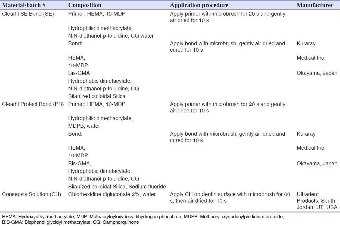
The resin composite (Filtek Z250, 3M, USA) was placed on the cured adhesives using a cylindrical split mold with a height of 2.5 mm and surface diameter of 2 mm in two increments of 1 and 1.5 mm. Each increment was light-cured for 40 s at 600 mW/cm2 with a light-curing unit (VIP Junior, Bisco, Schaumburg, IL, USA). Half of the specimens (n = 10) in each group were stored in tap water at 37°C for 24 h and the other half (n = 10) were stored in tap water containing 0.4% sodium azide with a stable pH at 37°C for 6 months and additionally thermocycled 1,000 times (between 5° and 55°C with 20 s dwell times) during 6 months prior to bond strength testing.
The shear test was performed using universal testing machine (Instron Z020, Zwick, Roell, Germany). A knife-edge shearing rod at a crosshead speed 1 mm/min was used to load the specimens until fracture. Shear bond strength in MPa was calculated from the peak load at failure divided by the specimen's surface area.
One specimen from the control and experimental groups pretreated with CH and bonded with the self-etch adhesive (PB) were prepared for SEM examination. Approximately, 2-mm thick disks were prepared from each specimen with Accutom-50 cut-off machine (Struers, Denmark) prior to examination with scanning electron microscope (SEM, XL30, Philips, Netherland) operating at 17 kV.
All data were analyzed with three-way analysis of variance (ANOVA) for the effect of adhesive system, CH, and storage time. For each adhesive, a two-way ANOVA was done to evaluate the interaction effect of storage time and CH, and t-test was done to compare the effect of two factors. All tests were done at a 0.05 level of significance and all analyses were performed using SPSS version 11.5 software (Chicago, IL, USA).
After testing, the fracture modes were evaluated using a stereomicroscope (Carl Zeiss Inc, Oberkochen, Germany) at ×10 and classified according to the predominant mode of fracture including: 1) Adhesive, 2) cohesive in dentin, 3) cohesive in composite, and 4) mixed, a combination of adhesive and cohesive.
RESULTS
The mean strength and standard deviations in the eight experimental groups are summarized in Figure 1. Three-way ANOVA showed that bond strength was significantly influenced by the adhesive type (P < 0.001) and time (P = 0.004). The effect of CH, the interactions between adhesive type and time, between adhesive type and CH, and the interaction among all the three factors was not significant (P > 0.05). However, the interaction between CH and time was statistically significant (P < 0.001). This interaction was statistically significant for each adhesive, SE (P = 0.02) and PB (P = 0.006). The bond strength of PB was significantly lower than that of SE in two time periods (P = 0.001).
Figure 1.
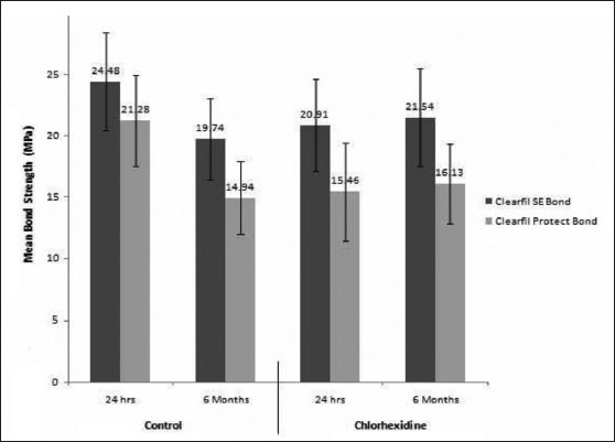
The mean shear bond strength for eight tested groups
The bond strength of two adhesives was significantly reduced after aging (P = 0.004). For two adhesives (SE, PB), CH had a significantly negative effect on the initial bond strength (P = 0.04 and 0.004, respectively), but bond strength was not altered over the 6 months of storage (P = 0.84 and 0.70, respectively). After a 6-month period, bond strength of two adhesives associated to CH did not vary with their control groups (P = 0.25 and 0.48). The frequencies of different failure modes are presented in Table 2. Representative SEM images of the dentin/restoration interface for the control and experimental groups are shown in Figures 2 and 3.
Table 2.
The frequency of fracture modes of eight groups
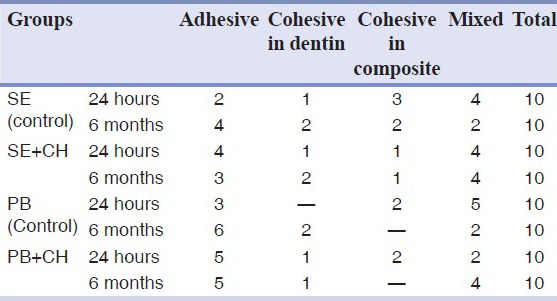
Figure 2.
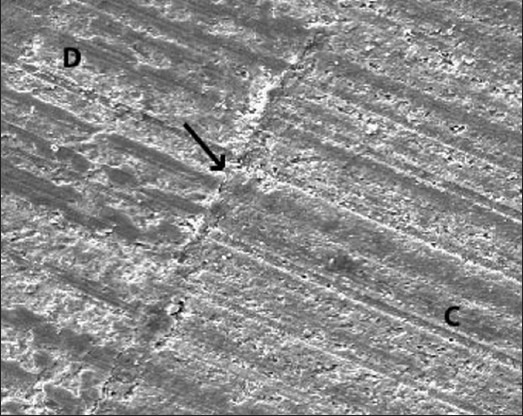
A representative scanning electron microscope (SEM) image of a bonded interface in control group (self-etch adhesive), where gap-free interface can be observed (×250). C = Composite, D = dentin
Figure 3.
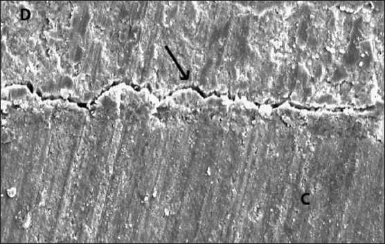
A representative SEM image of a bonded interface in experimental group (self-etch adhesive + chlorhexidine). Notice the gap between dentin and restoration (×250). C = Composite, D: dentin
DISCUSSION
In spite of a potential benefit for antibacterial activity, it is noteworthy that achieving effective durable bonding to dentin is still one of the most critical factors affecting long-term clinical success of a restoration. Hence, in the current study, the effect of two types of antibacterial agents on the bond strength of the two self-etch adhesives to dentin was tested in two time periods.
The present results revealed that although the CH application prior to the two tested adhesives significantly decreased the immediate bond strength, this bond was stable after aging in water; there was no significant difference between the control and experimental groups treated with CH after 6 months of storage. Therefore, our results cannot support complete acceptance of null hypothesis.
The adverse effect of CH solution on initial bonding performance of SE was previously reported by Ercan et al.[14] Also, Meiers and Shook[15] reported that CH application prior to acidic primer of Syntac significantly decreased its bond strength to dentin. However, in other studies, no adverse effect was found on sealing ability of SE[16,17] and bond strength of SE to dentin.[18,19] In a study by Siso et al.,[20] although there was no significant difference between the control and CH-treated groups, the latter group revealed a higher value of microleakage than that of the control group bonded by SE. It was reported that the application of CH prior to self-etch adhesives (Clearfil Tri S Bond and SE) did not affect immediate bond strength resin composite to dentin.[21,22]
The self-etch adhesives used in this study contain a functional acidic monomer, 10-MDP. It was demonstrated that dissociated CH cations could bind to phosphate groups and calcium of the hydroxyapatite.[23] The remaining cations might form bonds with phosphate anions of 10-MDP molecules of the self-etch adhesives after application of the adhesive on the surface treated with CH. This inhibitory effect on 10-MDP may impair the bonding ability of this functional acidic monomer (10-MDP) to dentinal calcium, reducing the bond strength to dentin.[24]
According to our results, a significant decrease in bond strength of the two adhesives was exhibited after aging. This finding is consistent with some studies.[25,26,27] However, the higher bond stability of PB was reported.[27,28]
Although self-etch adhesives are capable of simultaneous etching and priming, a discrepancy in the depth of demineralization and resin infiltration may occur in these adhesives. The incomplete resin infiltration could lead to creation of unprotected collagen fibrils.[29] The disintegration of these collagens could contribute to a decrease in the dentin bond strength of two self-etch adhesives observed in the current study after aging. Moreover, it has been shown that mild versions of self-etch adhesives can activate MMPs, resulting in degradation of suboptimal infiltrated collagen and loss of bond strength.[30]
The results of the present study revealed that although CH application compromised the immediate bond strength, it diminished loss of bond strength after aging. This result could be related to the preservative effect of CH on bonding interface. CH was applied on the smear layer-covered dentin, and then covered with acidic primer and adhesive resin may protect the unprotected collagen created during bonding procedures. The beneficial effect of 2% CH on stability of hybrid layer formed by an etch-and-rinse and a self-etch adhesives was reported in a recent study.[31]
Few studies have so far reported the effect of CH application prior to two-step self-etch adhesives on long-term bonding effectiveness to dentin. In a study by Campos et al.,[21] the preservative effect of CH 2% on bond strength of an all-in-one self-etch adhesive, Clearfil Tri S Bond, to dentin was reported during a 6-month aging period. However, Mobarak[22] has recently demonstrated that pretreatment with 2 or 5% CH is not able to diminish the loss of bond strength of SE to normal dentin over 2-year aging in artificial saliva under simulated intrapulpal pressure. Only 5% CH could preserve the bond strength to the affected dentin. These divergent results may be due to the difference in the CH concentration, aging condition, and aging duration time, bonding substrate, and testing method.
The results of the current study indicated that PB showed significantly lower bond strength to dentin than that of SE. This result was in agreement with those of two other studies.[32,33] The nonuniform distribution of NaF fillers may contribute to a relative poor mechanical property in some areas of bonding interface, resulting in lower bond strength of PB compared to SE.[32] Contrary to our results, some studies have suggested that incorporation of antibacterial monomer into primer did not adversely affect the dentinal bond strength of the adhesive.[6,8,34]
When evaluating the fracture modes of the eight tested groups in this study, the higher number of the adhesive fracture was detected in two groups of PB + CH and control group of PB (24 h). It seems that the higher number of the adhesive fracture was associated to the lower bond strength of these groups.
Further studies should investigate the interaction of CH with other two-step self-etch adhesives and the long-term effect of CH on bond strength of these adhesives to dentin.
CONCLUSION
Within the limitations of the current study, it may be concluded that although CH was capable of decreasing the loss of bond strength of the two-step self-etch adhesives to dentin over time, it compromised the initial bonding of the self-etch adhesives. Thus, further in vitro and in vivo studies should be conducted before the combined application of CH, and two-step self-etch adhesives can be recommended in clinical practice.
ACKNOWLEDGEMENTS
The authors thank the vice-chancellery of Shiraz University of Medical Sciences, for supporting the research (Grant # 89-5423). This article relevant thesis of Dr. A Alikhani. The authors would like to thank Dr. M Vossoughi for statistical analysis and Dr. N Shokrpour for editing the manuscript.
Footnotes
Source of Support: Vice Chancellore for research at Shiraz University of Medical Sciences
Conflict of Interest: The authors certify that they have no proprietary,financial,or other personal interest of any nature or kind in any product, service, and/or company that is presented in this article.
REFERENCES
- 1.De Munck J, Van Landuyt K, Peumans M, Poitevin A, Lambrechts P, Braem M, et al. A critical review of the durability of the adhesion to tooth tissue: Methods and results. J Dent Res. 2005;84:118–32. doi: 10.1177/154405910508400204. [DOI] [PubMed] [Google Scholar]
- 2.Eick S, Glockmann E, Brandl B, Pfister W. Adherence of Streptococcus mutans to various restorative materials in a continuous flow system. J Oral Rehabil. 2004;31:278–85. doi: 10.1046/j.0305-182X.2003.01233.x. [DOI] [PubMed] [Google Scholar]
- 3.Imazato S, Kuramoto A, Takahashi Y, Ebisu S, Peters MC. In vitro antibacterial effects of the dentin primer of Clearfil Protect Bond. Dent Mater. 2006;22:527–32. doi: 10.1016/j.dental.2005.05.009. [DOI] [PubMed] [Google Scholar]
- 4.Tay FR, Pashley DH. Aggressiveness of contemporary self-etching systems. I: Depth of penetration beyond dentin smear layers. Dent Mater. 2001;17:296–308. doi: 10.1016/s0109-5641(00)00087-7. [DOI] [PubMed] [Google Scholar]
- 5.Tay FR, Carvalho R, Sano H, Pashley DH. Effect of smear layers on the bonding of a self-etching primer to dentin. J Adhes Dent. 2000;2:99–116. [PubMed] [Google Scholar]
- 6.Cal E, Türkün LS, Türkün M, Toman M, Toksavul S. Effect of an antibacterial adhesive on the bond strength of three different luting resin composites. J Dent. 2006;34:372–80. doi: 10.1016/j.jdent.2005.08.004. [DOI] [PubMed] [Google Scholar]
- 7.Imazato S, Kaneko T, Takahashi Y, Noiri Y, Ebisu S. In vivo antibacterial effects of dentin primer incorporating MDPB. Oper Dent. 2004;29:369–75. [PubMed] [Google Scholar]
- 8.Imazato S, Tay FR, Kaneshiro AV, Takahashi Y, Ebisu S. An in vivo evaluation of bonding ability of comprehensive antibacterial adhesive system incorporating MDPB. Dent Mater. 2007;23:170–6. doi: 10.1016/j.dental.2006.01.005. [DOI] [PubMed] [Google Scholar]
- 9.Türkün LS, Ateş M, Türkün M, Uzer E. Antibacterial activity of two adhesive systems using various microbiological methods. J Adhes Dent. 2005;7:315–20. [PubMed] [Google Scholar]
- 10.Gultz J, Do L, Boylan R, Kaim J, Scherer W. Antimicrobial activity of cavity disinfectants. Gen Dent. 1999;47:187–90. [PubMed] [Google Scholar]
- 11.Pashley DH, Tay FR, Yiu C, Hashimoto M, Breschi L, Carvalho RM, et al. Collagen degradation by host-derived enzymes during aging. J Dent Res. 2004;83:216–21. doi: 10.1177/154405910408300306. [DOI] [PubMed] [Google Scholar]
- 12.Sulkala M, Tervahartiala T, Sorsa T, Larmas M, Salo T, Tjäderhane L. Matrix metalloproteinase-8 (MMP-8) is the major collagenase in human dentin. Arch Oral Biol. 2007;52:121–7. doi: 10.1016/j.archoralbio.2006.08.009. [DOI] [PubMed] [Google Scholar]
- 13.Orsini G, Mazzoni A, Orciani M, Patignano A, Procaccini M, Falconi M, et al. Matrix metalloproteinase-2 expression induced by two different adhesive systems on human pulp fibroblasts. J Endod. 2011;37:1663–7. doi: 10.1016/j.joen.2011.07.009. [DOI] [PubMed] [Google Scholar]
- 14.Ercan E, Erdemir A, Zorba YO, Eldeniz AU, Dalli M, Ince B, et al. Effect of different cavity disinfectants on shear bond strength of composite resin to dentin. J Adhes Dent. 2009;11:343–6. [PubMed] [Google Scholar]
- 15.Meiers JC, Shook LW. Effect of disinfectants on the bond strength of composite to dentin. Am J Dent. 1996;9:11–4. [PubMed] [Google Scholar]
- 16.Geraldo-Martins VR, Robles FR, Matos AB. Chlorhexidine's effect on sealing ability of composite restorations following Er:YAG laser cavity preparation. J Contemp Dent Pract. 2007;8:26–33. [PubMed] [Google Scholar]
- 17.Türkün M, Türkün LS, Kalender A. Effect of cavity disinfectants on the sealing ability of nonrinsing dentin-bonding resins. Quintessence Int. 2004;35:469–76. [PubMed] [Google Scholar]
- 18.de Castro FL, de Andrade MF, Duarte SL, Júnior, Vaz LG, Ahid FJ. Effect of 2% chlorhexidine on microtensile bond strength of composite to dentin. J Adhes Dent. 2003;5:129–38. [PubMed] [Google Scholar]
- 19.Mobarak EH, El-Korashy DI, Pashley DH. Effect of chlorhexidine concentrations on micro-shear bond strength of self-etch adhesive to normal and caries-affected dentin. Am J Dent. 2010;23:217–22. [PubMed] [Google Scholar]
- 20.Siso HS, Kustarci A, Göktolga EG. Microleakage in resin composite restorations after antimicrobial pre-treatments: Effect of KTP laser, chlorhexidine gluconate and Clearfil Protect Bond. Oper Dent. 2009;34:321–7. doi: 10.2341/08-96. [DOI] [PubMed] [Google Scholar]
- 21.Campos EA, Correr GM, Leonardi DP, Barato-Filho F, Gonzaga CC, Zielak JC. Chlorhexidine diminishes the loss of bond strength over time under simulated pulpal pressure and thermo-mechanical stressing. J Dent. 2009;37:108–14. doi: 10.1016/j.jdent.2008.10.003. [DOI] [PubMed] [Google Scholar]
- 22.Mobarak EH. Effect of chlorhexidine pretreatment on bond strength durability of caries-affected dentin over 2-year aging in artificial saliva and under simulated intrapulpal pressure. Oper Dent. 2011;36:649–60. doi: 10.2341/11-018-L. [DOI] [PubMed] [Google Scholar]
- 23.Say EC, Koray F, Tarim B, Soyman M, Gülmez T. In vitro effect of cavity disinfectants on the bond strength of dentin bonding systems. Quintessence Int. 2004;35:56–60. [PubMed] [Google Scholar]
- 24.Hiraishi N, Yiu CK, King NM, Tay FR. Effect of chlorhexidine incorporation into a self-etching primer on dentine bond strength of a luting cement. J Dent. 2010;38:496–502. doi: 10.1016/j.jdent.2010.03.005. [DOI] [PubMed] [Google Scholar]
- 25.van Landuyt KL, De Munck J, Mine A, Cardoso MV, Peumans M, Van Meerbeek B. Filler debonding & subhybrid-layer failures in self-etch adhesives. J Dent Res. 2010;89:1045–50. doi: 10.1177/0022034510375285. [DOI] [PubMed] [Google Scholar]
- 26.Nishiyama N, Tay FR, Fujita K, Pashley DH, Ikemura K, Hiraishi N, et al. Hydrolysis of functional monomers in a single-bottle self-etching primer--correlation of 13C NMR and TEM findings. J Dent Res. 2006;85:422–6. doi: 10.1177/154405910608500505. [DOI] [PMC free article] [PubMed] [Google Scholar]
- 27.Donmez N, Belli S, Pashley DH, Tay FR. Ultrastructural correlates of in vivo/in vitro bond degradation in self-etch adhesives. J Dent Res. 2005;84:355–9. doi: 10.1177/154405910508400412. [DOI] [PubMed] [Google Scholar]
- 28.Nakajima M, Okuda M, Ogata M, Pereira PN, Tagami J, Pashley DH. The durability of a fluoride-releasing resin adhesive system to dentin. Oper Dent. 2003;28:186–92. [PubMed] [Google Scholar]
- 29.Carvalho RM, Chersoni S, Frankenberger R, Pashley DH, Prati C, Tay FR. A challenge to the conventional wisdom that simultaneous etching and resin infiltration always occurs in self-etch adhesives. Biomaterials. 2005;26:1035–42. doi: 10.1016/j.biomaterials.2004.04.003. [DOI] [PubMed] [Google Scholar]
- 30.Tay FR, Pashley DH, Loushine RJ, Weller RN, Monticelli F, Osorio R. Self-etching adhesives increase collagenolytic activity in radicular dentin. J Endod. 2006;32:862–8. doi: 10.1016/j.joen.2006.04.005. [DOI] [PubMed] [Google Scholar]
- 31.Lafuente D. SEM analysis of hybrid layer and bonding interface after chlorhexidine use. Oper Dent. 2012;37:172–80. doi: 10.2341/10-251-L. [DOI] [PubMed] [Google Scholar]
- 32.Sidhu SK, Omata Y, Tanaka T, Koahiro K, Spreafico D, Semeraro S, et al. Bonding characteristics of newly developed all-in-one adhesives. J Biomed Mater Res B App Biomater. 2007;80:297–303. doi: 10.1002/jbm.b.30597. [DOI] [PubMed] [Google Scholar]
- 33.Yildirim S, Tosun G, Koyutürk AE, Sener Y, Sengün A, Ozer F, et al. Microtensile and Microshear Bond strength of an antibacterial self-etching system to primary tooth dentin. Eur J Dent. 2008;2:11–7. [PMC free article] [PubMed] [Google Scholar]
- 34.Sonoda H, Banerjee A, Sherriff M, Tagami J, Watson TF. An in vitro investigation of microtensile bond strengths of two dentine adhesives to caries-affected dentine. J Dent. 2005;33:335–42. doi: 10.1016/j.jdent.2004.09.009. [DOI] [PubMed] [Google Scholar]


