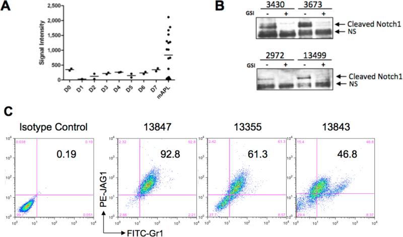
Figure 4. Jag1 and activated Notch signaling are found in murine APL samples.
A). Microarray expression data for Jag1 in murine Lin−Sca+ cells undergoing in vitro differentiation with G-CSF, and in 22 murine APL samples. Promyelocytes are highly enriched on days 2 and 3 of in vitro differentiation33 B) Western blot showing cleaved Notch1 in murine APL tumors treated for 48 hrs with either DMSO vehicle, or 2 μM compound E. The faster migrating band is non-specific (NS) and not sensitive to GSI treatment. 13499 and 3430 are GSI-sensitive tumors (Figure 7, Supplemental Table 1); 2972 and 3673 are insensitive. C). Intracellular flow cytometry detection of Jag1 in representative murine APL samples. Numbers correspond to percentage of cells co-expressing Gr-1 and Jag1.

