Abstract
The task of the visual system is to translate light into neuronal encoded information. This translation of photons into neuronal signals is achieved by photoreceptor neurons (PRs), specialized sensory neurons, located in the eye. Upon perception of light the PRs will send a signal to target neurons, which represent a first station of visual processing. Increasing complexity of visual processing stems from the number of distinct PR-subtypes and their various types of target neurons that are contacted. The visual system of the fruit fly larva represents a simple visual system (larval optic neuropil, LON) that consists of 12 PRs falling into two classes: blue-senstive PRs expressing Rhodopsin 5 (Rh5) and green-sensitive PRs expressing Rhodopsin 6 (Rh6). These afferents contact a small number of target neurons, including optic lobe pioneers (OLPs) and lateral clock neurons (LNs). We combine the use of genetic markers to label both PR-subtypes and the distinct, identifiable sets of target neurons with a serial EM reconstruction to generate a high-resolution map of the larval optic neuropil. We find that the larval optic neuropil shows a clear bipartite organization consisting of one domain innervated by PRs and one devoid of PR axons. The topology of PR projections, in particular the relationship between Rh5 and Rh6 afferents, is maintained from the nerve entering the brain to the axon terminals. The target neurons can be subdivided according to neurotransmitter or neuropeptide they use as well as the location within the brain. We further track the larval optic neuropil through development from first larval instar to its location in the adult brain as the accessory medulla.
Keywords: Drosophila, larval visual system, serial EM analysis, photoreceptors, sensory systems
Introduction
The Photoreceptor neurons (PRs) of the eye transform light induced signals into neuronal information, which is transmitted to the higher order neurons in visual information processing. Depending on which sensory receptor gene is expressed in a PR, the neuron will become sensitive to a certain spectrum of light. In the case of PRs sensory receptor genes are rhodopsins. In the mammalian visual system integration of visual sensory information already occurs in the retina, while in insects PRs connect to target neurons in the optic ganglia in the brain. In the fruit fly Drosophila melanogaster the adult compound eye is composed of about 800 unit-eyes, called ommatidia. Each ommatidium comprises eight PRs, which are subdivided into six outer PRs (R1–R6) and two inner PRs (R7 and R8). Outer PRs are required for motion detection and dim-light vision and express the broad-wavelength sensitive photoreceptor protein Rhodopsin1. The two inner PRs are required for color discrimination and color vision and express an array of four different rhodopsins genes (rh3, rh4, rh5 or rh6). Axonal projections of the PRs terminate in the optic ganglia of the adult brain (Borst, 2009; Sanes and Zipursky, 2010). The adult insect brain is traditionally subdivided into the central brain and the optic lobe. The optic lobe consists of four neuropil compartments; the outer two, lamina and medulla, receive input from retinal photoreceptors; the inner two, lobula and lobula plate, connect the medulla to the visual centers (“optic foci”) of the central brain. In line with this functional diversification outer and inner PRs connect to target neurons in distinct optic neuropils the lamina (outer PRs) and medulla (inner PRs), respectively (Morante and Desplan, 2004).
Compared to the rather complex organization of the adult visual system the larval visual system of is comparably simple. The larval eye (Bolwig’s organ; BO) is only composed of 12 PRs can be further divided into two PR-subtypes (Daniel et al., 1999; Green et al., 1993). Eight PRs express the green-sensitive Rh6, while the remaining four PRs express the blue-sensitive Rh5 (Sprecher and Desplan, 2008; Sprecher et al., 2007). PRs of the larval eye extend their axons into the larval optic neuropil (LON), a small neuropil compartment and first center for visual information processing in the larval brain. Larval PRs provide two essential functions for animal behavior: first in rapid light avoidance and second for circadian rhythm control (Mazzoni et al., 2005). Neurons of the clock circuit are marked by the cycling expression of the circadian regulator genes timeless (tim), period (per), cycle (cyc) and clock (clk). Among these clock neurons the lateral neurons (LNs) have been shown to connect to the LON. Contact between PRs and LNs is established during embryogenesis. Development and maintenance of dendritic arborizations of LNs into the LON requires the PRs (Malpel et al., 2002). LNs have further been shown to be important for the circadian modulation of photophobic behavior (Mazzoni et al.,2005; Keene et al., 2011)
In addition to the LNs two clusters of neurons have been reported to innervate the LON: a set of serotonergic neurons and the optic lobe pioneer neurons (OLPs) (Rodriguez Moncalvo and Campos, 2005; Tix et al., 1989). There are four distinct clusters of serotonergic neurons described in the larval brain containing a total of 22 neurons. The neurons innervating the LON have been assigned to the contra lateral SP2 cluster (Mukhopadhyay and Campos, 1995). The OLPs develop as part of the optic lobe placode as a cluster of three cells near the insertion of the stalk. OLPs innervate the LON starting during embryogenesis and are maintained into the adult fly. In detail analysis of their innervation pattern and functional relevance of these LON neurons are missing.
In order to gain a more detailed and complete view on the development and anatomy of LON-innervating neurons we have initiated a project where we combine electron microscopy with confocal microscopy. We integrate anatomical findings of a 3D reconstructed serial TEM (transmission electron microscopy) stack with molecular and genetic markers data gathered from confocal microscopic stacks. This paper presents the first in a series analyzing neuronal ultrastructure and connectivity of the LON. We provide here an overview of the developmental changes of the LON from first instar larva to the adult fly. We introduce different elements of the LON and their ultrastructure and connectivity within the LON, focusing in particular on the terminal arborization of the photoreceptor neurons providing sensory input to the LON.
MATERIAL AND METHODS
Drosophila strains and genetics
Flies and larvae were kept in a 12hr:12hr light-dark (LD) cycles at 25°C. For wild type, we used Oregon R, Canton S, w1118, yw122. We used the following fly strains: rh5-Gal4, rh6-Gal4, rh5-LacZ, rh6-LacZ, rh5-GFP, rh6-GFP (Cook et al., 2003), Nrv-Gal4, dvGlut-Gal4, Tdc2-Gal4, GMR-Gal4, pdf-Gal4, Cha-Gal4, UAS-CD8::GFP (Bloomington Stock Center). For first larval instar (L1) we dissected larvae between 0 and 12h post hatching; for 3rd instar (L3), we used wandering larvae (96–120h) post hatching.
Immunohistochemistry and antibodies
Dissection and analysis of the brain was done as previously described (Sprecher et al., 2007). The following primary antibodies were used: rabbit anti-Rh6 1:10,000 (Tahayato et al., 2003), rabbit anti-Serotonin 1:500 (Sigma), sheep anti-GFP 1:1000 (Biogenesis), mouse anti-Fasciclin II 1:10 (FasII; 1D4; DSHB), anti-Neuroglian 1:10; (BP104; Nrg; DSHB); m a 22C10 1:10, rat anti-Elav 1:30 (7E8A10; DSHB), mouse anti-Repo 1:50 (8D12; DSHB), rat anti-DNcadherin 1:10 (DN-Ex8; DNcad; DSHB), mouse anti-DEcadherin 1:10 (DCAD2; DEcad; DSHB), mouse anti-Bruchpilot 1:10 (NC82; Brp; DSHB), mouse anti-PDF 1:50 (PdfC7; DSHB), mouse anti-Chaoptin 1:10 (24B10; Chp; DSHB), mouse anti-βGAL 1:20 (40-1a; DSHB). Secondary antibodies used for confocal microscopy were Alexa-488, Alexa-555, and Alexa-647 antibodies generated in goat (Molecular probes), all at 1:300–1:500 dilution.
Laser confocal microscopy and image processing
Leica TCS SP2 and Sp5 microscopes were used for all imaging. Optical sections ranged from 0.2–1.5 micrometer and were recorded in ‘line average mode’ with a picture resolution of 512 × 512 or 1024 × 1024 pixels. Captured images from optical sections were processed using Leica confocal Software (LCS). Complete series of optical sections were imported and processed using ImageJ as previously described (Sprecher et al., 2006).
Serial TEM acquisition and processing
First instar larval brains were dissected and then fixed and embedded in epon resin as described in Johnson et al. (2009). A block containing a single brain was trimmed with a glass knife, and 60-nm serial sections were cut on a Leica UC8 ultratome. Ribbons of sections were collected onto single-slot copper grids with pioloform membranes. Sections were contrasted with 8% uranyl acetate and in Reynold’s lead citrate (Johnson et al., 2009). Serial sections were imaged automatically with the software Leginon (Suloway et al., 2005) driving an FEI Tecnai 12 electron microscope. A collection of partially overlapping images was acquired for each serial section. Images were automatically contrast-corrected and automatically montaged within and across sections using the open source software TrakEM2 (Cardona et al. 2010). Each neuron and glial cell was manually reconstructed in 3D with computer-assisted segmentation tools provided by TrakEM2. Reconstructed neurons were rendered in 3D with the software ImageJ 3D Viewer (Schmid et al., 2010). TrakEM2 is part of the FIJI software package, which can be accessed and downloaded here: http://pacific.mpi-cbg.de/wiki/index.php/Fiji.
RESULTS
The optic neuropil of the early larva (LON) is formed by the sensory terminals of the larval eye (Bolwig’s organ), as well as endings of several small populations of primary neurons that transmit visual input to the central brain. In this early larval stage the LON is enclosed by the epithelial optic anlage that is situated at the ventro-lateral surface of the brain (Fig. 1A, B). The optic anlage comprises an outer part (OOA), a curved hemicylindrical structure, and an inner part (IOA), located medially adjacent to the outer optic anlage (Fig. 1A, B, D). In preparations labeled with a synaptic marker, the LON appears as a synapse-rich, narrow process extending from the ventro-lateral surface of the central brain (Fig. 1C). The twelve sensory PR axons of the larval eye enter the outer optic anlage hemicylinder laterally and terminate in the LON (Fig. 1D, E). Larval PRs and their axons form two classes: one class of four cells expresses rh5; the other class of eight cells expresses rh6. As shown in Fig. 1E and described in more detail below, axon terminals of these two classes occupy largely non-overlapping territories in the LON.
Fig. 1.
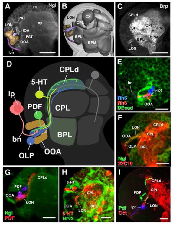
Elements of the larval optic neuropil (LON). All panels show schematic or confocal images representing frontal section of a first instar larval brain hemisphere; lateral to the left, dorsal up. A: Labeling with anti-Neuroglian (Ngl), showing neuronal cell bodies in cortex (co) and nerve processes, forming central neuropil (np). Z-projection of a confocal stack (3 μm). Added to the section is a 3D digital model (anterior view) of outer optic anlage (OOA, beige), inner optic anlage (IOA, gray), LON, purple, and Bolwig’s nerve (bn, magenta), primary axon tract (PAT). B: 3D digital model of brain hemisphere (posterior view), showing brain surface (light gray), neuropil compartments (dark gray: BPL, baso-posterior lateral compartment; CPL centro-posterior lateral compartment; BPM baso-posterior medial compartment; CX calyx), and visual system (coloring as in A). C: Labeling with anti-Bruchpilot (Brp), a marker for synapses, outlines neuropil compartments, including LON. Z-projection of a confocal stack (3 μm). CPLd dorsal subdivision of CPL; CPI centro-poterior intermediate compartment; CPM; central-posterior medial compartment). D: Schematic representation of neuropil compartments of central brain and of larval visual system, including main neuronal elements contributing to LON: larval photoreceptors (lp, red), bn (red); optic lobe pioneers (OLP, blue), PDF neurons (PDF; green), serotonergic neurons (5-HT; yellow), OOA (brown). E: Z-projection of a confocal stack (17 μm) showing larval photoreceptor projections. Rh5 and Rh6 are differentially labeled by rh5-GFP (blue) and rh6-LacZ (red). Anti-Drosophila E-cadherin (DEcad, green) labels the OOA surrounding LON. Note that terminals (lpt) of Rh5-positive axons are located more medially in LON than terminals of Rh6 terminals. F: Labeling with antibody 22C10 (red) and anti-Ngl (green), Z-projection of a confocal stack (22 μm). 22C10 marks (among many other, central brain neurons) the OLP, whose cell bodies are located laterally in the OOA. OLP fibers project through LON towards central brain. G: GFP driven by pdf-Gal4 labels the PDF neurons (red; cell bodies rendered in purple to set them apart from dendrites), anti-Ngl (green). Z-projection of a confocal stack (20 μm). PDF cell bodies are located in brain cortex adjacent to OOA; profuse dendritic branches are formed in the LON, and axons project out of the LON towards the CPLd compartment of the central brain. H: Labeling with anti-Serotonin (5-HT; red), showing pair of serotonergic neuronal cell bodies with neurites projecting towards LON. Z-projection of a confocal stack (20 μm). GFP driven by Nervana2-Gal4 (Nrv2; green) labels glial cells. I: Z-projection of a confocal stack (29 μm) showing a third instar larval brain labeled with anti-PDF (Green), Rh6-projections with anti-Rh6 (blue) and GFP driven by Tdc2-Gal4 labels (Oct; red). Subset of octopaminergic neurons projects into LON. Scale bars: 10μm (A, C, E, F, G, I), 15 μm (H).
We found that at least two populations of neurons are postsynaptic to larval PRs (see below). One population is comprised of three optic lobe pioneer neurons (OLP) per hemisphere, the other the four neurons expressing the neuropeptide pigment dispersing factor (PDF) per hemisphere. These four neurons belong the circadian clock circuit and are termed lateral neurons (LNs). Cell bodies of the OLPs, which flank the optic anlage laterally (Fig. 1F), are derived from the same embryonic placode (called optic lobe placode (Green et al., 1993)) that also gives rise to optic anlage itself (Chang et al., 2003). OLP axons fasciculate with PR axons as they enter the optic neuropil from laterally (Fig. 1F) (Tix et al., 1989; Campos et al., 1995; Nassif et al., 1998). OLP axons exit the LON medially and enter the central brain neuropil.
The LNs form part of a central brain lineage located in the dorsal brain cortex (Fig. 1G). LNs cell body fibers project ventrally through an opening in the outer optic anlage (Fig. 1D, G); after entering the optic neuropil and giving off short, dendritic side branches (Malpel et al., 2002), fibers turn medially, exit the LON and continue to the CPLd compartment of the central brain (Fig. 1G, H).
A third group of neurons contributing to the LON are serotonergic cells located in the dorsal brain cortex. The larval brain comprises four clusters of serotonergic neurons, among which one cluster consisting of two neurons have been shown to extend their dendritic arbors into the LON (Rodriguez Moncalvo and Campos, 2005). These innervations into the LON have been suggested to stem from the contralateral SP2 cluster (Monastirioti, 1999; Mukhopadhyay and Campos, 1995). These neurons branch profusely in the central brain, in particular the CPLd and BLP compartments (Fig. 1I). In addition, two fibers continue ventrally and approach the LON. In the early larva, the serotonergic fibers terminate near the entry point to the LON; towards later larval stages, they continue growing into the LON. Interestingly the development of proper dendritic arbors into the LON dependents on the activity and presence of the larval PRs, more precisely on the larval Rh6-PR subtype (Rodriguez Moncalvo and Campos, 2005). Moreover the LON is also innervated by arborizations stemming from octopaminergic/tyraminergic neurons (Fig 1H), which until now have not been further characterized.
Developmental relationship of the larval and adult optic lobe neuropil
Most of the development of the adult optic lobe takes place during metamorphosis. However, small primordia of the adult visual neuropils start to appear in the late larva when the respective neurons, generated from the outer and inner optica anlage (OOA and IOA), are born and send out short, unbranched neurites (Ngo et al., 2010). In the late larva, the lamina (La) and medulla neuropils (Mp: proximal medulla; Md: distal medulla) represent two curved sheets surrounding a central “plug” which constitutes the primordia of the lobula (Lo) and lobula plate (Lp) (Fig. 2A–C). All of these neuropil primordia are formed by axons and growth cones, but lack synapses, as evidenced by the absence of label when using synaptic markers such as anti-Brp alone (data not shown). Functional maturation, which includes the formation of synapses, takes place during the pupal period. Thus, in the larva, the small LON remains the only functional neuropil compartment guiding visual behavior.
Fig. 2.
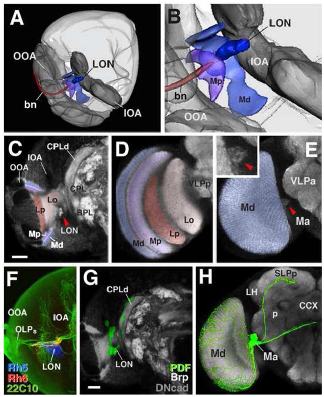
Topology of larval optic neuropil (LON) throughout development. A, B: 3D digital model of mid third instar larval brain hemisphere, postero-lateral view. Optic anlagen (outer optic anlage: OOA; inner optic anlage: IOA) have grown and form crescent-shaped epithelia at the ventro-lateral brain surface (rendered in gray). Space in between OOA and IOA is filled by neurons of medulla (cell bodies not shown) whose fibers have started to form the neuropil of the adult medulla (proximal medulla, Mp, purple; distal medulla, Md, light blue); bolwig nerve (bn, brown). The LON is accreted to the medial edge of the medulla neuropil. C, D: Frontal single confocal section of late third instar larval (C) and adult (D) brain hemisphere (lateral to the left). Different optic neuropils are rendered in different colors (Md, blue; Mp, purple; Lo, lobula faint pink; Lp, lobula plate, red). In C, Neurons are labeled by anti-DN-cadherin (DNcad; faint gray). The IOA and OOA are DNcad negative and stand out as dark spaces. Labeling of synapses with anti-Brp is added to global neuronal staining with anti-DNcad,; this results in bright signal demarcating larval neuropil compartments containing mature synapses, including the larval optic neuropil (red arrowhead). Neuropil compartments stained with anti-DN-cad (BPL, baso-posterior lateral compartment; CPL centro-posterior lateral compartment; CPLd centro-posterior lateral dorsal compartment; VLPp, ventrolateral protocerebrum, posterior subdivision). E: frontal single confocal section of adult brain at a more anterior level than section shown in (D). Shown in this panel is anterior surface and medial rim of the distal medulla, to which the accessory medulla (Ma), the descendant of the LON, is attached (red arrowhead; enlarged view shown in inset). Neuropil compartment stained with anti-DN-cad (VLPa ventrolateral protocerebrum, anterior subdivision). F: Z-projection of a confocal stack (39 μm) of late third instar larval brain hemisphere. Neurons are labeled by 22C10 antibody (green). Larval photoreceptor terminals are differentially labeled by rh5-GFP (blue), rh6-LacZ (red) and demarcate the larval optic neuropil. Note that 22C10-positive cell bodies of optic lobe pioneers (OLPs) have been “pulled away” from their original location (which was right next to the LON; see Fig. 1D, F) by the expanding optic lobe. G, H: Frontal single confocal section of late larval (G) and adult (H) brain hemisphere labeled with anti-DN-cad. As explained for panel (C), anti-Brp was added in G to enhance staining of larval compartments containing mature synapses, including the LON. Labeling of PDF neurons (pdf-Gal4 > UAS-GFP; green) illustrates the topological relationship between larval and adult visual system. PDF-neuronal dendrites branch in larval LON and adult accessory medulla (red arrowhead). Axons project to larval CPLd compartment (G), which grows during metamorphosis to become the adult superior lateral protocerebrum, posterior subdivision (SLPp in H). PDF neurons add additional dendrites branching profusely throughout adult medulla (orange arrowhead); axons added during metamorphosis project contralaterally through the great commissure (CCX central complex; p peduncle; LH, lateral horn).
The LON is located in the center of the growing adult optic neuropils, in between the lobula and medulla (Fig. 2A–C). As reported previously (Helfrich-Forster et al., 2002; Malpel et al., 2002), the LON is incorporated into the adult optic lobe as the accessory medulla (Ma), a distinctly visible protrusion of the (main) medulla neuropil (Fig. 2E, inset red arrowhead). Likewise, the Rh5-PRs of the larval eye persist into the adult stage. The larval Rh6-PR subtype undergoes apoptotic cell death while the Rh5-PRs switch expression of rh5 to rh6 (Sprecher and Desplan, 2008). The terminal projections of the transforming PRs are maintained and even increase in complexity during metamorphosis.
Target neurons, such as the OLPs and LNs, also survive metamorphosis (Helfrich-Forster, 1997; Helfrich-Forster et al., 2002; Tix et al., 1989). Similar to their terminal dendrites, which become part of the adult medulla, cell bodies of the OLPs come to lie in the medulla cortex. This shift of the cell bodies away from their (dendritic) area of termination already occurs in the larva; in the late third instar larva, the OLP somata, embedded in the expanding mass of newly produced medulla neurons, are “dragged” away from the LON (compare Fig. 1 and Fig. 2F). Whereas the adult pattern of connectivity of OLPs is not yet known, that of the LNs has been documented in several previous reports (Fig. 2G, H), (Helfrich-Forster et al., 2002). During pupariation four additional LNs develop. The larval LNs will become the adult sLNvs (small LNvs), while the adult specific LNvs are bigger in cell size therefore called l-LNvs (large LNvs). In the adult the LNvs still send their axon terminals to a dorsal domain of the adult brain that spans part of the superior lateral and superior medial protocerebum (SLP, SMP). In addition, they have formed a second, commissural branch that reaches the contra-lateral optic lobe (Fig. 2H). Dendrites of the s-LNvs still innervate the accessory medulla; in addition during metamorphosis neurites stemming from the l-LNvs innervate the medulla (Fig. 2H).
Pattern of PR axonal termination reveal a bipartite organization of the LON
The series of consecutive EM sections on which the following analysis is based included the neuropil of one brain hemisphere (Fig. 3A). The plane of sectioning was tilted dorso-posterior to ventro-anterior (Fig. 3C) (Cardona et al., 2010). The LON is included in 110 sections at the posterior end of the series (delineated by the double line in Fig. 3C). The large electron-lucent terminal boutons of the PRs, enclosed within the epithelial cells of the outer optic anlage, are a prominent hallmark of the LON (Fig. 3A, B). The LON is clearly set apart from the central brain neuropil, to which it is connected by a thin stalk that contains not more than 15 neurites (Fig. 3E). The stalk of the LON is surrounded by a layer of neuropilie glia (arrowheads in Fig. 3E). Bundles of tightly packed axons flank the LON stalk (Fig. 3E); these bundles represent the primary axon tracts (PATs) which carry the axons of distinct lineages of primary neurons located in the dorsolateral and baso-lateral brain cortex (DPL and BL lineages) (Younossi-Hartenstein et al., 2006).
Fig. 3.
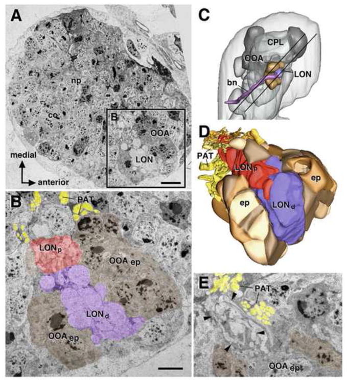
Topology of larval optic neuropil (LON) as part of series of contiguous transmission electron microscopy (TEM) images. A: Montage of TEM images of one L1 larval brain hemisphere (co cortex; np neuropil; OOA outer optic anlage). B: High magnification of section of larval visual system, boxed in (A). Shown are epithelial outer optic anlage (OOAep; rendered brown), distal LON (LONd; purple), defined by presence of large diameter photoreceptor terminals (arrows), proximal LON (LONp; red), axon tracts of primary lineages flanking optic anlage (primary axon tract; PAT; yellow). C: 3D digital model of L1 larval brain hemisphere, lateral view. Neuropil compartments (dark gray; CPL centro-posterior lateral compartment) and larval visual system (rendered in same colors as in Fig. 1A, B) are shown as landmarks. Parallel lines demarcate the brain “slice” included in the TEM stack that was used for the analysis of the larval optic neuropil in this paper. D: 3D digital model of LON and OOA, reconstructed from TEM stack. The model is shown at an orientation that is used most frequently in the following figures of this paper: the view is from dorso-posterior (indicated by red arrow in (C), exposing the LONd and LONp, enclosed by the hemicylindrical outer optic anlage. E: TEM image of proximal LON (boundaries indicated by arrowheads) at higher magnification. Scale bars: 5μm (A), 2 μm (B, E).
TEM images lack genetic and molecular markers commonly used in confocal microscopy. However we have been able to identify neurites within or connecting to the LON based on shared anatomical landmarks and properties neurons between TEM and confocal data. These landmarks include cell body location, location where neurites enter the neuropil, neurite size, neurite projection and arborization pattern. Specifically we have tracked a neurite of the LNs, as well as the OLP neurites from the cell body (soma) throughout the entire stack where they exit the LON through the connecting stalk between LON and central brain. The particularity of the OLPs is that they are in first instar larvae directly adjacent to the optic lobe epithelium, thus easily identifiable. The LNs enter the LON by penetrating the optic lobe epithelium and can therefore also be easily identified (see below). The LON stalk represents the main connection between the central brain neuropil and the LON. Neurons that connect to the LON through the stalk such as the octopaminergic or serotonergic neurons cannot be properly identified in the TEM stack.
The LON has a bipartite organization, comprising a distal domain (LONd) where most PRs axons terminate (purple in Fig. 3B, D), and a smaller, proximal domain (LONp) that almost exclusively consists of neurites of target neurons of the PRs (see below). We found a total of 12 terminal PR axons that terminate in the LON. Terminal PR axons are typical representatives of the class of globular neurites (Cardona et al., 2010), which are characterized by large (1–2μm), rounded swellings (“globuli” or “boutons”). One PR terminal is shown in Fig. 4. Note that the axon, which has a diameter of 0.2–0.3μm while traveling in the Bolwig’s nerve, forms three serially arranged boutons in the distal LON. Boutons are rich in ribosomes and mitochondria, but have few microtubules, in contrast to the slender axon segments in between the boutons, which contain dense arrays of microtubules (arrow in Fig. 4F). Output synapses (sy) of the PR terminals are located exclusively on the boutons, facing the center of the distal domain of the LON (Fig. 4B, C). Given that only the small group of Rh5-PRs reach the proximal domain of the LON, this domain receives but a few PR output synapses. Ultrastructurally, synapses can be recognized by the synaptic ribbon (“T-bar”), membrane density, and concentration of vesicles (Fig. 4D). Synaptic density is relatively low, with an average of 3.1 synapses per bouton, or 6.5 synapes per PR terminal. Our analysis provides no evidence for a difference between the numbers of presynaptic sites among individual PR terminals; however, for a valid statistical analysis, more specimens of brains would be required.
Fig. 4.

Ultrastructural characteristics of larval photoreceptor terminals (lpt). A: 3D digital model of larval optic neuropil (LON, gray) and outer optic anlage (OOA, brown), dorso-posterior view. One photoreceptor terminal is shown (green). B: Model of LON and the same photoreceptor terminal as in A. Note varicose and bouton-shaped (bt) thickenings of photoreceptor terminal. Output synapses (sy) of this terminal are shown in magenta, bolwig nerve (bn). C: Statistics of the twelve lpt analyzed. D: TEM image of two adjacent bt of photoreceptor terminals, with synapse (sy; note characteristic T-bar and synaptic density, arrowhead). Other profiles in this image include optic lobe pioneer (OLP) and (uncharacterized) target neuron (tn). E, F: TEM images of photoreceptor terminal (shaded green), showing thin connector (cn) between adjacent boutons (bt). Arrowhead in F (magnified view of connector) points at array of microtubules. Note also profile of electron-dense glial cell (gl) adjacent to photoreceptor terminal and the epithelial cell (ep). Bars: Scale bars: 0.5μm (D), 1.5 μm (E), 0.2 μm (F).
Topographically ordered projection of the PR axons in the LON
The projection pattern of the PRs in the adult optic lobe is organized in a highly stereotyped, topographically ordered fashion. Each of the roughly 800 ommatidia of the eye is represented by one column in the medulla neuropil. This ordered projection allows for the formation of central images of the visual stimuli. By contrast, the larval eye consists of a single cluster of densely packed PRs and likely functions to detect differences in light intensity. Nevertheless, PR axon terminals follow a topographically ordered pattern, whereby the neighborhood relationships of PR axons in the Bolwig’s nerve are mirrored in their site of termination in the LON (Fig. 5). First, the Rh5-PR axons and Rh6-PR axons are segregated in the nerve, with the former being located centrally and medially, and the latter grouped peripherally around them (Fig. 5A–F). In the LON, Rh5-PR axons project posteriorly, and continue further medially than the Rh6-PR axons (Fig. 5A–F). Thus, Rh5-PR axons are the only afferents that reach into the proximal domain of the LON. A topographically ordered projection can also be discerned among the Rh6-PR axons. Axons located medially in the nerve (arbitrarily numbered 1, 4 and 11 in Fig. 5) terminate in the anterior and more distal LON (Fig. 5B, C, I, J). Lateral axons (3, 6, 10) terminate in the posterior LON (Fig. 5B, C, M, N) and axons at intermediate positions (2, 5, 12) are preferentially terminating in the center of the LON (Fig. 5L). Comparably within the larval eye the four Rh5-PRs form a clearly distinguishable cluster and are not intermingled with Rh6-PRs (Sprecher et al., 2007).
Fig. 5.
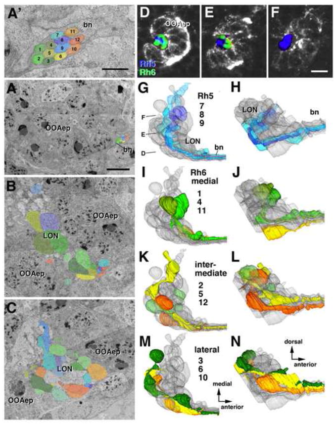
Topographically ordered projection of larval photoreceptor terminals in larval optic neuropil (LON). A–C: TEM sections of larval photoreceptor terminals and LON at different anterior-posterior levels, indicated by lines in panel H. The twelve individual photoreceptor axons are rendered in different colors and arbitrarily annotated by numbers, shown in (A′). A, A′: Bolwig’s nerve (bn) as it contacts the outer optic anlage (OOAep). Presumed Rh5-positive axons (shades of blue and purple) occupy a centro-medial position within nerve; Rh6-positive axons (shades of green to orange) are peripherally. B: Section of central part of LON. C: Section of posterior part of LON. D–F: Single sagittal confocal sections of early larval visual system (OOAep labeled by anti-DE-cadherin antibody). Planes of sections are indicated in panel G. Rh5-positive and Rh6-positive photoreceptor axons are differentially labeled by rh5-GFP and rh6-GFP. Note that Rh5-positive terminal axons (blue) occupy a posterior location in LON (arrowhead in E), and terminate mainly in proximal LON (F), which does not receive terminal axons of Rh6-positive axons (green). G–N: 3D digital models of LON (gray) and individual photoreceptor axon terminals, rendered in same colors as used in TEM images in A–C). Models of left column (G, I, K, M) present dorsal view; right column offers lateral views. G, H: three presumed Rh5-positive photoreceptor terminal axons, recognizable by their posterior position in LON (arrowhead in H) and termination in proximal (i.e., medial) LON (arrow in G). I, J: Rh6-positive receptor axons 1, 4, 11, traveling medially in Bolwig’s nerve (A′), have terminal boutons in anterior LON. K, L: Rh6-positive axons 2, 5, 12, located at intermediate levels in nerve, terminate preferentially in central domain of LON. M, N: Lateral Rh6-positive axons 3, 6, 10 terminate in posterior LON. Scale bars: 0.5μm (A′), 1 μm (A), 2 μm (B, C, D), 3 μm (D, E, F).
Synaptic connectivity of target neurons and larval PRs in the LON
Based on their characteristic point of entry into the LON, two populations of the PR target neurons can be recognized in the EM-stack, the OLPs and LNs. OLPs are the only neurons whose cell bodies are located at the lateral (external) surface of the optic lobe (arrowheads in Fig. 6A; Fig. 6F), which may be reflective of the fact that they are developmentally closely related to the outer optic anlage (Chang et al., 2003). The Bolwig’s nerve contacts the three OLP cell bodies at their medial surface (green arrowhead Fig. 6B; Fig. 6C); OLP neurites and PR axons fasciculate and enter the LON from laterally (Fig. 6B, C, F, G). The OLP neurites give off multiple, short, dendritic branches in the distal domain of the LON where they contact the PR terminal boutons (Fig. 6A, F). A significant fraction of PR terminal synapses (approximately 25%) included OLP branches as postsynaptic partners; in several cases, the same PR terminal contacted at least two OLPs.
Fig. 6.
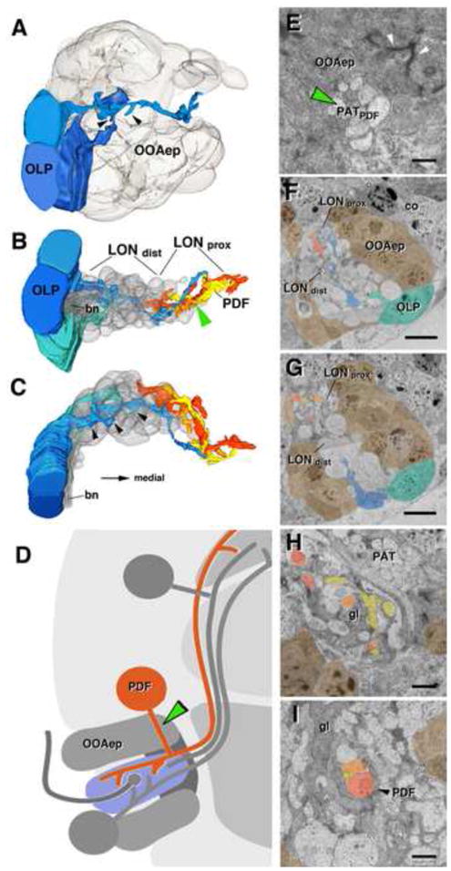
Projection neurons of the larval optic neuropil (LON). A–C: 3D digital models of LON, showing optic lobe pioneers (OLP) in shades of blue, and PDF-dendritic branches (PDF) in shades of orange, yellow and red; cellbodies of the outer optic anlage epithelium (OOAep) are translucent grey. The same colors are used in panels E–I, which show TEM sections of LON at different levels. Cell bodies of OLPs are located laterally adjacent to OOAep, flanking the entering Bolwig’s nerve (bn) (A, F, G). Note multiple short, dendritic branches of OLP fibers, preferentially in distal LON (LONdist; arrowheads in A–C). PDF neuritis are found preferentiall in the proximal LON (LONprox) and LONdist. Cell bodies of PDF neurons, shown schematically in (D), are located dorsally adjacent to the OOA. Cell body fibers form a bundle (PATpdf) that penetrates through the OOA epithelium (green arrowhead in D and E; represents a section of the OOA epithelium near its outer, apical boundary; note sub-apical adherens junctions shown by white arrowheads). Cortex neuron cell body (co) flanking the OOAep (in E). PDF-neuronal fibers form bundle in proximal LON (arrowhead in I) and extend branches throughout this compartment (B, C, F, G, H), glia cells (gl), PAT primary lineage related axon tract (I, H). Scale bars: 2 μm (F, G), 1μm (H, I, E).
The second easily identifiable set of LON neurons is the LNs. The somata of these neurons form part of the cortex and are located dorso-anteriorly of the outer optic anlage (Fig. 6D). LN axons form a bundle that penetrates the outer optic anlage epithelium and enters the proximal LON (Fig. 6B, D, green arrowhead in E). Here they form a T-junction, with one (axonal) process turning medially and exiting the LON towards the central brain, and the other (dendritic) process turning laterally (Fig. 6D). The processes of the four LNs fasciculate together (Fig. 6H, I), before profusely branching throughout the proximal domain of the LON (Fig. 6B, C, G, H). LN neurites within the LON are preferentially postsynaptic. Given that most LN neurites are found in the proximal LON, only a small number of PR output synapses (which are concentrated in the distal LON; see above) directly target LN neurons.
Non-neuronal elements of the LON
The LON is surrounded by the horseshoe-shaped outer optic anlage, which is formed by approximately 45 epithelial cells (43 in the specimen used here). outer optic anlage epithelial cells are columnar or cuboideal, and face with their apical surface outward, as evidenced by the fact that the zonula adherens, a membrane specialization surrounding the apical neck of epithelial cells, is located at the outer surface of the outer optic anlage, away from the LON (Fig. 7E, arrowheads). The outer optic anlage epithelium is in direct contact with the somata of brain neurons (apically; Fig. 7A, arrow) and the PR terminal boutons (basally; Fig. 7H, arrow), without any intervening glial layer.
Fig. 7.
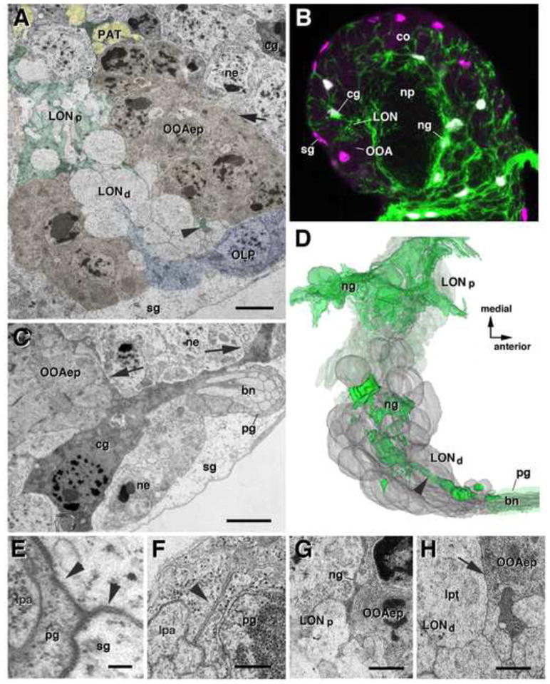
Glial element of the larval optic neuropil (LON). A: TEM section showing outer optic anlage epithelium (OOAep) and LON (LONd: distal LON; LONp: proximal LON). OOA epithelium is shaded brown; glial processes in green; optic lobe pioneers (OLP) in blue; primary axon tracts (PAT) in yellow. Arrow points at interface between apical membranes of OOA epithelial cells and neurons (ne), which lacks any glial layer. Surface glia (sg) surrounds brain surface, including OOA and optic lobe pioneers (OLP). Note cortex glial cell (cg) directly adjacent to OOA and LON, cortex neuron cell bodies (ne) (A, C). B: Single confocal section of first instar larval brain hemisphere, labeled with Nrv2-Gal4 driven GFP (green; glial cell bodies and processes) and anti-Repo (glial nuclei, magenta).. sg have repo-positive nuclei, but do not express the Nrv2 driver. Neuropil glia (ng) surrounds central brain neuropil (np), cortex (co). C: TEM section showing lateral part of the OOAep and brain cortex. Electron-dense cortex cg emits processes in between neurons of cortex (arrowheads). Note also Bolwig’s nerve (bn), surrounded by peripheral glial sheath (pg), which contacts both surface glia (sg) and cortex glia. D: 3D digital model of LON (gray) and glial processes present in LON (green). Note relative scarcity of glial lamellae in distal LON, compared to proximal LON. Arrowhead in D (and A) points at straight glial process that extends from the entry point of the bn to the center of the LON. E–H: TEM images showing details of glial structure at high magnification. E: septate junctions (arrowheads) between pg (surrounding larval photoreceptor axons, lpa) and sg. F: Auto-septate junction (arrowhead) formed by mesaxonal peripheral glia around photoreceptor axons of bn. G: Glial lamella (ng) between OOAep and neurites of LONp. H: Interface between LONd, containing boutons of larval photoreceptor terminals (lpt), and OOA epithelium. Note absence of glial sheath (arrow).
Glial processes, formed by one or two cell bodies located close to the outer optic anlage (Fig. 7B), are numerous in the proximal LON, but very sparse in the distal LON. Glial processes can be recognized by their sheath-like (lamelliform) processes that wrap individual neurites, bundles of neurites, or neuronal cell bodies. One distinguishes four main classes of glia: surface glia, surrounding the external surface of the CNS; cortex glia, forming sheaths around the neuronal cell bodies in the cortex, but also in the neuropil; neuropil glia (ng), exclusively extending processes around and within the neuropil; and peripheral glia (pg), forming sheaths around the peripheral nerves. Surface glia (sg) and peripheral glia have specialized septate junctions that form the blood-brain barrier.
A layer of surface glia extends over the lateral surface of the outer optic anlage and LON (Fig. 7A, B). The Bolwig’s nerve is wrapped by a peripheral glial sheath forming characteristic septate junctions (arrowheads in Fig. 7E, F). At the point where the nerve enters the LON, the peripheral glia sheath contacts the brain surface glia and then stops. Septate junctions connect the seam where peripheral and surface glia contact each other (arrowhead in Fig. 7E). Once inside the distal LON, PR terminal arborizations are almost completely devoid of glia (Fig. 7D, H). There is a conspicuous, bar-shaped glial process that extends all the way from the Bolwig’s nerve entry point into the center of the LON (arrowhead in Fig. 7A, D). We could not determine the location of the cell that forms this bar, as well as the other scattered glial processes within the distal LON.
In contrast to the distal LON, the proximal LON is rich in glial processes, forming a sheath around the stalk of the LON, as well as around individual small fascicles (like that of the LNs) within the proximal LON (Fig. 7A, B, D). As mentioned above, confocal images and the EM stack used here show typically two glial cell bodies closely associated with the optic lobe which, based on their location, would be classified as cortex glia (Fig. 7B, C). They can be recognized immuno-histochemically by the expression of Nrv2 and Repo (Fig. 7B). From these cells emanate lamellae that interlace among neuronal somata, from sheaths around the primary axon tracts at the junction between LON and central neuropil, and penetrate into the proximal LON (Fig. 7A, B).
DISCUSSION
Comparison of the larval and adult visual system
The numerical simplicity of the larval visual system as compared to the adult visual system is striking. Contrasting the over 6000 PRs of the compound eye the larval eye only harbours about 12 PRs. Interestingly both eyes can vary in the exact number of PRs displaying a certain degree of plasticity (Green et al., 1993; Ready et al., 1976; Sprecher et al., 2007). Axonal projections of certain PR-subtypes however are strictly stereotypically organized (Morante and Desplan, 2008). In the adult optic ganglia the outer PRs project to the lamina neuropil and inners to the medulla. Projections of Rh5- versus Rh6-PRs in the LON also occupy certain restricted domains. Interestingly axonal termini of PRs in the retina maintain the spatial information in a retinotopic map. Even though it seems unlikely that the larva perceives spatial information from one eye the projections into the LON are topically organized. Interestingly the two PR-subtypes show a clustered organization within the larval eye, so that at least at this level a spatial organization is observed. The clear bi-partite organization of the LON resembles the overall architecture of the adult optic ganglia. The distal part of the LON is innervated by PRs comparably to the lamina and “outer medulla”, while the proximal part of the LON lacks PR innervation, comparable to the lobula complex and deeper layers of the medulla (Borst, 2009). Thus both larval and adult visual neuropils consist of PR-innervated and PR-devoid components.
Network components of the larval visual network
The LON is innervated by an intriguingly small number of interneurons. Currently only 10 interneurons have been identified and described, which connect to the LON consisting of the four PDF-expressing LNs, the 5th LN (non PDF expressing), the two serotonergic neurons and the three OLPs. Here we show that the OLPs are post-synaptically to the PRs and project into the proximal LON domain as well as the adjacent central brain. These neurons are therefore likely to act as projection neurons or local interneurons possibly already allowing an integration of visual information within the LON. The LNs are postsynaptic in the LON and likely directly innervated by the PRs.
They project into dorsal domains of the central brain neuropil. PDF-expressing LNs topological and the 5th LN show distinct projection patterns (Keene et al., 2011). Moreover the octopamincergic/tyraminergic system also innervates the LON. These innervations stem from two octopaminergic cells in the SOG, each innervating the LONs of both hemispheres (M. Selcho, personal communication). Since for most network components of the LON a more detailed analysis is missing it will be of great interest to learn which components are directly innervated by the PRs, which are the local circuits, which act as projection neurons and if there is input from other neurons than the PRs. The neurites forming the LON stalk are very limited in number. Since the stalk is the main connection between the LON to the central brain we estimate the total number of LON innervating neurons not to be much larger than 15. This in-depth neuroanatomical analysis of other LON-innervating neurons, the projection pattern of LON neurons into central brain neuropil compartments will further extend our understanding of how visual information is processed.
Implication of neuroanatomy on visual behavior
The larval eye has been shown to have important functions for rapid light-dependent behaviors as well as on the entrainment of the circadian rhythm. We have recently shown that for rapid light avoidance only the blue-sensitive Rh5-PRs are required, while both PR-types are sufficient for resetting the circadian rhythm (Keene et al., 2011). A set of three neurons of the clock circuit is also essential for this behavior: the 5th LN and the two dorsal neurons 2 (DN2s). While the two DN2s do not innervate the LON the 5th LN shows large arborizations in the LON making it a likely candidate for directly acting downstream of the PRs for light avoidance. Since both PRs are cholinergic it is likely that the functional divergence is based on connectivity. It is therefore of utmost importance to investigate the distinct population of target neurons in more detail identifying differential innervation patterns of the two PR subtypes to their target neurons. Interestingly an alternative light-perception pathway has been recently identified, which does not make use of the larval eye (Diaz and Sprecher, 2011; Xiang et al., 2010). This pathway makes use of class IV multidendritic neurons tilling the body wall of the larva. How these distinct photosensory pathways converge to mediate phototactic behavior remains unknown. The serotonergic system has been shown to act in order to inhibit the response to light in certain behavioral paradigms. Genetic ablation of serotonergic neurons increases light-response in a “light on- light off” assay. Beside the function for behavior it has also been shown that certain network components are essential for the development of the circuit. The establishment of proper dendritic arbors of the serotonergic neurons into the LON dependents on the activity and presence of the larval PRs, more precisely on the larval Rh6-PR subtype (Rodriguez Moncalvo and Campos, 2009).
Highlights.
In this study we describe the organization of the larval optic neuropile.
We complement confocal microscopy with serial EM reconstruction.
We follow the LON and its neurons from early larval stages into the adult.
We characterize the sensory endings of photoreceptors in the larval optic neuropile and the LON-innervating neurons.
For the first time we conclusively show synaptic between larval PRs and two set of target neurons.
Footnotes
Publisher's Disclaimer: This is a PDF file of an unedited manuscript that has been accepted for publication. As a service to our customers we are providing this early version of the manuscript. The manuscript will undergo copyediting, typesetting, and review of the resulting proof before it is published in its final citable form. Please note that during the production process errors may be discovered which could affect the content, and all legal disclaimers that apply to the journal pertain.
References
- Borst A. Drosophila’s view on insect vision. Curr Biol. 2009;19:R36–47. doi: 10.1016/j.cub.2008.11.001. [DOI] [PubMed] [Google Scholar]
- Cardona A, Saalfeld S, Preibisch S, Schmid B, Cheng A, Pulokas J, Tomancak P, Hartenstein V. An integrated micro- and macroarchitectural analysis of the Drosophila brain by computer-assisted serial section electron microscopy. PLoS Biol. 2010;8 doi: 10.1371/journal.pbio.1000502. [DOI] [PMC free article] [PubMed] [Google Scholar]
- Campos AR, Lee KJ, Steller H. Establishment of neuronal connectivity during development of the Drosophila larval visual system. J Neurobiol. 1995 Nov;28(3):313–29. doi: 10.1002/neu.480280305. [DOI] [PubMed] [Google Scholar]
- Chang T, Shy D, Hartenstein V. Antagonistic relationship between Dpp and EGFR signaling in Drosophila head patterning. Dev Biol. 2003;263:103–13. doi: 10.1016/s0012-1606(03)00448-2. [DOI] [PubMed] [Google Scholar]
- Cook T, Pichaud F, Sonneville R, Papatsenko D, Desplan C. Distinction between color photoreceptor cell fates is controlled by Prospero in Drosophila. Dev Cell. 2003;4:853–64. doi: 10.1016/s1534-5807(03)00156-4. [DOI] [PubMed] [Google Scholar]
- Daniel A, Dumstrei K, Lengyel JA, Hartenstein V. The control of cell fate in the embryonic visual system by atonal, tailless and EGFR signaling. Development. 1999;126:2945–54. doi: 10.1242/dev.126.13.2945. [DOI] [PubMed] [Google Scholar]
- Diaz NN, Sprecher SG. Photoreceptors: unconventional ways of seeing. Curr Biol. 2011;21:R25–7. doi: 10.1016/j.cub.2010.11.063. [DOI] [PubMed] [Google Scholar]
- Green P, Hartenstein AY, Hartenstein V. The embryonic development of the Drosophila visual system. Cell Tissue Res. 1993;273:583–98. doi: 10.1007/BF00333712. [DOI] [PubMed] [Google Scholar]
- Helfrich-Forster C. Development of pigment-dispersing hormone-immunoreactive neurons in the nervous system of Drosophila melanogaster. J Comp Neurol. 1997;380:335–54. doi: 10.1002/(sici)1096-9861(19970414)380:3<335::aid-cne4>3.0.co;2-3. [DOI] [PubMed] [Google Scholar]
- Helfrich-Forster C, Edwards T, Yasuyama K, Wisotzki B, Schneuwly S, Stanewsky R, Meinertzhagen IA, Hofbauer A. The extraretinal eyelet of Drosophila: development, ultrastructure, and putative circadian function. J Neurosci. 2002;22:9255–66. doi: 10.1523/JNEUROSCI.22-21-09255.2002. [DOI] [PMC free article] [PubMed] [Google Scholar]
- Johnson EL, 3rd, Fetter RD, Davis GW. Negative regulation of active zone assembly by a newly identified SR protein kinase. PLoS Biol. 2009;7:e1000193. doi: 10.1371/journal.pbio.1000193. [DOI] [PMC free article] [PubMed] [Google Scholar]
- Malpel S, Klarsfeld A, Rouyer F. Larval optic nerve and adult extra-retinal photoreceptors sequentially associate with clock neurons during Drosophila brain development. Development. 2002;129:1443–53. doi: 10.1242/dev.129.6.1443. [DOI] [PubMed] [Google Scholar]
- Mazzoni EO, Desplan C, Blau J. Circadian pacemaker neurons transmit and modulate visual information to control a rapid behavioral response. Neuron. 2005;45:293–300. doi: 10.1016/j.neuron.2004.12.038. [DOI] [PubMed] [Google Scholar]
- Monastirioti M. Biogenic amine systems in the fruit fly Drosophila melanogaster. Microsc Res Tech. 1999;45:106–21. doi: 10.1002/(SICI)1097-0029(19990415)45:2<106::AID-JEMT5>3.0.CO;2-3. [DOI] [PubMed] [Google Scholar]
- Morante J, Desplan C. Building a projection map for photoreceptor neurons in the Drosophila optic lobes. Semin Cell Dev Biol. 2004;15:137–43. doi: 10.1016/j.semcdb.2003.09.007. [DOI] [PubMed] [Google Scholar]
- Morante J, Desplan C. The color-vision circuit in the medulla of Drosophila. Curr Biol. 2008;18:553–65. doi: 10.1016/j.cub.2008.02.075. [DOI] [PMC free article] [PubMed] [Google Scholar]
- Mukhopadhyay M, Campos AR. The larval optic nerve is required for the development of an identified serotonergic arborization in Drosophila melanogaster. Dev Biol. 1995;169:629–43. doi: 10.1006/dbio.1995.1175. [DOI] [PubMed] [Google Scholar]
- Nassif C, Noveen A, Hartenstein V. Embryonic development of the Drosophila brain. I Pattern of pioneer tracts. J Comp Neurol. 1998 Dec 7;402(1):10–31. [PubMed] [Google Scholar]
- Ngo KT, Wang J, Junker M, Kriz S, Vo G, Asem B, Olson JM, Banerjee U, Hartenstein V. Concomitant requirement for Notch and Jak/Stat signaling during neuro-epithelial differentiation in the Drosophila optic lobe. Dev Biol. 2010;346:284–95. doi: 10.1016/j.ydbio.2010.07.036. [DOI] [PMC free article] [PubMed] [Google Scholar]
- Ready DF, Hanson TE, Benzer S. Development of the Drosophila retina, a neurocrystalline lattice. Dev Biol. 1976;53:217–40. doi: 10.1016/0012-1606(76)90225-6. [DOI] [PubMed] [Google Scholar]
- Rodriguez Moncalvo VG, Campos AR. Genetic dissection of trophic interactions in the larval optic neuropil of Drosophila melanogaster. Dev Biol. 2005;286:549–58. doi: 10.1016/j.ydbio.2005.08.030. [DOI] [PubMed] [Google Scholar]
- Rodriguez Moncalvo VG, Campos AR. Role of serotonergic neurons in the Drosophila larval response to light. BMC Neurosci. 2009;10:66. doi: 10.1186/1471-2202-10-66. [DOI] [PMC free article] [PubMed] [Google Scholar]
- Sanes JR, Zipursky SL. Design principles of insect and vertebrate visual systems. Neuron. 2010;66:15–36. doi: 10.1016/j.neuron.2010.01.018. [DOI] [PMC free article] [PubMed] [Google Scholar]
- Schmid B, Schindelin J, Cardona A, Longair M, Heisenberg M. A high-level 3D visualization API for Java and ImageJ. BMC Bioinformatics. 2010;11:274. doi: 10.1186/1471-2105-11-274. [DOI] [PMC free article] [PubMed] [Google Scholar]
- Sprecher SG, Desplan C. Switch of rhodopsin expression in terminally differentiated Drosophila sensory neurons. Nature. 2008;454:533–7. doi: 10.1038/nature07062. [DOI] [PMC free article] [PubMed] [Google Scholar]
- Sprecher SG, Pichaud F, Desplan C. Adult and larval photoreceptors use different mechanisms to specify the same Rhodopsin fates. Genes Dev. 2007;21:2182–95. doi: 10.1101/gad.1565407. [DOI] [PMC free article] [PubMed] [Google Scholar]
- Sprecher SG, Urbach R, Technau GM, Rijli FM, Reichert H, Hirth F. The columnar gene vnd is required for tritocerebral neuromere formation during embryonic brain development of Drosophila. Development. 2006;133:4331–9. doi: 10.1242/dev.02606. [DOI] [PubMed] [Google Scholar]
- Suloway C, Pulokas J, Fellmann D, Cheng A, Guerra F, Quispe J, Stagg S, Potter CS, Carragher B. Automated molecular microscopy: the new Leginon system. J Struct Biol. 2005;151:41–60. doi: 10.1016/j.jsb.2005.03.010. [DOI] [PubMed] [Google Scholar]
- Tahayato A, Sonneville R, Pichaud F, Wernet MF, Papatsenko D, Beaufils P, Cook T, Desplan C. Otd/Crx, a dual regulator for the specification of ommatidia subtypes in the Drosophila retina. Dev Cell. 2003;5:391–402. doi: 10.1016/s1534-5807(03)00239-9. [DOI] [PubMed] [Google Scholar]
- Tix S, Minden JS, Technau GM. Pre-existing neuronal pathways in the developing optic lobes of Drosophila. Development. 1989;105:739–46. doi: 10.1242/dev.105.4.739. [DOI] [PubMed] [Google Scholar]
- Xiang Y, Yuan Q, Vogt N, Looger LL, Jan LY, Jan YN. Light-avoidance-mediating photoreceptors tile the Drosophila larval body wall. Nature. 2010;468:921–6. doi: 10.1038/nature09576. [DOI] [PMC free article] [PubMed] [Google Scholar]
- Younossi-Hartenstein A, Nguyen B, Shy D, Hartenstein V. Embryonic origin of the Drosophila brain neuropil. J Comp Neurol. 2006;497:981–98. doi: 10.1002/cne.20884. [DOI] [PubMed] [Google Scholar]


