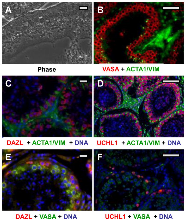
Figure 2. Localization of SPG antigens in adult canine testis.
(A) Phase image of tissue after deparaffinization. (B–D) Duel label probes in which putative SPG antigens (red) and somatic cell antigens (green) were marked. Putative SPG antigens (red) were probed with either rabbit (B) or goat (C,D) primary antibodies as indicated, followed by the appropriate Alexa-594 conjugated donkey secondary antibody. Somatic cells (green) were labeled with mixed mouse antibodies against alpha actin and vimentin, followed by an anti-mouse Alexa 488 conjugate. (E,F) Duel label probes for two SPG antigens. Note that the UCHL1 staining intensity in F is much lower than in D, showing only the most strongly positive cells. Bars = 150 um.

