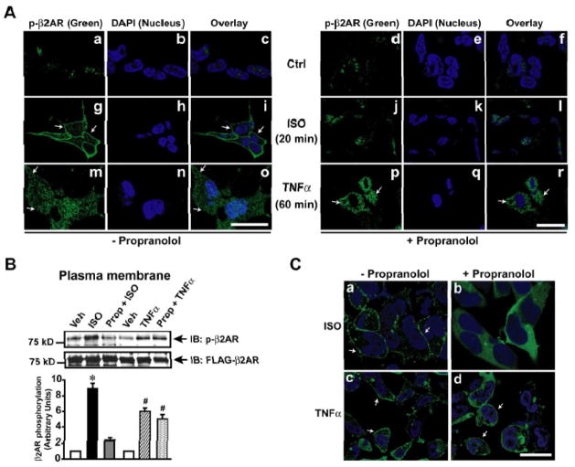
Figure 4.
TNFα mediated βAR desensitization is agonist independent. (A) FLAG-β2AR phosphorylation was visualized by confocal microscopy using anti-phospho-β2AR antibody (green) following ISO stimulation or TNFα treatment (60 minutes) in the presence and absence of β-blocker propranolol. Nucleus was visualized by DAPI (blue) staining. Scale bar: 10 μm. (B) Immunoblots of phospho-β2AR and FLAG-β2AR following TNFα treatment of HEK-FLAG-β2AR cells in the presence or absence of propranolol. Densitometric analysis of the same is shown on the lower panel. (n=4), *p< 0.001 versus Veh, #p< 0.001 versus Veh. (C) β-Arrestin (green) recruitment to the plasma membrane was visualized by confocal microscopy using double stable cells expressing GFP-β-Arrestin and HA-β2AR following ISO or TNFα in the presence or absence of β-blocker propranolol. Scale bar: 10 μm

