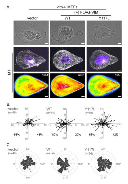
Figure 5. VIF are essential for MT organization and cell polarity.
VIF (+FLAG-Vimentin WT), the ULF mutant (+FLAG-Vimentin Y117L) or a vector control was expressed in vim-/- MEFs and the cells then were seeded on teardrop shape micropatterns. Immunofluorescence/phase images of MT and nuclei (blue) were collected and cell population heat maps of MT were generated in Image J (A). Cell polarity was analyzed by plotting vectors for nucleus center and centrosome center calculated in Image J (B) (see Materials and Methods; also Figure 4). The vectors were converted into angles and polar coordinate plots were generated for each system (C) (see Materials and Methods; also Figure 4). n = number of cells used to generate the data. Scale bar = 5 μm.

