Abstract
While preconditioning is induced before stroke onset, ischemic postconditioning (IPostC) is performed after reperfusion, which typically refers to a series of mechanical interruption of blood reperfusion after stroke. IPostC is known to reduce infarction in wild type animals. We investigated if IPostC protects against brain injury induced by focal ischemia in T-cell-deficient nude rats and to examine its effects on Akt and the mammalian target of rapamycin (mTOR) pathway. Although IPostC reduced infarct size at 2 days post-stroke in wild type rats, it did not attenuate infarction in nude rats. Despite the unaltered infarct size in nude rats, IPostC increased levels of phosphorylated Akt (p-Akt) and Akt isoforms (Akt1, Akt2, Akt3), and p-mTOR, p-S6K and p-4EBP1 in the mTOR pathway, as well as GAP-43, both in the peri-infarct area and core, 24 hours after stroke. IPostC improved neurological function in nude rats 1–30 days after stroke and reduced the extent of brain damage 30 days after stroke. The mTOR inhibitor rapamycin abolished the long-term protective effects of IPostC. We determined that IPostC did not inhibit acute infarction in nude rats but did provide long-term protection by enhancing Akt and mTOR activity during the acute post-stroke phase.
Keywords: Stroke, Ischemic postconditioning, T cells, Akt, mTOR
Introduction
Ischemic postconditioning (IPostC) refers to brief reperfusion interruptions that reduce infarction after cerebral ischemia and mitigate neurological deficits (Zhao 2009, Zhao et al. 2006). This contrasts with ischemic preconditioning (Zhao et al. 2003) and has demonstrated comparable protective effects (Zhao 2009, Zhao 2011). The underlying protective mechanisms of IPostC are associated with its ability to attenuate production of free radicals (Zhao 2009, Zhao et al. 2006), to inhibit apoptotic cell signaling pathways (Xing et al. 2008) and to promote cell-survival signaling pathways (Wang et al. 2008) such as the Akt pathway(Pignataro et al. 2008) (Zhao 2009, Zhao et al. 2006, Gao et al. 2008b).
T cells are active in adaptive immunity and play a central role in cell-mediated immunity. Neutrophils and macrophages have been thought to participate in innate immunity and contribute to brain injury induced by stroke but T cells have also been recently shown to have detrimental effects in the ischemic brain (Kleinschnitz et al. 2010, Yilmaz et al. 2006, Liesz et al. 2009, Hurn et al. 2007). T cells, like macrophages and neutrophils, infiltrate the ischemic brain after stroke (Iadecola & Anrather 2011) yet T cell deficits reduce infarct sizes (Hurn et al. 2007). T cell subsets play various roles in stroke. Deficits of either CD4 or CD8 T cells result in smaller infarct sizes (Kleinschnitz et al. 2010, Yilmaz et al. 2006) while deficits of regulatory T cells (Treg) lead to enlarged and delayed infarct sizes (Liesz et al. 2009). Our laboratory recently showed reduced-infarction in Th1-deficient mice and increased infarction in Th2-deficient mice (Gu et al. 2012). Because IPostC protects the ischemic brain by blocking the inflammatory response, and T cells mediate inflammation, we hypothesized that T cells are crucial for the protective effects of IPostC. Speculating that IPostC reduces infarction by blocking T cell function, we predicted that IPostC would not reduce infarct sizes in T-cell-deficient nude rats.
As we have reviewed, the protective mechanisms of IPostC include multiple cell-signaling pathways such as the Akt pathway (Zhao 2009, Zhao 2011, Zhao 2013). IPostC has been shown to promote Akt activity and, conversely, inhibition of Akt blocks the protective effects of IPostC (Gao et al. 2008b, Pignataro et al. 2008). Akt directly and indirectly activates mTOR activity, which then promotes the downstream molecules 4E-BP1 and S6K to enhance cell differentiation, growth, and survival (Sabbah et al. 2011, Martelli et al. 2010). The mTOR pathway's involvement in IPostC has not been reported.
Our study investigated the acute and long-term protective effects of IPostC in T-cell-deficient nude rats. We examined the acute effects of IPostC on Akt activity, including Akt phosphorylation and protein levels of the Akt isoforms Akt 1, Akt 2 and Akt 3 as well as phosphorylation of mTOR, 4E-BP1 and S6K. Whether or not mTOR inhibition blocks the protective effects of IPostC was also studied.
Methods
Animal experiments were conducted according to the protocols approved by the Stanford Institutional Animal Care and Use Committee and the NIH Guidelines for Care and Use of Laboratory Animals. Animals were housed under a 12:12 hour light/dark cycle with food and water available ad libitum.
Focal cerebral ischemia and postconditioning
Focal ischemia was induced by 30 minutes of transient bilateral common carotid arteries (CCAs) occlusion and permanent distal middle cerebral artery (MCA) occlusion in male Sprague–Dawley rats (230-250g, Charles River Laboratories International, Wilmington, MA, USA) and T-cell-deficient rats (230-250g, RNU rats, Charles River Laboratories International, Wilmington, MA, USA)(Zhao et al. 2006). Rats were anesthetized by 5% isoflurane and maintained by 2–3% isoflurane. Core body temperature was monitored with a rectal probe and kept at 37°C throughout the experiment. A ventral midline incision was made and the 2 CCAs were isolated. A 2 cm vertical scalp incision was made midway between the left eye and ear. The temporalis muscle was bisected and a 2 mm burr hole was made at the junction of the zygomatic arch and squamous bone. The distal MCA was exposed and cauterized above the rhinal fissure at the intersection of the lateral vein and MCA. The CCAs were occluded for 30 minutes with suture tightening. IPostC was conducted immediately after reperfusion by 3 cycles of 30 second reperfusions and 10 second occlusions of the bilateral CCAs as described (Zhao et al. 2006).Blood gas, heart rate, respiratory rate and temperature were monitored throughout the surgery and kept within physiological ranges.
General histology and infarct size measurement
Infarction was measured by 2,3,5-triphenyl-2H-tetrazolium chloride (TTC) or cresyl violet staining. Wild-type and nude rats sacrificed at 2 days or 30 days (with or without rapamycin pre-treatment) after stroke were perfused transcardially with cold 0.9% saline followed by 4% paraformaldehyde (PFA) in PBS (pH 7.4). Brains from wild-type and nude rats sacrificed 2 days after stroke were sectioned into 5 coronal blocks rostral (level 1) to caudal (level 5) and stained with 1% TTC solution. Brains from nude rats sacrificed 30 days (with or without rapamycin pre-treatment) after stroke were post-fixed in 4% PFA, 20% sucrose for 24 hours and sectioned into 30μm slices and mounted onto glass slides using a cryostat. Slices were stained with cresyl violet. The damaged or lost areas of the infarcted cortex, as indicated by TTC or cresyl violet were measured by a technician blinded to the animal's condition, normalized to the contralateral cortex and expressed as a percentage, as described previously (Zhao et al. 2006, Gao et al. 2008a, Gao et al. 2008b).
Behavioral testing
The home cage, vibrissa-elicited limb use and postural reflex tests were used to quantify motor asymmetry caused by a unilateral cortical stroke and performed by a technician blinded to the experimental conditions (Gao et al. 2008b, Ren et al. 2008, Zhao et al. 2005). Nude rats were divided into 4 groups: (1) sham surgery without ischemia; (2) ischemia only for 30 minutes (control ischemia); (3) ischemia plus postconditioning without rapamycin pre-treatment; (4) ischemia plus postconditioning with rapamycin pre-treatment. Rats were handled for 3 days before stroke, and baseline tested on the day before surgery. All behavior tests were performed at 1, 2, 3, 7, 14, 21 and 30 days post-stroke by 2 technicians blinded to the experimental conditions. The methods used in the home cage, vibrissa-elicited limb use, postural reflex and vibrissa-elicited forelimb placement tests were detailed in our previous studies (Gao et al. 2008b, Ren et al. 2008, Zhao et al. 2005).
Protein preparation of in vivo experiments for western blotting
Ischemic nude rat brains corresponding to the peri-infarct area and ischemic core were harvested at 1, 5, 9 and 24 hours after stroke onset in order to investigate the effects of in vivo stroke on protein expression of pAkt, Akt, Akt isoforms, pmTOR, mTOR, pS6K, S6K, p4EBP1 and 4EBP1, including pmTOR and pS6K expression, as well as GAP-43, after rapamycin pre-treatment. The area of peri-infarction refers to the ischemic tissue spared by postconditioning while the ischemic core refers to the infarcted region in the ischemic brain treated with IPostC, as described (Gao et al. 2008b). Brain tissue from animals that underwent sham surgery without ischemia was also prepared for western blotting. Whole cell protein was extracted from the fresh brain tissue, and a western blot was performed as described with modification (Zhao et al. 2005, Gao et al. 2008b). Briefly, brain tissue was cut into small pieces and homogenized in a glass homogenizer using 7 volumes of the cold cell extraction buffer (Catalog# FNN0001, Invitrogen, OR, USA), containing 1mM PMSF and a protease inhibitor cocktail (1:20, Catalog# P-2714, Sigma, St. Louis, MO, USA). The homogenate was centrifuged at 13000 rpm for 20 minutes at 4°C, and the supernatant was removed for protein detection.
Western blot procedures and antibodies
Twenty micrograms of protein were loaded into each lane and subjected to SDS-PAGE using 4–15% Ready Gel (Catalog #L050505A2; Bio-Rad, Hercules, CA) at 200V for 45 minutes. Protein bands were transferred to polyvinylidene fluoride membranes (Millipore, Bedford, MA, USA) at 100 V for 2 hours. Membranes were incubated overnight with primary antibodies at 4°C followed by Alexa Fluor 488 donkey anti-rabbit or anti-mouse IgG secondary antibody (1:5000, Invitrogen, Eugene, OR, USA) for 1 hour in a dark room. Table 1 lists each primary antibody, manufacturer, catalog number and detection method used. Membranes were scanned using Typhoon Trio (GE Healthcare). Optical densities of all protein bands were analyzed using IMAGEQUANT 5.2 software (GE Healthcare). Samples from sham surgery were used as controls for experimental samples. All samples were run on the same gel.
Table 1.
Antibodies, their concentrations and manufacturers used in the present studies
| Antibodies | Source | Dilutions | Manufacturer | Catalog# | Application |
|---|---|---|---|---|---|
| P-Akt (Ser473) | Rabbit | 1:1000 | Cell Signaling | 9271 | WB |
| Akt | Rabbit | 1:1000 | Cell Signaling | 9272 | WB |
| Akt1 | Rabbit | 1:1000 | Cell Signaling | 2938 | WB |
| Akt2 | Rabbit | 1:1000 | Cell Signaling | 3063 | WB |
| Akt3 | Rabbit | 1:500 | Cell Signaling | 3788 | WB |
| P-mTOR (Ser2448) | Rabbit | 1:200/1:1000 | Cell Signaling | 2971 | IF/WB |
| mTOR | Rabbit | 1:1000 | Cell Signaling | 2983 | WB |
| P-S6K p70 (Ser371) | Rabbit | 1:500 | Cell Signaling | 9208 | WB |
| S6K p70 | Rabbit | 1:500 | Cell Signaling | 9202 | WB |
| P-4EBP1 | Rabbit | 1:500 | Cell Signaling | 9456 | WB |
| 4-EBP1 | Rabbit | 1:500 | Cell Signaling | 9452 | WB |
| GAP-43 | Rabbit | 1:500 | Cell Signaling | 5307 | WB |
| β-actin | Mouse | 1:3000 | Sigma | A-5441 | WB |
IF=Immunofluorescence, WB=Western Blots
Drug delivery
To study whether rapamycin, an mTOR inhibitor, abolishes the long term protection of postconditioning in nude rats, rapamycin (Calbiochem, Billerica, MA, USA) was dissolved in PBS to a final concentration of 0.1 mM. 10 μl of rapamycin, or vehicle, was infused into the ventricular space ipsilateral to the ischemia, 1hour before ischemia onset, as described (Zhao et al. 2005), at the coordinate point from bregma: anteroposterior, 0.92 mm; mediolateral, 1.5 mm; dorsoventral, 3.5 mm. Infarct size and behavior tests were performed 30 days after stroke as described above.
Immunofluorescence staining and confocal microscopy
For immunofluorescent staining of brain sections, sham nude rats, ischemic nude rats, ischemic nude rats with postconditioning and ischemic nude rats pre-treated by rapamycin and with postconditioning were anesthetized and perfused transcardially with 0.9% saline followed by 4°C PFA in PBS (pH 7.4), then fixed with 4% PFA for 24 hours. Free floating 30 um sections were cut on a cryostat and stored in antifreeze solution at -20°C. Sections were incubated in blocking solution containing 5% horse serum (Sigma, St. Louis, MO, USA) and 0.3% Triton X-100 in PBS for 2 hours at RT followed by incubation in primary antibodies (Table 1) at 4°C overnight. The next day, sections were washed with PBS and incubated for 1 hour at RT (light shielded) in secondary antibody (Alexa Fluor 647 donkey anti-rabbit or anti-mouse IgG, 1 : 200, Invitrogen, Eugene, OR, USA) and cover-slipped with 1 drop of Vectashield mounting medium with 4', 6-diamidino-2-phenylindole (DAPI, Vector Laboratories, Burlingame, CA, USA). Immunofluorescent staining was examined using a Zeiss confocal microscope (Zeiss LSM 510, Thornwood, NY, USA).
Statistical analysis
GraphPad Prism 5.0 software (GraphPad Software, La Jolla, CA) was used for statistical analyses. For infarction analyses, one-way ANOVA was used followed by the fisher least significant difference post hoc test. For western blots, two-way ANOVA was used to compare the optical densities of all protein levels of the peri-infarct area versus the core at the stated time points, followed by the fisher least significant difference post hoc test. Two-way ANOVA was used to analyze various protein bands between postconditioning, postconditioning with rapamycin pre-treatment and control ischemia. For behavioral tests, one-way repeated measures ANOVA was used to compare a test at different time points in the same group, and two-way ANOVA was used to compare tests between postconditioning, postconditioning with rapamycin pre-treatment, control ischemia and sham groups, followed by the fisher least significant difference post hoc test. Tests were considered significant at P values < 0.05. Data are presented as mean±SEM.
Results
IPostC reduced infarct sizes in wild-type but not in nude rats measured 2 days post-stroke
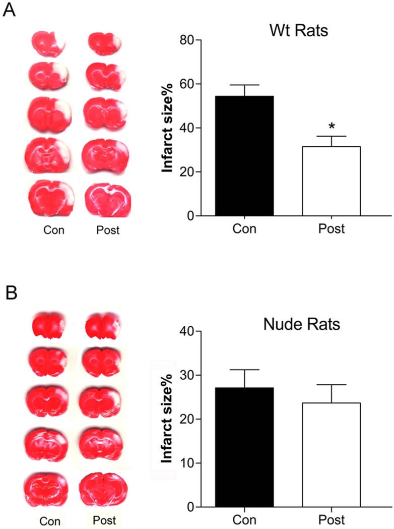
As we have reported previously (Gao et al. 2008b, Ren et al. 2008, Zhao et al. 2006), we confirmed in this study that IPostC reduced infarct sizes in wild-type rats measured 2 days post-stroke (Figure 1). In a result differing from that in wild-type rats, the cortical infarction in nude rats induced by stroke was 27.1±4.3% in control ischemia vs 23.7±4.2% in postconditioning (N=6). A statistical analysis predicts that a total of 89 animals is required to reach P<0.05, thus we conclude that there is no significant difference between the two groups (Figure 1). In the control group without IPostC, nude rat infarction was smaller than that in wild-type rats (P=0.023, N=6).
Figure 1. IPostC reduced acute infarct sizes in wild-type rats but not in nude rats.
Stroke was induced by transient occlusion of bilateral CCA for 30 min combined with permanent distal MCA occlusion. Infarct sizes were measured 2 days post-stroke by TTC staining. A. Representative TTC staining for infarction and average infarct sizes in wild-type rats, * vs control. B. Same for nude rats. P < 0.05. N = 8/group.
Akt and mTOR activities were enhanced by IPostC
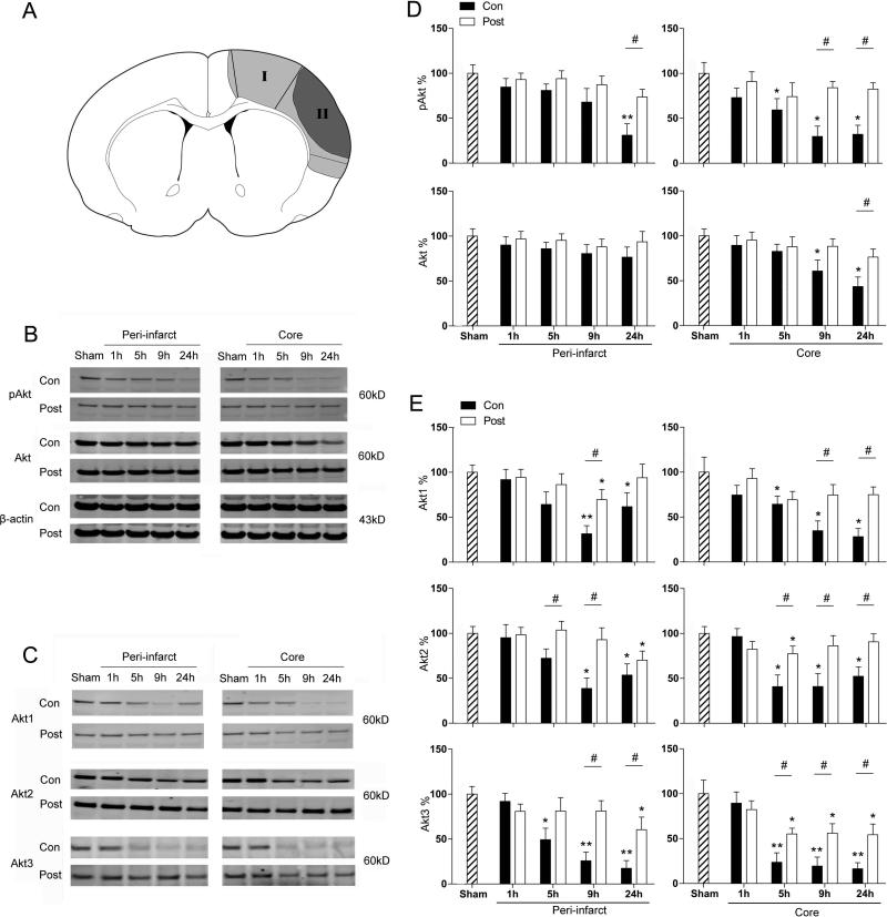
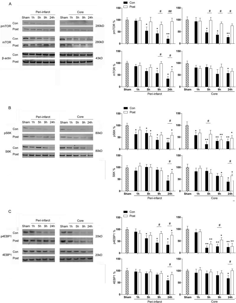
Despite the unaltered infarct size, we hypothesized that IPostC may improve pathological outcomes of neuronal survival at molecular levels. Both peri-infarct region and ischemic core were dissected for Western blotting (Fig. 2). Indeed, the results showed that protein levels of p-Akt (phosphorylated Akt) and Akt isoforms (Akt1, Akt2, Akt3) in the Akt pathway were reduced after stroke, and IPostC significantly increased these protein levels at certain time points, in both the ischemic core and peri-infarct region (Figure 2). Similarly, p-mTOR, p-S6K, p-4EBP1 and total proteins of mTOR, S6K and 4E-BP1 in the mTOR pathway were also reduced by stroke both in the peri-infarct area and in the core after stroke (P<0.05), and IPostC attenuated these reductions (Figure 3).
Figure 2. The effects of IPostC on protein levels of p-Akt, Akt and Akt isoforms.
Rat brains were harvested at 1, 5, 9, and 24h post-stroke for western blotting. A. A diagram shows that peri-infarct area and ischemic core were dissected for western blot. The peri-infarct area (I) is defined as the ischemic region spared by IPostC, whereas the ischemic core (II) is defined as the infarcted ischemic region receiving IPostC. B. Representative protein bands of pAkt, Akt and beta-actin in rat brains after stroke with and without postconditioning. C. Representative protein bands of Akt isoforms with bar graphs that represent average protein levels. D and E. The bar graphs represent average protein levels from ischemic brains.* and ** vs sham, P < 0.05 and 0.01, respectively. # is P < 0.05, between the 2 indicated groups. N = 6–8/group.
Figure 3. The effects of IPostC on protein levels in the mTOR pathway.
A. Representative protein bands of pmTOR, mTOR and beta-actin. The bar graphs show the statistical results. B. Representative protein bands and bar graphs for statistical results of pS6K, S6K and beta-actin. C. Representative protein bands and bar graphs for statistical results of p4EBP1, 4EBP1 and beta-actin. * and ** vs sham, P < 0.05 and 0.01, respectively; #, ##, between indicated 2 groups, P < 0.05. 0.01, respectively. N = 6–8/group
IPostC attenuated neurological deficits and long-term brain injury, which were blocked by mTOR inhibition
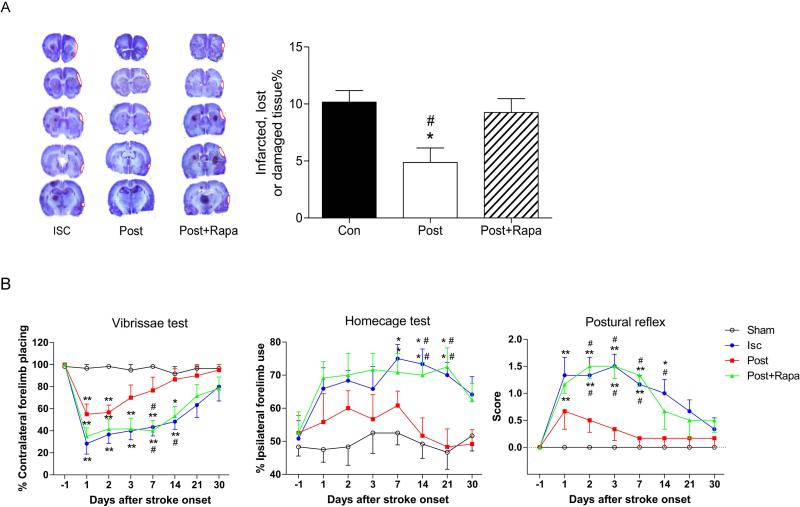
Since IPostC improved protein expression of the Akt/mTOR cell survival signaling pathways, we tested our hypothesis that IPostC improves neurological function and inhibits the effects of stroke on brain injury up to 30 days post-stroke. The results of cresyl violet staining showed that IPostC reduced brain damage size from 10.2±1.4% to 4.9±1.6% (N=6, P<0.05) measured 30 days post-stroke (Figure 4). The results of behavioral tests showed that IPostC improved neurological function in nude rats when measured from 1 to 30 days after stroke (N=6, P<0.05). In addition, injection of the mTOR inhibitor rapamycin abolished the long-term protective effects of postconditioning (Figure 4).
Figure 4. mTOR inhibition blocked long-term protective effects of postconditioning on brain injury and behavioral deficits after stroke.
A. Representative Cresyl violet staining for brain injury measured 30 days after stroke. Average infarct sizes are shown in the bar graph. Rapamycin, the mTOR inhibitor, was shown to reverse the protective effects of postconditioning. B. Results of vibrissae, home cage and postural reflex tests. Postconditioning promoted behavioral recovery, which was blocked by rapamycin. N = 6–8/group.
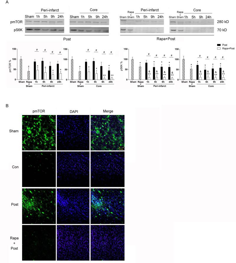
Western blot results confirmed that rapamycin injection reduced protein levels of pmTOR and pS6K in brains with or without stroke plus IPostC (Figure 5A). Confocal microscopy showed that pmTOR expression in the peri-infarct area receiving IPostC was reduced by rapamycin (Figure 5B).
Figure 5. Effects of rapamycin on protein levels of pmTOR and pS6K.
A. Western blots show that rapamycin reduced protein levels of pmTOR and pS6K, represented by protein bands of pmTOR and pS6K in both the peri-infarct area and the ischemic core. Animals receiving IPostC were treated with or without rapamycin. The sham group indicates that animals received sham surgery only; the Rapa+Sham group indicates that animals received sham surgery plus rapamycin injection. Rapamycin injection reduced protein levels of both pmTOR and mS6K in the group receiving sham surgery alone with or without stroke and IPostC.* and ** vs sham, P < 0.05 and 0.01, respectively; & vs rapa + sham, P < 0.05; # P < 0.05, between indicated 2 groups. B. Confocal microscopy shows representative immunofluorescent staining of pmTOR and pS6K. Imaged in a peri-infarct region 24 hours post-stroke with IPostC, with and without rapamycin. Scale bar, 60μM.
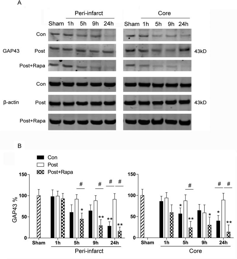
As we have found that IPostC did not alter acute infarction but still improved neurological function, we further measured protein levels of presynaptic growth associated Protein 43 (GAP43), which is associated with brain plasticity and neurological function. The results showed that protein levels of GAP43 were immediately decreased after stroke, which were maintained by IPostC treatment. However, rapamycin administration abolished the protective effect of IPostC (Fig.6).
Fig. 6. The acute effects of rapamycin injection on protein levels of GAP43 after stroke.
A. Representative protein bands of GAP43. Protein of β-actin was probed to show even loading of proteins for western blotting. B. Bar graphs show average protein optical densities, which were normalized to sham groups, and expressed as percentages. The protein levels were decreased post-stroke, but IPostC maintained its levels. Rapamycin administration abolished the protective effects of IPostC. *, ** vs sham, P<0.05, 0.01, respectively; #, between indicated two groups, P<0.05. N=6-8/group.
Discussion
We found that IPostC did not reduce infarct sizes measured 2 days post-stroke in nude rats but attenuated neurological deficits and inhibited long-term brain injury measured 30 days post-stroke. Our data implies that T cells are a factor involved in the protective effects of IPostC. Despite unaltered infarct sizes, we showed that IPostC attenuated reductions in pAkt levels, Akt isoform proteins, as well as pmTOR, mTOR, p4E-BP1, and pS6K, as well as GAP43 in the acute ischemic brain. Because rapamycin, an mTOR pathway inhibitor, blocked the long-term protective effects of IPostC on brain injury and neurological deficits, we conclude that IPostC provides protection by promoting mTOR activity in nude rats.
In another set of experiments, we have proven that mTOR also plays a critical role in brain damage after stroke and contributes to the protective effects of IPostC in wild type rats (unpublished data). As consistent with the current study, we found that IPostC attenuated stroke-induced reductions in protein phosphorylation in the mTOR pathway as measured from 1hour to 24hours after stroke. Both rapamycin and mTOR shRNA injection worsened infarction and inhibited protection by IPostC. In addition, S6K gene transfer protected against brain injury. Furthermore, we found that mTOR inhibition played a critical role in long-term protection of IPostC, as rapamycin abolished the long-term protection afforded by IPostC on neurological functions and injury size as measured 3 weeks post-stroke. Lastly, we examined if early modulation of mTOR activity had long-term effects on protein levels of p-mTOR, p-S6K and p-4EBP1 in the mTOR pathway. We found that IPostC improved protein levels in the mTOR pathway at least 1 week after stroke, but rapamycin administration resulted in significant reductions in these proteins both at 1 and 3 weeks with IPostC. Taken together, mTOR appears to play similar roles in the protective effects of IPostC in wild type rats compared with nude rats. Nevertheless, the relationship between T cells and changes in Akt and mTOR pathways after stroke remain elusive. Since T cells are involved in inflammation after stroke (Jin et al. 2010, Kleinschnitz et al. 2010), and inflammation may affect Akt and mTOR activity (Guha & Mackman 2002, Weichhart et al. 2011), T cells may affect the Akt and mTOR pathways via inflammatory response. Future studies are required to compare changes in these cell signaling pathways between nude rats and wild type rats.
In this current study, our primary purpose is to address whether IPostC has similar protective effects in nude rats whose T cells are not present, as T cells have been shown to contribute to brain injury (Gu et al. 2012, Xiong et al. 2013). We and others have previously shown that IPostC reduced infarct sizes measured 2 to 3 days after stroke in wild-type rats, and this protective effect lasted 1 month (Gao et al. 2008b, Ren et al. 2008, Zhao et al. 2006). In the T cell-deficient nude rats in our current study, however, IPostC did not reduce infarct sizes measured at 2 days, but reduced brain injury size measured at 30 days post-stroke. These results suggest that T cells are crucial to modulating the acute protective effects of IPostC, and that cell types other than T cells may mediate the long-term protective effects of IPostC.
We and others have previously shown that Akt contributes to the protective effects of IPostC in wild-type rats, as IPostC promotes pAkt levels and PI3K/Akt inhibitors block the protective effects of IPostC (Pignataro et al. 2008, Gao et al. 2008a). Although in this study IPostC improved Akt activity measured 24 hours after stroke, this activity apparently did not reduce acute infarct size, but more likely contributed to improved neurological function and long-term outcomes. As the mTOR pathway is downstream of the Akt pathway, we further examined the effects of IPostC on critical proteins in the mTOR pathway. Although some studies have reported on mTOR's involvement in ischemic injury, results regarding mTOR's role in neuroprotection are controversial. For example, inhibition or deletion of PTEN was shown to promote axonal outgrowth or inhibit neuronal death by enhancing mTOR activity(Mao et al. 2012, Shi et al. 2011), and t-PA increased neuronal survival by activating mTOR pathways, resulting in improved energy availability (Wu et al.). mTOR activity was also shown to promote astrocyte survival after cerebral ischemia (Pastor et al. 2009). However, mTOR has also been shown to promote post-ischemic long-term potentiation, which causes increased neuronal death, and mTOR inhibition by rapamycin reduced neuronal damage (Ghiglieri et al. 2010). With regard to the neuroprotective effects of mTOR, we have shown for the first time that IPostC increases protein activity in the mTOR pathway, including pmTOR, p4E-BP1 and pS6K proteins in nude rats. Because injection of the mTOR inhibitor rapamycin abolished the protective effects of IPostC on long-term brain injury size and neurological deficits, we conclude that mTOR is a crucial protein for the long-term protective effects of IPostC.
As IPostC improved neurological function measured one day post-stroke, we further hypothesized that it improves functional protein related with neuroprotection. GAP43 is expressed on the presynaptic terminals, and is a known growth and plasticity protein (Carmichael 2003). Its overexpression has been associated with improved brain recovery and learning ability following treatment with neuroprotectants after stroke (Yoon et al. 2012). We found that IPostC improved protein levels of GAP43, and rapamycin administration abolished the protective effects of IPostC on GAP43 levels. These results further strengthened why IPostC did not reduce infarction yet improved neurological function.
The detrimental effects of T cells in brain injury induced by stroke have been well documented (Kleinschnitz et al. 2010, Yilmaz et al. 2006, Liesz et al. 2009, Hurn et al. 2007). We recently demonstrated that T cell subsets are responsible for distinct effects in brain injury after stroke. We showed that deficits of CD4 and CD8 T cells resulted in smaller infarct sizes, while the lack of Treg cells had no effect on acute infarction measured at 2 days post-stroke (Gu et al. 2012). We also showed that while Th1 cell deficiency was neuroprotective, Th2 cell deficiency resulted in enlarged infarction (Gu et al. 2012). We recently reported that the detrimental effects of T cells are ischemic model dependent (Xiong et al. 2012). In the distal MCA occlusion (MCAo) model, in contrast to the MCA suture occlusion model, the distal MCA was permanently occluded and the bilateral common carotid arteries (CCAs) were transiently occluded for 60 minutes (Xiong et al. 2012). This model is similar to the model used in our study, except that the bilateral CCA occlusion was 30 minutes. In the MCA suture occlusion model the MCA was transiently occluded for 100 minutes by the insertion of a monofilament suture (Xiong et al. 2012). T cell deficient nude rats showed an approximate 50% reduction in infarct size in the MCA suture occlusion model when compared to wild-type rats, whereas lack of T-cells had no effect in the distal MCAo model. In our study, however, when the time of bilateral CCAo occlusion was reduced to 30 minutes T cell deficient nude rats also had smaller infarctions than wild-type rats. Therefore, the protective effects of T cell deficiency are determined by ischemic models as well as ischemic durations. Our study indicates that T cells inhibition may be responsible for part of the acute protective effects of IPostC because IPostC did not reduce acute infarction in nude rats as in wild-type rats. Future studies should address why and how T cells are involved in IPostC, and why IPostC has long-term protective effects on brain injury.
In summary, T cells appear to have a crucial role in the protective effects of IPostC against stroke-induced brain injury. In the absence of T cells, IPostC did not reduce acute infarction but did improve neurological deficits for at least 30 days, at which time brain injury size was also reduced. It is plausible that this protection is associated with increased Akt and mTOR activity. Our study provides innovative insights into the underlying protective mechanisms of IPostC. Further study is required to address how T cells enhance the protective effects of IPostC and the associated cell-signaling pathways.
Acknowledgements
The authors thank Ms. Cindy H. Samos and Greta Beekhuis for manuscript assistance. This study was supported by AHA (Western State Affiliate) grant in aid 10GRNT4200024 and NIH grant 1R01NS 064136 (HZ).
Abbreviations
- IPostC
Ischemic postconditioning
- mTOR
mammalian target of rapamycin
- p-Akt
phosphorylated Akt
- CCA
common carotid arteries
- MCA
Middle Cerebral Artery
- MCAo
distal MCA occlusion
Footnotes
There is no conflict of interest to be disclosed.
References
- Carmichael ST. Plasticity of cortical projections after stroke. The Neuroscientist : a review journal bringing neurobiology, neurology and psychiatry. 2003;9:64–75. doi: 10.1177/1073858402239592. [DOI] [PubMed] [Google Scholar]
- Gao X, Ren C, Zhao H. Protective effects of ischemic postconditioning compared with gradual reperfusion or preconditioning. J Neurosci Res. 2008a;86:2505–2511. doi: 10.1002/jnr.21703. [DOI] [PubMed] [Google Scholar]
- Gao X, Zhang H, Takahashi T, Hsieh J, Liao J, Steinberg GK, Zhao H. The Akt signaling pathway contributes to postconditioning's protection against stroke; the protection is associated with the MAPK and PKC pathways. J Neurochem. 2008b;105:943–955. doi: 10.1111/j.1471-4159.2008.05218.x. [DOI] [PMC free article] [PubMed] [Google Scholar]
- Ghiglieri V, Pendolino V, Bagetta V, Sgobio C, Calabresi P, Picconi B. mTOR inhibitor rapamycin suppresses striatal post-ischemic LTP. Exp Neurol. 2010;226:328–331. doi: 10.1016/j.expneurol.2010.09.012. [DOI] [PubMed] [Google Scholar]
- Gu L, Xiong X, Zhang H, Xu B, Steinberg GK, Zhao H. Distinctive effects of T cell subsets in neuronal injury induced by cocultured splenocytes in vitro and by in vivo stroke in mice. Stroke. 2012;43:1941–1946. doi: 10.1161/STROKEAHA.112.656611. [DOI] [PMC free article] [PubMed] [Google Scholar]
- Guha M, Mackman N. The phosphatidylinositol 3-kinase-Akt pathway limits lipopolysaccharide activation of signaling pathways and expression of inflammatory mediators in human monocytic cells. J Biol Chem. 2002;277:32124–32132. doi: 10.1074/jbc.M203298200. [DOI] [PubMed] [Google Scholar]
- Hurn PD, Subramanian S, Parker SM, Afentoulis ME, Kaler LJ, Vandenbark AA, Offner H. T- and B-cell-deficient mice with experimental stroke have reduced lesion size and inflammation. J Cereb Blood Flow Metab. 2007;27:1798–1805. doi: 10.1038/sj.jcbfm.9600482. [DOI] [PMC free article] [PubMed] [Google Scholar]
- Iadecola C, Anrather J. The immunology of stroke: from mechanisms to translation. Nat Med. 2011;17:796–808. doi: 10.1038/nm.2399. [DOI] [PMC free article] [PubMed] [Google Scholar]
- Jin R, Yang G, Li G. Inflammatory mechanisms in ischemic stroke: role of inflammatory cells. J Leukoc Biol. 2010;87:779–789. doi: 10.1189/jlb.1109766. [DOI] [PMC free article] [PubMed] [Google Scholar]
- Kleinschnitz C, Schwab N, Kraft P, et al. Early detrimental T-cell effects in experimental cerebral ischemia are neither related to adaptive immunity nor thrombus formation. Blood. 2010;115:3835–3842. doi: 10.1182/blood-2009-10-249078. [DOI] [PubMed] [Google Scholar]
- Liesz A, Suri-Payer E, Veltkamp C, Doerr H, Sommer C, Rivest S, Giese T, Veltkamp R. Regulatory T cells are key cerebroprotective immunomodulators in acute experimental stroke. Nat Med. 2009;15:192–199. doi: 10.1038/nm.1927. [DOI] [PubMed] [Google Scholar]
- Mao L, Jia J, Zhou X, et al. Delayed administration of a PTEN inhibitor BPV improves functional recovery after experimental stroke. Neuroscience. 2012;231:272–281. doi: 10.1016/j.neuroscience.2012.11.050. [DOI] [PMC free article] [PubMed] [Google Scholar]
- Martelli AM, Chiarini F, Evangelisti C, Grimaldi C, Ognibene A, Manzoli L, Billi AM, McCubrey JA. The phosphatidylinositol 3-kinase/AKT/mammalian target of rapamycin signaling network and the control of normal myelopoiesis. Histol Histopathol. 2010;25:669–680. doi: 10.14670/HH-25.669. [DOI] [PubMed] [Google Scholar]
- Pastor MD, Garcia-Yebenes I, Fradejas N, Perez-Ortiz JM, Mora-Lee S, Tranque P, Moro MA, Pende M, Calvo S. mTOR/S6 kinase pathway contributes to astrocyte survival during ischemia. J Biol Chem. 2009;284:22067–22078. doi: 10.1074/jbc.M109.033100. [DOI] [PMC free article] [PubMed] [Google Scholar]
- Pignataro G, Meller R, Inoue K, Ordonez AN, Ashley MD, Xiong Z, Gala R, Simon RP. In vivo and in vitro characterization of a novel neuroprotective strategy for stroke: ischemic postconditioning. J Cereb Blood Flow Metab. 2008;28:232–241. doi: 10.1038/sj.jcbfm.9600559. [DOI] [PubMed] [Google Scholar]
- Ren C, Gao X, Niu G, Yan Z, Chen X, Zhao H. Delayed postconditioning protects against focal ischemic brain injury in rats. PLoS One. 2008;3:e3851. doi: 10.1371/journal.pone.0003851. [DOI] [PMC free article] [PubMed] [Google Scholar]
- Sabbah DA, Brattain MG, Zhong H. Dual inhibitors of PI3K/mTOR or mTOR-selective inhibitors: which way shall we go? Curr Med Chem. 2011;18:5528–5544. doi: 10.2174/092986711798347298. [DOI] [PubMed] [Google Scholar]
- Shi GD, OuYang YP, Shi JG, Liu Y, Yuan W, Jia LS. PTEN deletion prevents ischemic brain injury by activating the mTOR signaling pathway. Biochem Biophys Res Commun. 2011;404:941–945. doi: 10.1016/j.bbrc.2010.12.085. [DOI] [PubMed] [Google Scholar]
- Wang JY, Shen J, Gao Q, Ye ZG, Yang SY, Liang HW, Bruce IC, Luo BY, Xia Q. Ischemic postconditioning protects against global cerebral ischemia/reperfusion-induced injury in rats. Stroke. 2008;39:983–990. doi: 10.1161/STROKEAHA.107.499079. [DOI] [PubMed] [Google Scholar]
- Weichhart T, Haidinger M, Katholnig K, et al. Inhibition of mTOR blocks the anti-inflammatory effects of glucocorticoids in myeloid immune cells. Blood. 2011;117:4273–4283. doi: 10.1182/blood-2010-09-310888. [DOI] [PubMed] [Google Scholar]
- Wu F, Wu J, Nicholson AD, et al. Tissue-type plasminogen activator regulates the neuronal uptake of glucose in the ischemic brain. J Neurosci. 32:9848–9858. doi: 10.1523/JNEUROSCI.1241-12.2012. [DOI] [PMC free article] [PubMed] [Google Scholar]
- Xing B, Chen H, Zhang M, Zhao D, Jiang R, Liu X, Zhang S. Ischemic postconditioning inhibits apoptosis after focal cerebral ischemia/reperfusion injury in the rat. Stroke. 2008;39:2362–2369. doi: 10.1161/STROKEAHA.107.507939. [DOI] [PubMed] [Google Scholar]
- Xiong X, Gu L, Zhang H, Xu B, Zhu S, Zhao H. The protective effects of T cell deficiency against brain injury are ischemic model-dependent in rats. Neurochem Int. 2012 doi: 10.1016/j.neuint.2012.11.016. [DOI] [PMC free article] [PubMed] [Google Scholar]
- Xiong X, Gu L, Zhang H, Xu B, Zhu S, Zhao H. The protective effects of T cell deficiency against brain injury are ischemic model-dependent in rats. Neurochem Int. 2013;62:265–270. doi: 10.1016/j.neuint.2012.11.016. [DOI] [PMC free article] [PubMed] [Google Scholar]
- Yilmaz G, Arumugam TV, Stokes KY, Granger DN. Role of T lymphocytes and interferon-gamma in ischemic stroke. Circulation. 2006;113:2105–2112. doi: 10.1161/CIRCULATIONAHA.105.593046. [DOI] [PubMed] [Google Scholar]
- Yoon KJ, Oh BM, Kim DY. Functional improvement and neuroplastic effects of anodal transcranial direct current stimulation (tDCS) delivered 1 day vs. 1 week after cerebral ischemia in rats. Brain research. 2012;1452:61–72. doi: 10.1016/j.brainres.2012.02.062. [DOI] [PubMed] [Google Scholar]
- Zhao H. Ischemic postconditioning as a novel avenue to protect against brain injury after stroke. J Cereb Blood Flow Metab. 2009;29:873–885. doi: 10.1038/jcbfm.2009.13. [DOI] [PMC free article] [PubMed] [Google Scholar]
- Zhao H. The Protective Effects of Ischemic Postconditioning against Stroke: From Rapid to Delayed and Remote Postconditioning. Open Drug Discov J. 2011;5:138–147. doi: 10.2174/1877381801002010138. [DOI] [PMC free article] [PubMed] [Google Scholar]
- Zhao H. Hurdles to Clear Before Clinical Translation of Ischemic Postconditioning Against Stroke. Transl. Stroke Res. 2013;4:63–70. doi: 10.1007/s12975-012-0243-0. [DOI] [PMC free article] [PubMed] [Google Scholar]
- Zhao H, Sapolsky RM, Steinberg GK. Interrupting reperfusion as a stroke therapy: ischemic postconditioning reduces infarct size after focal ischemia in rats. J Cereb Blood Flow Metab. 2006;26:1114–1121. doi: 10.1038/sj.jcbfm.9600348. [DOI] [PubMed] [Google Scholar]
- Zhao H, Shimohata T, Wang JQ, Sun G, Schaal DW, Sapolsky RM, Steinberg GK. Akt contributes to neuroprotection by hypothermia against cerebral ischemia in rats. J Neurosci. 2005;25:9794–9806. doi: 10.1523/JNEUROSCI.3163-05.2005. [DOI] [PMC free article] [PubMed] [Google Scholar]
- Zhao ZQ, Corvera JS, Halkos ME, Kerendi F, Wang NP, Guyton RA, Vinten-Johansen J. Inhibition of myocardial injury by ischemic postconditioning during reperfusion: comparison with ischemic preconditioning. Am J Physiol Heart Circ Physiol. 2003;285:H579–588. doi: 10.1152/ajpheart.01064.2002. [DOI] [PubMed] [Google Scholar]


