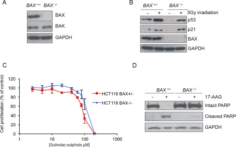
Figure 1. Validation of the isogenic model for BAX knockout in HCT116 human colon cancer cells.
(A) BAX is expressed in HCT116 BAX +/− but not in HCT116 BAX −/− cells. The cells were collected during logarithmic growth and analyzed for the presence of BAX and BAK by immunoblotting. (B) HCT116 BAX +/− and BAX −/− cells both showed induction of the p53 pathway in response to DNA damage. Cells were exposed to 5Gy irradiation and collected 4 hours after exposure. Expression of p53 and p21 was determined by immunoblotting. (C) BAX knockout does not affect sensitivity to sulindac sulfide when measured by 96 hours SRB cell proliferation assay following exposure to increasing concentrations of compound. Data are presented as mean ± SEM, N=3. (D) BAX knockout prevents apoptosis as determined by PARP cleavage in HCT116 BAX +/− and BAX −/− cells exposed to 2.5 × GI50 sulindac sulfide (HCT116 BAX +/− 233μM, HCT116 BAX −/− 273μM as determined by 96 hours SRB assay) or the equivalent concentration of drug vehicle. Cells were harvested after 48 hours and the expression of intact and cleaved PARP analyzed by immunoblotting. GAPDH was included as a loading control in panels A, B and D. Blots are representative of at least two independent experiments.

