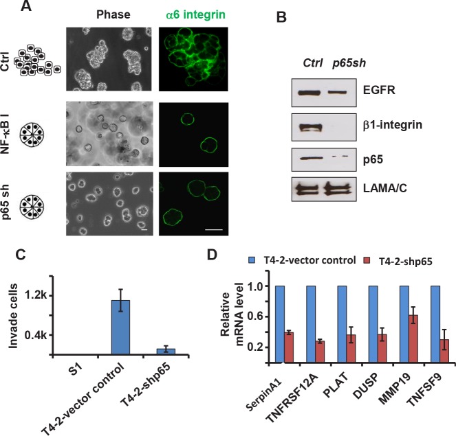
Figure 4. Inhibition of the NFkB pathway reverses the malignant phenotypes of T4-2 cells in 3D culture.
A) Phase and immunofluorescence images of T4-2 cells. Cells were either treated with NFkB inhibitor (Wedelolactone, 10 μM) and cultured in 3D for four days orinfected with p65 shRNA lentivirus before plating in 3D culture. Blocking the NFkB pathway reprograms the cells to form polarized spheroids structure. Green: alpha6-integrin. Scale: 25μm. B) Immunoblotting analysis of beta1-integrin, EGFR and p65 expression in control and p65 shRNA-expressing T4-2 cells. C) Transwell invasion analysis of control and p65 shRNA-expressing T4-2 cells. Graph displays average ± SEM; n=3. D) Quantification of mRNA levels of NFkB target genes by real-time PCR. Control and p65 shRNA-expressing T4-2 cells were cultured in 3D for 4 days before RNA extraction.

