Abstract
Previously, anatomists considered paranasal sinuses as a mysterious region of the human skull. Historically, paranasal sinuses were first identified by ancient Egyptians and later, by Greek physicians. After a long period of no remarkable improvement in the understanding of anatomy during the Middle Ages, anatomists of the Renaissance period-Leonardo da Vinci and Vesalius-made their own contribution. Nathaniel Highmore's name is also associated with the anatomy of paranasal sinuses as he was first to describe the maxillary sinus.
Keywords: Paranasal sinuses, Anatomy, History
The first fundamental principle that medical teachers explain to their students is that, in order to treat an illness, a physician should go back to basics. In other words, to recognize specific pathologic phenomena causing symptoms, physicians should precisely know the manner in which an organism works, namely, its physiological function. However, physiology is inextricably linked to anatomy, that is, the human body structure. Therefore, everything in the art of medicine-from examining a plain radiograph to operating-requires a thorough knowledge of anatomy. An integral part of anatomy, the importance of which, unfortunately, is many times underestimated, is its history. Undoubtedly, nowadays the approach to medicine is becoming more practical, technical, and specialized, and doctors are generally unaware of its evolution over the years. For this reason, all physicians should at least have an overview of the history of the anatomical region they deal with; this will provide them with an intimate knowledge of their science.
The paranasal sinuses are, indubitably, a region of special interest for many medical specialists, including those working in the fields of maxillofacial surgery, otorhinolaryngology, and dentistry. The paranasal sinuses, which are carefully hidden from the eye inside the bones of the skull, have repeatedly puzzled anatomists of the past. Presumably, because they are closely related to vital organs of the human body, such as the brain, eye, nose, and mouth, many peculiar theories about their function have been developed since years. The history of paranasal sinuses, definitely, begins from the history of the words "sinus" and "antrum." The Latin word "sinus" stands for a curve, hollow in land, or a bay or gulf. It can also mean the innermost part of something [1]. Therefore, obviously, the etymology of the word is connected with the structure of the region. The same discipline applies also to the word "antrum," which, combined with Highmore's name-"Highmore's antrum"-is attributed to the maxillary sinus. The word "antrum" derives from the Greek word "άντρον," which means a hollow in land, cave, or grotto, and even a place inhabited by nymphs or other Greek deities, or a place dedicated to them [2].
Paranasal sinuses were first identified inside the bones of the skull by ancient Egyptians. Medical writings dating back from 3700 to 1500 BC provide evidence that Egyptians were familiar with the structure of the maxillary bones [3], which means that they might also have been aware of the maxillary sinuses. According to the Edwin Smith Papyrus of about 1600 BC, ancient Egyptians were also interested in the treatment of nasal injuries and nasal fractures [4]. Nonetheless, the most astonishing testimony is that, while mummifying a human body, Egyptians used special instruments to remove the brain through the nasal cavity, probably removing it through the ethmoid cells [5]. For this reason, ancient Egyptians are considered the pioneers of sinus surgery.
After Egyptians, ancient Greek physicians, like Hippocrates, Galen, and Celsus, may have also recognized the paranasal sinuses as part of the structure of the skull. However, they didn't described them in detail in their works. Specifically, Hippocrates, in his writings, gave instructions on how to treat nasal polyps [6], and he provided detailed information on how to reconstruct nose injuries by repositioning the dislocated bones [4]. Moreover, he indicated that, in the process of producing voice, the air that humans breathe out passes through empty cavities inside the head and echoes [7]. These empty cavities, as referred to by Hippocrates, seem to be the paranasal sinuses. Later, Aulus Celsus, in Book VI and VII of his medical treatise 'De Medicina,' provided a description of the surgical anatomy of the nose and the olfactory nerves passing through the cribriform plate of the ethmoid bone [8].
In contrast to the exploratory mood of ancient physicians and their efforts to base science on proof and experiment, physicians of the Middle Ages were clearly influenced by their adhesion to theological beliefs, which characterized not only the science of the era but social and political life as well. In particular, parts of the human body with unknown function were attributed with intricate and mysterious features and properties, while, in the same way, inexplicable diseases were misinterpreted. A prime example of this would be the functions attributed to the paranasal sinuses. Specifically, there were two basic theories. According to the first one, it was believed that the paranasal sinuses served as cavities containing oils that lubricated the orbit and facilitated eye movements. The second theory has been saved thanks to the Spanish physician Sansovino, who claimed that the paranasal sinuses were called "la cloaca del cerebro" because they were thought to be cavities that drained the brain of its "malignant spirits [9]."
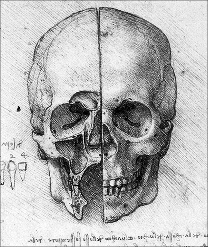
A few years later in history, the brilliant mind of Leonardo da Vinci, the "homo universalis," set new foundations for science, especially medicine. Da Vinci was born in 1452. By combining the skills of a talented artist and a nimble-witted scientist, he created anatomical drawings of the human body, including the skull and the paranasal sinuses, which, however, were discovered many years later by the scientific community [10]. In one of his most well-known drawings, a frontal cross-section of one-half of a human skull is depicted, in which the frontal and maxillary sinuses are represented (Fig. 1) [11]. What is impressive is the fact that he recognized the close relationship of the maxillary sinus with the teeth of the upper jaw. This can be perfectly understood by the accurate representation of the projection of the teeth into the floor of the maxillary sinus [12]. What is more, in the writings accompanying his drawings, Leonardo assumed that the cavity inside the maxillary bone, namely the maxillary sinus, contained a humor whose purpose was to nourish the roots of the teeth [13].
Fig. 1.
Leonardo da Vinci's depiction of a skull. The image is one of da Vinci's anatomical drawings of a human skull. The left half of the skull is sectioned to reveal the frontal sinus and the maxillary sinus. Of note is the close relation of the two sinuses to the orbit and the teeth of the upper jaw, as understood by Leonardo. Reprinted from Leonardo Da Vinci's drawings [11].
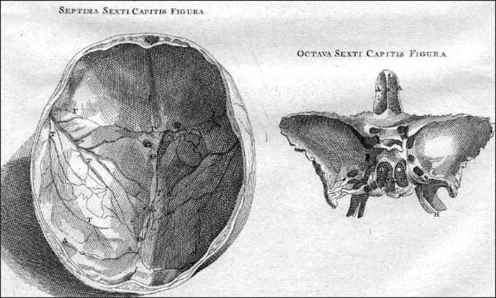
The great anatomist of the Renaissance, Andreas Vesalius (1514-1564), gave an inadequate description of the paranasal sinuses in his work 'De Humani Corporis Fabrica' written in 1543. Moreover, despite the large number of anatomical drawings in his book, Vesalius scarcely provided any illustrations of the paranasal sinuses. Specifically, there is no image of the maxillary sinus, although Vesalius recognized its presence; on the other hand, the frontal sinuses are shown only in a transverse cross-section that depicted the calvaria. Nonetheless, there is a drawing of the sphenoid bone in which the sphenoid sinuses are depicted to be separated by the sphenoid septum (Fig. 2). In terms of the function of the paranasal sinuses, Vesalius stated that these empty cavities reduced the weight of the bone and contributed to the formation of the voice [14, 15].
Fig. 2.
Vesalius' images of the skull and the sphenoid bone. The images constitute anatomical drawings from Vesalius' work 'De Humani Corporis Fabrica.' Most of the illustrations in this book were created by Jan Stephan van Calcar, an Italian artist, one of Titian's students. The left half of the picture shows a transverse cross-section of the skull, depicting the calvaria whereas the right half shows the sphenoid bone. The right-hand image shows the frontal sinus as well as the two sphenoid sinuses, which are separated by the sphenoid septum. Reprinted from Andreas Vesalius, De Humani Corporis Fabrica [13].
Nathaniel Highmore is the anatomist whose name is perhaps most associated with the history of the paranasal sinuses, especially the maxillary sinus. Born in 1613, Highmore, in order to fulfill his dreams, did not follow his family tradition, which dictated that some of the family members should join the clergy. At the age of 17, he entered Queen's College, and the next year Trinity College, Oxford. There, he spent the next 10 years of his life studying science, philosophy, classics, and finally medicine, achieving his medical degree in 1641. Highmore, besides successfully practicing medicine in Sherborne, was also interested in anatomy too [16]. In particular, he had good knowledge of the anatomy of the dog and sheep, and he is known to have dissected an ostrich. Using the experience he had gained from his dissections, he wrote a treatise on anatomy entitled 'Corporis Humani Disquisitio Anatomica,' which was dedicated to his friend Harvey, whom he deeply admired [17]. Highmore died in 1685 and was buried in Purse Caundle near Sherborne. After some research, Dr. Courcy Prideaux found his grave, along with an inscription to his memory, which may be translated as: "Here have been laid (to rest) in hope of the Resurrection to the Life Eternal, the remains of Nathaniel Highmore, Doctor of Medicine, a man of great learning, who died 21st March A.D. 1685, in the 71st year of his age [18]."
Highmore's name is specially connected with the anatomy of the maxillary sinus because it is believed that he was first to describe and draw it. This is why for many years, the maxillary sinus was known as "Highmore's antrum." Even today, the maxillary sinus is referred to as "Highmore's antrum" in medical schools, although Highmore was not actually the first anatomist to discover it. This may be explained by the fact that Leonardo da Vinci's drawings were discovered only in 1901 [9], when Highmore's name had already consolidated itself in the anatomical nomenclature.
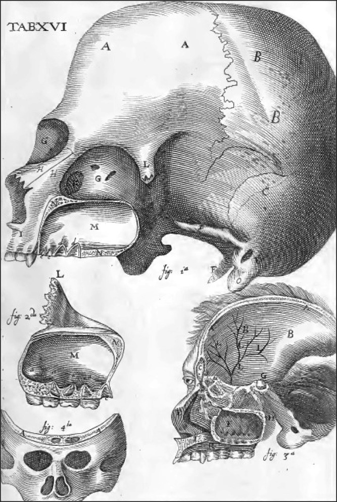
Nonetheless, the contribution of Nathaniel Highmore should not be underestimated. In his treatise, Highmore included anatomical drawings depicting the maxillary sinus as well as the frontal sinus and the ethmoid (Fig. 3) [13]. In the text that accompanies the drawings, Highmore described the close relationship of the maxillary sinus with the orbit and the teeth of the upper jaw, noticing that their roots tend to project into the inside of the sinus. Moreover, he discussed the density of the bone walls and observed that the maxillary sinus was mostly empty and was only occasionally filled with mucus. According to him, this mucus was a humor of the head, which drained into the maxillary sinus. Highmore also supplemented his anatomical text with an eventful story of one of his female patients. Specifically, after the extraction of a canine tooth and frightened of the effusion of pus passing through the slight opening to the maxillary sinus caused by the extraction, the patient tried to identify the source of the pus by pushing a slate pencil through the opening. Alarmed by the view of the pencil disappearing about two inches inside her head, she tried the same with a feather. As it was more flexible, the feather penetrated the opening even further. The patient-terrified that the feather had penetrated her brain-consulted Highmore, who explained to her the presence of the maxillary sinus by showing her his drawings [19].
Fig. 3.
Illustrations from Nathaniel Highmore's book 'Corporis Humani Disquisitio Anatomica.' The maxillary sinus and the projection of the teeth of the upper jaw into the floor of the sinus are clearly shown. The cross-section of the skull in the bottom right of the picture reveals the frontal sinus, sphenoid sinus, ethmoid cells and maxillary sinus. Reprinted from Nathaniel Highmore, Corporis humani disquisitio anatomica, p. 227 [19].
It is remarkable that even Highmore was confused about the function of the maxillary sinus and even more about the origin of the mucus that he had sometimes observed in it. The first to identify that the mucus was not produced by the brain, but was a product of the paranasal structures themselves, was Schneider, in 1660 [9, 13]. Another anatomist who contributed to the understanding of the anatomy of the paranasal sinuses was Emil Zuckerkandl from Austria who described the nose and the paranasal sinuses in great detail in 1870 [9]. Furthermore, at the beginning of the twentieth century, Harris Peyton Mosher of Harvard University dissected many cadavers so that he could study the anatomy of the paranasal sinuses [6]. He is also well known for his accurate anatomical description of the ethmoid sinuses. Noting their close relation to the skull base and the orbit, he claimed that intranasal ethmoidectomy, as a surgical procedure, was "the easiest way to kill a patient" [8, 20].
Since Mosher, our understanding of the histology, embryology, and surgery of the paranasal sinuses has developed exponentially, thanks to an accurate knowledge of their anatomy. To this end, all scientists of the past who improved our understanding of the paranasal sinuses' anatomy made their own contributions, regardless of its importance. Being aware of their felicitous observations, their hypotheses, and even their mistakes, broadens the horizon of a physician's mind and equips him/her with creativity and critical spirit.
References
- 1.Nall GH. Macmillan's elementary Latin-English dictionary. London: Macmillan and Co. Ltd; 1964. [Google Scholar]
- 2.Gazi A. Dictionary of the Greek language. Athens: Mati; 1839. [Google Scholar]
- 3.Formby ML. The maxillary sinus. Proc R Soc Med. 1960;53:163–168. [PMC free article] [PubMed] [Google Scholar]
- 4.Lascaratos JG, Segas JV, Trompoukis CC, Assimakopoulos DA. From the roots of rhinology: the reconstruction of nasal injuries by Hippocrates. Ann Otol Rhinol Laryngol. 2003;112:159–162. doi: 10.1177/000348940311200209. [DOI] [PubMed] [Google Scholar]
- 5.Tange RA. Some historical aspects of the surgical treatment of the infected maxillary sinus. Rhinology. 1991;29:155–162. [PubMed] [Google Scholar]
- 6.Leopold D. A history of rhinology in North America. Otolaryngol Head Neck Surg. 1996;115:283–297. doi: 10.1016/S0194-5998(96)70041-4. [DOI] [PubMed] [Google Scholar]
- 7.Hippocrates . Collected writings. Vol. 4 Athens: Cactus; 1992. [Google Scholar]
- 8.Kaluskar SK. Evolution of rhinology. Indian J Otolaryngol Head Neck Surg. 2008;60:101–105. doi: 10.1007/s12070-008-0050-9. [DOI] [PMC free article] [PubMed] [Google Scholar]
- 9.Nogueira JF, Jr, Hermann DR, Américo Rdos R, Barauna Filho IS, Stamm AE, Pignatari SS. A brief history of otorhinolaryngolgy: otology, laryngology and rhinology. Braz J Otorhinolaryngol. 2007;73:693–703. doi: 10.1016/S1808-8694(15)30132-4. [DOI] [PMC free article] [PubMed] [Google Scholar]
- 10.Tsoucalas G, Gentimi F, Kousoulis AA, Karamanou M, Androutsos G. Joseph Gensoul and the earliest illustrated operations for maxillary sinus carcinoma. Eur Arch Otorhinolaryngol. 2013;270:359–362. doi: 10.1007/s00405-012-2123-3. [DOI] [PubMed] [Google Scholar]
- 11.The drawings of Leonardo da Vinci [Internet] [cited 2013 Dec 23]. Available from: http://www.drawingsofleonardo.org/
- 12.Jose AM. Anatomy and Leonardo da Vinci. Yale J Biol Med. 2001;74:185–195. [PMC free article] [PubMed] [Google Scholar]
- 13.Feldmann H. The maxillary sinus and its illness in the history of rhinology. Images from the history of otorhinolaryngology, highlighted by instruments from the collection of the German Medical History Museum in Ingolstadt. Laryngorhinootologie. 1998;77:587–595. doi: 10.1055/s-2007-997031. [DOI] [PubMed] [Google Scholar]
- 14.Garrison D, Hast M. On the fabric of the human body: an annotated translation of the 1543 and 1555 editions of Andreas Vesalius' De Humani Corporis Fabrica [Internet] Illinois: Northwestern University; 2003. [cited 2013 Apr 28]. Available from: http://vesalius.northwestern.edu/flash.html. [Google Scholar]
- 15.Garrison DH, Hast MH. Andreas Vesalius on the larynx and hyoid bone: an annotated translation from the 1543 and 1555 editions of De humani corporis fabrica. Med Hist. 1993;37:3–36. doi: 10.1017/s0025727300057665. [DOI] [PMC free article] [PubMed] [Google Scholar]
- 16.Wells WA. Nathaniel Highmore, seventeenth century pioneer in anatomy and embryology. Laryngoscope. 1948;58:583–597. doi: 10.1288/00005537-194806000-00009. [DOI] [PubMed] [Google Scholar]
- 17.Stephen L, Lee S. Dictionary of national biography. Vol. 26 New York: Macmillan and Co; 1891. [Google Scholar]
- 18.Prideaux de C. A note on Nathaniel Highmore, M.D. [1613-1685], and his memorial tablet in Purse Caundle Church, Dorset. Proc R Soc Med. 1914;7:106–8. [PMC free article] [PubMed] [Google Scholar]
- 19.Highmore N. Corporis humani disquisitio anatomica. The Hague: Samuel Brown; 1651. [Google Scholar]
- 20.Mosher HP. The anatomy of the sphenoidal sinus and the method of approaching it from the antrum. Laryngoscope. 1903;13:177–214. [Google Scholar]


