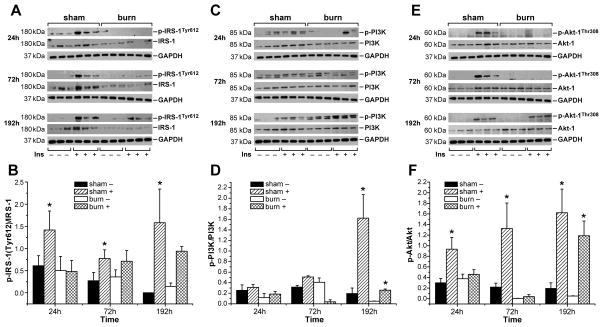
Figure 4. Burn injury is associated with impaired activation of IRS-1 at its tyrosine binding site and inhibits PI3K/Akt signaling after insulin administration.
Accumulation of hepatic phospho-IRS-1(Tyr612)/IRS-1(A) phospho PI3K/PI3K (C) and phospho-Akt/Akt (E) was determined in both, sham burned and burned animals before (-) and 1 min after insulin injection (+) at 24, 72 and 192 h post-burn or post-sham procedure. GAPDH served as control loading protein. Histograms depict intensities of the phosphorylated protein bands divided by the intensity of the total form of the respective protein band (B, D, F). Results shown represent three different animals per group, as indicated in the main text. Bars represent means; error bars correspond to S.E.M. Asterisks denote statistical significance: p < 0.05 for every comparison between groups.

